The hardware method of treating eye diseases rightfully occupies a leading position. Devices for improving vision have become the main attribute of human health. The procedure using such devices allows you to painlessly cope with disorders in just 10-15 procedures. This treatment is based on stimulation of the optic nerves and training of the extraocular muscles.
Complete solutions
NPL Medoptika produces ophthalmological equipment, ophthalmological devices for treatment, prevention and vision testing. Our devices allow you to perform eye training and restore vision for such eye diseases as myopia (nearsightedness), presbyopia (farsightedness), amblyopia (lazy eye syndrome), and strabismus. The Rucheek device (Medoptika TAK-6) is used to train eye accommodation during accommodation spasm and asthenopia (computer eye fatigue). Eye exercises on Rucheyka provide eye stimulation for refractive errors (myopia, astigmatism, hypermetropia) and amblyopia. Using the Rucheek eye simulator is effective for progressive myopia in children (school myopia). Accommodation training on Rucheyka implements the well-known exercise “Mark on glass” to restore vision. The Light Pen device is an amblyotrainer, an eye simulator, in which vision training is carried out according to the “see - circle” principle. The Mosaic kit and the Relief computer program are designed to restore vision with pattern stimulation - a special exercise for the eyes in the treatment of amblyopia. It is important to begin treatment of amblyopia in children as early as possible, accompanying this treatment with independent vision training at home. The Rainbow kit and the Gamma computer program are intended for the treatment of strabismus. Visual exercises are carried out using red-blue glasses to separate the visual fields of the eyes. In Rainbow, coloring of a picture is performed - just like in a biviseo trainer. The active action of the leading hand additionally stimulates the organ of vision, so the Rainbow can be used for both strabismus and amblyopia. In the Gamma program, fusion exercises are performed, as in the synoptophore - the main device for the treatment of strabismus. At the same time, Gamma is also available for independent use. The optotype tables (Album No. 1 and Set No. 4) are intended for vision testing. They include tables of letters, figures, rings, the E sign (observation distances of 1, 3, 4 and 5 meters), texts for near (30 cm) and for the monitor (60 cm). By monitoring visual acuity, you can set the permissible visual load and monitor the dynamics of changes in vision during the treatment of myopia, amblyopia or farsightedness. The devices have contraindications (mainly increased convulsive excitability and disturbances of the vestibular apparatus). Before starting independent use, you should consult an ophthalmologist.
Advantages and essence of hardware treatment
Painless eye procedures using devices make it possible to use this method of improving vision for children.
The advantages of treatment with this method are that the eyes are not exposed to harmful medications and they do not use “grandmother’s methods” at home, but use progressive and tested eye devices that improve vision without harming it. This method consists of carrying out therapy without surgery. The method does not pose a threat to the human body and does not cause pain. A special feature that can be noted is that without any risk to health, hardware treatment is also performed on children. Before prescribing this type of physiotherapy, a diagnosis is carried out, after confirmation of which, this procedure can be carried out to treat the eyes. To make the superiority of this therapy clearer, it is worth citing four arguments:
- Quite fast recovery.
- High percentage of efficiency.
- Safe to use for adults and children.
- Affordable cost of the procedure.
Indications for use
The diagnostic and treatment methods implemented through the Forbis device are indicated for patients with the following disorders:
- Strabismus of accommodative, partially accommodative and non-accommodative types, with close to symmetrical or symmetrical eye position, which is achieved by optical correction or surgery;
- Small or variable strabismus angle no more than 10 degrees according to Hirschberg;
- Unstable binocular vision, asthenopia, diplopia;
- Decompensated heterophoria;
- Violation of accommodation (spasm or weakness in myopia), paresis;
- Nystagmus.
A necessary condition for diagnosing visual functions and treating identified anomalies using the Forbis device is the presence of simultaneous vision and the possibility of bifoveal fusion. In this case, the device is used in the mode of constant illumination of test objects. In order to improve visual acuity, the device is switched to a pulsed illumination mode for test objects. It is advisable to use the same pulsed illumination mode when correcting amblyopia of a squinting eye.
Prevention of myopia
To prevent myopia, it is recommended to adhere to the following rules:
- Correct lighting mode - visual stress only in good lighting, using directional light, a table lamp 60-100 W;
- It is recommended to alternate visual strain with active, active rest;
- Monitoring the condition of the spine, preventing cervical osteochondrosis;
- Gymnastics for the eyes - if accommodation is weakened, after 20-30 minutes of training it is recommended to perform gymnastics for the eyes;
- Rational nutrition enriched with vitamins.
Clinical manifestations and course of the disease
The first signs of myopia appear between the ages of 7 and 12 years and progress until age 20 in women and 22 years in men.
In the future, as a rule, vision stabilizes, but may deteriorate further. As the anterior-posterior axis of the eye increases, the retinal cells located in the zone of maximum light sensitivity are thinned out like the mesh of a stocking that is pulled over the leg, which can lead to other diseases.
For example, age-related macular degeneration usually develops after 55 years, and it joins severe myopia at a much earlier age. It is customary to distinguish three degrees of myopia: weak - up to 3.0 diopters; average - 6.0 diopters; high - over 6.0 diopters.
As the degree of myopia increases, the deficiency of blood supply to the eye increases, which leads to degeneration of the retina, iris, cornea, and the possible early occurrence of cataracts. According to the clinical course, myopia is divided into non-progressive and progressive.
The progression of myopia can occur slowly and end with the completion of growth of the body. Sometimes myopia progresses continuously and reaches high degrees (up to 30 - 40 diopters), accompanied by a number of complications and a significant decrease in vision. This myopia is called malignant myopic disease.
Non-progressive myopia is manifested by decreased distance vision and can be easily corrected. Temporarily progressive myopia also has a favorable course. Constantly progressive myopia is always a serious disease, which is the main cause of disability associated with pathology of the organ of vision.
In some cases, due to a number of reasons, including hereditary predisposition, poor visual hygiene, weakening of the body as a result of poor nutrition, various diseases, overwork, etc., myopia progresses. It is associated with a pathological increase in the length of the eyeball and can lead to deterioration in the nutrition of eye tissue, thinning, dystrophy, tears and detachment of the retina, and opacification of the vitreous body. In this regard, it is very important to notice the first signs in a timely manner and contact an ophthalmologist.
The progression of myopia can lead to irreversible changes both in the central parts of the retina and in the periphery of the fundus. If an ophthalmologist detects peripheral retinal dystrophies, which pose a potential risk of retinal detachment, patients undergo laser coagulation - preventive strengthening of the retina in areas of dystrophy.
It is not difficult to recognize the development of myopia. If you see that your child is squinting or frowning, trying to see something, is forced to bring the text close enough to his eyes, or you yourself feel these manifestations, this is already a sufficient reason to consult a doctor.
Causes
The cause of myopia is a modified shape of the eyeball. It becomes more like an oval than a circle. As a result, a refraction error occurs, causing light rays to pass through the eyeball and focus in front of the retina. This is the reason for the “blurring” of distant objects. In normal vision, light is focused directly on the retina. Myopia can be congenital or acquired (appears during the period of growth of the body).
Based on research from the Moscow Research Institute of Eye Diseases named after. Helmholtz identified two main pathogenetic links of myopia: a discrepancy between the capabilities of the weakened accommodative apparatus of the eyes and visual load and a weakening of the strength properties of the sclera (the outer shell of the eye), and its stretching under the influence of intraocular pressure. According to the first mechanism, a more favorable form of myopia is formed, which remains only an optical defect of the eye; according to the second, myopia is formed as a serious disease, prone to progression and complications.
In the origin of both forms of myopia, the genetic factor is of significant importance, with the first form of myopia being inherited predominantly in an autosomal dominant manner, and the second in an autosomal recessive manner. Hereditary myopia appears somewhat earlier in girls than in boys.
This is apparently due to endocrine changes in the body during puberty. Transient (transitory) myopia can occur during pregnancy, as well as with increased blood sugar levels. The mechanism of development of myopia in childhood can be presented as follows.
For example, while reading, a child tilts his head forward, which puts a lot of stress on the spine, especially if the position and posture are incorrect, resulting in a displacement of the cervical vertebrae and a reflex tension in the neck muscles. This muscle spasm causes disruption of blood flow in the vertebral arteries, which provide “nutrition” to the ocular nerve centers (the so-called oculomotor nuclei of the brain), which “guide” the eye muscles. Weakness of the eye muscles occurs and the focusing (accommodative) ability of the eye is impaired (in addition, paretic strabismus may develop).
With weakened accommodative ability, intense visual work at close range becomes too much for the eyes. In these cases, the body is forced to change the optical system of the eyes in such a way as to adapt it to work at close range without straining accommodation.
This is achieved mainly through a directed moderate lengthening of the anteroposterior axis of the eye during the period of its growth and formation of refraction. Myopia occurs (distance vision deteriorates), which, although somewhat progressive, usually does not reach more than 3.0-4.0 diopters. If a person works for a long time, leaning close to an object, the temporary stretching of the eye becomes permanent, and myopia from functional (temporary, associated with weakness of the focusing muscles) turns into permanent, organic (due to the elongated shape of the eyeball).
Reviews from doctors and buyers
Svetodar has undergone clinical trials that have proven its safety and effectiveness in several leading medical institutions in Russia and has positive reviews from ophthalmologists who used it, including academician, professor Sidorenko E.I., academician, doctor of medical sciences. Dembsky L.K., Ph.D., doctor of the highest category Nikolaeva G.V. and others. You can read their reviews on the website ZDOROVIE-GLAZA.RU
Use of the APEK device for myopia
The use of the APEC device is a highly effective method of treating and preventing myopia in both adults and children. The device is effective for almost any degree of myopia.
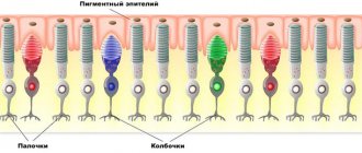
The color flow from the emitters of the device passes through all the media of the eye and hits the retina, causing rhythmic excitation in the retinal neurons, restoring their activity and helping to improve vision.
Orange color radiation improves blood circulation in the choroid of the eye and metabolic processes in the retina, and blue photostimulation relieves swelling and inflammation in the retina and prevents the progression of myopia.
Green color normalizes intraocular pressure, relieves eye fatigue, and increases visual acuity. Point sources of radiation shifted to the periphery with a pulse of complex shape - smoothly increasing and quickly decaying, with a sequential effect on both eyes, trigger the muscular system of the eyes, training the accommodative apparatus in a favorable manner, contributing to the restoration and consolidation of the normal shape of the eyeball.
Intrapulse modulation with a frequency corresponding to the alpha rhythm of the brain restores the disturbed rhythm of the visual analyzer, eliminates the imbalance between the hemispheres of the brain, as if erasing the negative program, which helps to normalize the psycho-emotional state.
Pulsed photostimulation of the retina with orange, green and blue colors helps strengthen it and improve the trophism (nutrition) of the internal structures of the eye, which helps slow the progression of myopia and prevent the development of complications and loss of vision.
