White spots in the eyes usually appear against the background of various pathological changes in the structures of the visual organs. This phenomenon may be a consequence of various diseases.
So, ophthalmological diseases have a disappointing tendency towards rejuvenation - now almost a third of all patients with impaired vision are preschool children and adolescents. The rest are represented by middle-aged and elderly people. The high-risk group includes those who have congenital eye diseases, engage in specific work, and are regularly exposed to the harmful effects of toxic fumes, gases and chemical compounds.
You may be interested in:How to use contact lenses: selection options, wearing rules and care
People often call a white dot in the eye a thorn. This pathology has long been surrounded by all sorts of myths with mystical origins. However, in reality, the defect often leads to deterioration of vision, even to complete blindness. Especially when it comes to a white dot in the pupil of the eye. This pathology poses a significant danger not only to vision, but to the entire health of the visual system.
Causes of a white spot in the eye
There may be several prerequisites for the development of such a problem. To identify them, it is necessary to undergo an ophthalmological examination.
The localization of the white spot in the eye can be different: the pupils, cornea, vitreous body and other structures. There are not so many reasons for the occurrence of pathology. Taking into account the location, it is possible to determine the presence of predisposing factors, which often relate to the functioning of the brain, heart, blood vessels, and nervous system.
You may be interested in: Stye begins on the eye: what to do, how to treat it
Treating eyesores
Small cloud-like opacities and defects in the periphery do not require surgical intervention, but they need to be monitored over time.
Conservative therapy is indicated to stabilize the condition and includes:
instillation of Dionin in increasing concentrations during the initial stages of the disease);- corticosteroids: Hydrocortisone ointment, Dexamethasone eye drops, etc.;
- vitamins B12, A, C, E;
- physiotherapy: electrophoresis with potassium iodide, Papain, Lidase, Aloe extract or Vitreous body, course intramuscular injections are possible;
- for ulceration - ointments that enhance reparation processes, for example, with Solcoseryl, Actovegin, etc.
- drugs for the prevention of secondary glaucoma (Pilocarpine hydrochloride);
- pathogenetic therapy (depending on the etiological factor: antituberculosis, antiviral, antibiotics, etc.).
- diet: an excess of fatty foods under certain conditions can lead to an increase in cholesterol levels and its deposition on the scar tissue of the cataract, which will aggravate the condition. On the contrary, food containing large amounts of vitamins A, , , B, gives the body strength to fight the disease.
Surgical treatment of ocular leukoma
Keratoplasty is an operation that is performed if conservative therapy is not indicated and the patient’s quality of life suffers. Using a laser, the affected segment is excised layer by layer, followed by rehabilitation therapy.
, corneal keratoprosthesis helps restore vision in case of leukoma . The biomaterial is removed from a deceased donor, and keratoprosthesis with an artificial corneal implant is also possible.
note
There are special banks in which ophthalmic transplants are stored. All of them have been tested for infections and are safe for humans.
Progress of the operation:
- Under anesthesia, the affected area of the cornea is removed using a special laser.
- A donor graft is placed in its place and secured with sutures.
- Full or partial prosthetics are possible, which is decided on an individual basis.
- After the operation, all physical activity is excluded, the sutures are removed after 6 months, and final scarring occurs after 12 months.
- Hospitalization takes 24-48 hours.
The precision of the excimer laser avoids bleeding, astigmatism and keratoconus.
Goals of surgery for eyesores:
- increasing visual acuity by restoring the shape and transparency of the cornea;
- slowing down the progression of pathology in the future.
If visual acuity is high enough (0.2-0.4), you should refrain from surgery, since there remains a danger of clouding of the graft due to incompatibility and loss of vision.
A cataract complicated by glaucoma or impaired light perception is a contraindication for corneal transplantation.
The prognosis for recovery after surgery is positive; for leukomas caused by burns or extensive trauma, it is doubtful.
Mishina Victoria, doctor, medical columnist
33, total, today
( 62 votes, average: 4.13 out of 5)
Cataract: symptoms, diagnosis, treatment and prevention
Angular conjunctivitis - inflammation of the mucous membrane of the eyes
Related Posts
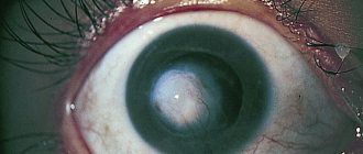
Leukoma
In its normal state, the cornea has a convex shape and an absolutely transparent texture. A disease such as leukoma leads to the modification of healthy tissue into connective tissue. The area with the transformed tissue ceases to function and changes fibrously, which leads to the appearance of a shapeless scar.
Leukoma is a white spot, sometimes with a milky tint, that is located on the surface of the eye. The closer the growth is to the pupil, the faster vision declines. With abnormal tissue transformation, constant growth of scars is observed.
Cause
There are main reasons that can cause formation on the inside of the eyelid:
- Mechanical damage to the mucous membrane. These could be scratches, cuts, blows. As a result of these actions, fluid accumulates inside the cavity of the mucous membrane. A small pimple forms, which may resolve over time. If this does not happen, the doctor will remove it painlessly.
- Allergy. Most often, this condition occurs when using low-quality cosmetics. The condition is easily eliminated with the help of antihistamines and the abolition of the cause that caused it.
- Infection. It may be the result of a complication from a viral disease or due to the introduction of multiplying bacteria during poor-quality cosmetic procedures in the eye area.
- Gastrointestinal disease. Many digestive tract problems are accompanied by a rash on the body. One of the places for such formation is the inner side of the eyelids.
- Inflammation, irritation. Such a reaction can be caused by negative environmental factors that affect the sensitive area of the mucous membrane of the eyes and eyelids. Such factors include smoke, smog, and chemicals.
- Hormonal fluctuations. After adolescence or menopause, the amount of hormones in the human body changes dramatically. This causes pimples to form on various parts of the body.
- Aging process. If a person is over 60 years old, inevitable aging of the conjunctiva, cornea, and eyelid skin occurs. Pimples filled with clear fluid may form.
The doctor must identify the cause of the condition in order to know whether to provide treatment or not. Some pimples resolve on their own over time. If they are caused by a pathological process, treatment is required.
Floaters before eyes
This phenomenon is a consequence of destruction in the structures of the vitreous body. In its normal state, it has a transparent texture and jelly-like consistency.
The vitreous body is located throughout the eye cavity, helps maintain a healthy spherical shape, and controls the elasticity of muscle fibers. Often the disease is associated with existing vascular pathologies. In this case, the colorless structure of the vitreous body changes to connective tissue and gradually becomes cloudy.
The main causes of the defect include the following factors:
- neck osteochondrosis;
- excessive physical activity;
- dystonia of vegetative-vascular origin;
- atherosclerotic processes;
- hypertension;
- avitaminosis.
Retinal detachments, traumatic brain injuries, eye injuries, and hemorrhages are all conditions that can cause floaters and whiteheads to appear on the surface of the cornea.
Causes and symptoms
When transparent, colored or dark spots appear before your eyes, interfering with your normal life, it is worth thinking about the possible root cause. Sometimes defects go away on their own, since their occurrence is associated with a temporary phenomenon. But often the cause is unpleasant diseases that require mandatory treatment. Therefore, it is extremely important to see a doctor.
Possible causes of stains:
- eye disease - the problem may be associated with damage to the vitreous body, cataracts, abnormalities of the ocular vessels, inflammation or infections;
- mechanical damage to the eyes - the defect is caused by light contact with a foreign object or dust particles, sometimes it develops against the background of a blow to the head;
- problems with the brain - the spot appears due to impaired cerebral circulation, traumatic brain injury, or brain surgery;
- deterioration of the cardiovascular system - low or high blood pressure, anemia and vascular disorders often lead to visual defects;
- disturbances in the functioning of the body - one of the reasons are pathologies of the liver or gastrointestinal tract, diabetes mellitus, vitamin deficiency, osteochondrosis and metabolic disorders;
- disruptions in the nervous system - often spots appear in those who have developed VSD disease associated with stress, overwork and lack of sleep;
- overwork – prolonged eye strain or physical exhaustion can cause spots or spots to appear;
- poisoning – the cause may be poisoning from low-quality products or coming out of a long binge;
- pregnancy - changes in the body during pregnancy lead to malfunctions of many functions and cause a number of minor disorders, including with the eyes.
Some spots are benign growths that do not require any intervention, or are the result of an allergic reaction. In such cases, treatment is not even required.
The symptoms of most defects are identical. A spot in the eye often interferes with looking at objects, causes blurred vision, contributes to decreased vision and can lead to loss of certain visual areas. It can move when you blink or move your eyes. At the same time, other symptoms cannot be ruled out.
The spots are often accompanied by special manifestations:
- inflammation of the eyelids;
- discharge from the eyes;
- increased tearfulness;
- itching sensation in the eyes;
- sensitivity to light;
- dizziness, headache.
Symptoms from the underlying disease that caused the spot may also occur. Sometimes the eye can hurt, and painkillers do not always give the desired effect. This often happens when it is damaged due to serious pathology.
Abnormal changes in the lens
The appearance of white spots in the eyes is often a harbinger of cataracts. Lens opacities range from cream to deep gray. Cataracts can be congenital or acquired. Most often, the pathology is diagnosed in older people. The problem can be eliminated conservatively and surgically. If the condition is advanced, the patient is scheduled for surgery, during which the damaged lens is removed and an intraocular lens is implanted.
Restructuring in corneal structures
A white spot on the cornea of the eye may not affect a person’s visual capabilities in any way. The normal transparent structure of the shell is replaced by cloudy tissue. Pathology can be generalized or local. With a pronounced abnormal process, vision gradually deteriorates.
Cloudiness of the cornea can be caused by many factors:
- syphilis;
- chronic form of conjunctivitis;
- keratitis;
- infectious diseases;
- tuberculosis of any type.
Any eye pathology of an inflammatory nature can lead to the appearance of white spots. The influence of toxins, chemical and thermal burns, various injuries - all this may well provoke the appearance of a cataract.
1.Pigmentation of the cornea
Pigmentation of the cornea of the eye (nevus)
It differs from a regular birthmark only in that it is located on the eyeball. Just like on the body, a mole on the eye can appear at any age and change in size and color throughout life. However, most often a child is born with a small pigmented spot on the iris of the eye. This phenomenon is asymmetrical. The spot may be round or have the shape of a sector with a center in the middle of the pupil; the mole is located on the cornea or on the white of the eye. Acquired pigment spots on the cornea are usually associated with changes in hormonal levels.
The pigment melanin is responsible for the color of the eyes, just like on the skin.
. The color of birthmarks in the eye can be brown, yellowish, black, or pink. It has been noticed that fair-skinned and fair-haired people are more likely to have eye moles.
Most often, pigmentation of the cornea of the eye is not dangerous
. However, you need to monitor and consult a doctor if significant changes occur in pigmentation in a short period of time. This may be a sign of degeneration of a benign formation into melanoma of the eye.
Sign up for a consultation
A must read! Help with treatment and hospitalization!
Retinal modification
White spots in the eye on the iris form due to insufficient blood supply to the tissues. Doctors call this condition retinal angiopathy. Pathology develops due to:
- any injuries - mechanical, chemical, thermal;
- vascular atherosclerosis;
- hypertension.
In addition, the defect can occur against the background of diabetes, hypotension and other diseases. Adverse habits can also lead to a deterioration in the blood supply to the eye. Along with the appearance of white spots in the eye, patients may complain of pain and blurred vision.
What is a white spot on the retina
Retina (fundus of the eye)
- This is the inner lining of the eyeball. It is part of the nervous system and is the first section of the visual analyzer. In the retina, light energy is converted into nerve impulses and the primary analysis of visual information occurs. The retina has a mesh structure, hence the name retina.
There are a large number of hereditary and acquired diseases and disorders that may involve the retina.
Some of them include:
Retinal pigmentary degeneration is a hereditary disease affecting the retina, resulting in loss of peripheral vision.
Macular dystrophy is a group of diseases characterized by loss of central vision due to death or damage to macular cells.
Rod-cone dystrophy is a group of diseases in which vision loss is caused by damage to the photoreceptor cells of the retina.
When the retina is detached, the latter is separated from the back wall of the eyeball.
Arterial hypertension and diabetes mellitus can cause damage to the capillaries supplying the retina with blood, leading to the development of hypertensive or diabetic retinopathy.
Retinoblastoma is a malignant tumor of the retina.
Macular degeneration is a pathology of blood vessels and a disturbance in the nutrition of the central zone of the retina.
RETINA ANOMALIES
Retinoblastoma
is a malignant tumor of the retina. This tumor occurs in the first months or first years of a child's life. This tumor is hereditary. In a quarter of cases, the tumor occurs in both eyes at once.
Retinoblastoma develops from remnants of embryonic retinal cells.
The tumor grows quickly, and after a few months it occupies the entire orbit. Blindness occurs quickly. One of the first signs of a tumor may be a specific yellow glow in the pupil. It is called "amaurotic or blind cat's eye." A tuberous protrusion of a yellowish-golden color is found in the fundus. Often the tumor grows into the sclera, periorbital fatty tissue, brain and very quickly metastasizes to other organs. Most often in the bones, liver, lungs.
Other manifestations of the problem
In addition to the white spot in the eye, patients may also complain of blurred vision, blurred vision, which is caused by impaired refraction of rays. In addition, a person with this problem may experience excessive lacrimation, a feeling of the presence of a foreign object, and conjunctival hyperemia.
If the white dot is localized in the center of the eye, visual acuity decreases.
Diagnostics
Which doctor should you contact if white spots appear in the eye? The first thing you need to do is go to an ophthalmologist. The doctor will conduct several clinical studies, starting with studying the symptoms, interviewing the patient and collecting the necessary medical history.
To identify the causes of the appearance of a white spot and prescribe further treatment at the New Look clinic, patients are offered a number of examinations:
- assessment of eyeball refraction;
- determination of the visual field;
- Ultrasound of the fundus;
- measurement of intraocular pressure;
- assessment of the condition of the ocular vessels;
- a thorough examination of the eyeball using a microscope;
- measuring the depth of corneal structures.
Among other things, it is recommended to diagnose hidden diseases, as well as determine the general condition of the visual system. Completing all stages of the examination is necessary to prescribe appropriate therapy and exclude other pathologies of internal organs.
How to treat
If a white dot that appears in the eye does not provoke obvious deterioration in vision, then special therapy is usually not prescribed. In general, treatment tactics are selected taking into account the reasons that provoked the development of the pathology. Specialists at the New Look clinic offer several ways to solve the diagnosed problem:
- for cataracts and destructive changes in the cornea, surgical intervention is most often recommended;
- for inflammatory processes, special anti-inflammatory drugs are prescribed in the form of eye drops;
- when connective scar tissue forms, absorbable drugs are prescribed, for example, Hypromellose, Actovegin, Korneregel.
In this medical center, surgical correction is performed using professional modern equipment. Today, operations are quite affordable and have a short recovery period, which is very convenient.
So you should not try to treat your eyes with all sorts of alternative means, as well as various drops, without an established diagnosis. Before starting therapy, you should consult an ophthalmologist.
Eleven eye symptoms that indicate health problems
If you wear contact lenses, an eye test can help determine the significance of a few white spots on the cornea (the clear layer at the front of the eyeball). They can be a signal of infection of the cornea, which happens when it is damaged by a foreign object.
If the infection is accompanied by pain and inflammation, see a doctor immediately. Just don't worry, this is a very common occurrence. Most likely, you will just have to stop using contact lenses for a few days, writes headinsider.net
If you notice that your eyes continue to twitch despite your best efforts, give yourself time to calm down. Diagnostics of the eyes suggests that severe stress is to blame. Run a bath, read a book, and head to bed early.
Fog before the eyes may indicate poor vision. But if the cloudiness doesn't go away, it could signal a more serious problem, such as diabetes. 73% of diabetic patients in a study by the American Association of Clinical Endocrinologists said they had experienced blurred vision at one time or another. These eye symptoms may also be an early warning sign of other eye diseases, such as cataracts or macular degeneration.
Most often, it is in older people that a white ring may appear around the iris of the eye. It may indicate high levels of cholesterol and triglycerides in the body. If you have such a ring, you should be aware of your increased risk of heart attack or stroke. If you have a white ring, consult your eye doctor or your healthcare provider. Eye diagnostics alone will not help here.
Persistent dryness of the eyes may indicate dry eye syndrome. This disease is caused by a lack of moisture in the eyes. You can buy over-the-counter moisturizing drops or see your doctor for a prescription. Dry eyes can also indicate an allergy.
If you suddenly notice a yellow spot or bulge in the corner of your eye, you may be spending too much time in the sun. This spot is called a pinguecula and it develops in the same way as other side effects of negative sun exposure. Rush to your doctor's appointment and buy sunglasses with ultra-high UV protection.
If your eye whites turn yellow, it can be scary at first. Most likely, this is due to the so-called bile spillage. When your liver is unable to cleanse the cells, a yellow component called bilirubin accumulates, causing yellow eyes.
Your skin may also take on a yellow tint. Bile spillage may indicate the presence of an infection in the liver, and eye diagnostics alone are not enough in this case. It is better to consult a doctor immediately.
Do you work on your laptop all day? Do you watch a lot of TV? Do you spend a lot of time on Viber? If you don't take your eyes off your screens for a long time, they may start to water. These eye symptoms are common and you just need to put your phone aside for a while.
If you notice a rupture of blood vessels in the eye, there is no need to sound the alarm. These eye symptoms are common among people whose eyes are under constant strain. Sometimes blood red eyes accompany a cold. For example, when you cough a lot or stay up late at night.
Do you know the feeling when suddenly some small particles move around you? Most likely yes. After all, this is an absolutely common phenomenon that happens to everyone from time to time. However, when you see these particles all the time, you should start to worry - this could indicate a retinal tear or even retinal detachment.
If your eyes are swollen and red, there is no itching or irritation, then most likely you are just very tired and need to get some sleep.
Author: headinsider.net
Prevention
The main measure to prevent the occurrence of white spots in the eyes is to generally strengthen the retinal tissue. To do this, you should systematically take multivitamin complexes, lead a healthy lifestyle and visit an ophthalmologist every year for a preventive examination. It is especially important to follow these rules for those who already have a history of chronic eye pathologies.
In fact, the health of the visual organs is often in the hands of the patient himself. If the pathology has already appeared, then you should not delay visiting a doctor - only he can carry out the necessary diagnostics and prescribe appropriate therapy.
Source