Papilledema is caused by increased intraocular pressure. Swelling on both sides is more common. It lasts up to two weeks. Unilateral swelling is rare. The appearance of swelling under one eye is associated with a disease of this particular organ. The pathology needs to be seriously treated. Treatment is preceded by diagnosis. It allows you to find out the causes of the phenomenon. Untreated papilledema leads to vision loss.
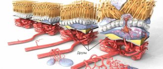
Anatomy of the optic nerve
The ON is the second pair of cranial nerves that are characterized by visual stimulation. It consists of 4 sections - intracranial, intracanalicular, intraocular and intraorbital.
The optic disc is the optic disc; it represents the junction of optical fibers. Its length is 1 mm, diameter is 1.75–2 mm. The optic disc is located in the nasal part of the fundus of the eye.
The second pair of cranial nerves is surrounded by three meninges. Its thickness is 3–3.5 mm, length — 3.5–5.5 cm. GN fibers vary in direction and caliber, there are thin and thick. The latter transmit light stimulation to the visual part of the cortex, the former are reflex, they are necessary for transmitting the light impulse to the parasympathetic nervous system.
Causes of papilledema
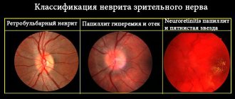
Neuritis
The cause and origin are largely unknown, but doctors are considering multiple sclerosis. Affected patients suffer from blurred vision, color saturation (especially red), and painful eye movement.
Symptoms are transitional, that is, they usually form independently. If the posterior part of the optic nerve is affected (retrobulbar neuritis), then inflammation and swelling of the optic nerve head may not be diagnosed during fundus examination. Other imaging methods are needed.
Optic neuropathy
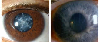
In the event of acute closure of the blood vessels of the eye, the ON is irreversibly damaged; without the supply of oxygen and nutrients through the blood, the ON neurons die. Because these cells cannot be re-formed, vision gradually decreases to the point of blindness. Upon examination, the ophthalmologist sees a pale, swollen optic nerve-papilla and its head, and slight bleeding. The patient notices severe visual impairment and limited field of vision.
Atherosclerosis or embolism due to heart disease (atrial fibrillation/endocarditis) are the most common causes. Arteritis of the temporal artery, autoimmune cystitis, causes optic neuropathy.
Pathologies of internal organs
Some diseases of internal organs lead to the development of pathology. For example, stroke, hypertension and kidney failure.
Each condition is characterized by certain signs; they are easy to distinguish when carrying out appropriate diagnostics.
Arteries of the temporal artery
This is an autoimmune disease leading to vascular inflammation (vasculitis). In approximately 30% of cases, the ophthalmic arteries are affected, which leads to weakened vision up to blindness.
The patient often suffers from severe headaches and has difficulty chewing food. On physical examination, the temporal artery is tender. If the eyes are affected, the specialist sees signs of a swollen optic disc.
Glaucoma
Glaucoma is a consequence of pathologically increased pressure in the eyes. It damages the optic nerve, or rather the nipple. The patient notices blurred vision and loss of the visual field.
Optic atrophy
Optic nerve atrophy indicates irreversible damage to optic nerve tissue. The primary form, caused by a congenital malfunction, differs from the secondary one. In the latter case, the cause is other diseases or mechanical factors. Possible factors:
- strong pressure on the optic nerve;
- circulatory disorders;
- inflammation;
- intoxication.
These conditions are characterized by swelling of the optic head, defects in the field of visual perception and deterioration of acuity.
Other reasons
Edema of the optic nerve develops due to increased pressure inside the skull associated with purulent abscesses in the cranial space, internal bleeding, and head injury. The cause of optic disc edema is craniosynostosis and meningitis, encephalitis.
Preventive measures
It is quite difficult to prevent the appearance of papilledema, since the pathological process develops against the background of diseases and disorders, including those that a person is not able to prevent. To prevent congestion in the brain, it is recommended to prevent the activity of bacterial microflora and parasites, regardless of the location of the latter.
When treating inflammatory pathologies, it is recommended to strictly follow medical prescriptions and avoid overdose of medications. This is especially true when antibacterial medications are used. It is also important not to interrupt treatment ahead of schedule, even if the symptoms of the disease do not bother you for several days.
For early diagnosis of edema, it is recommended to undergo an ophthalmological examination every 6 or 12 months. The disease occurs suddenly and in the initial stages of development does not provoke pronounced symptoms.
Despite the fact that there are no specific methods for preventing this disorder, the measures described above help reduce the risks of developing DLD.
The pathology develops against the background of increased intracranial pressure, which is caused by infections, inflammatory and other ailments. In case of PVD, the use of corticosteroids and diuretics is indicated. In advanced cases, the deviation is treated with surgery by shunting the affected optic nerves.
Classification
Papilledema can be bilateral or unilateral. In the first case, both organs of the optical system are affected, in the second - only one.
With unilateral swelling of the optic nerve, the symptoms are mild; even experienced ophthalmologists do not always notice the swelling.
The disease has an acute and chronic nature. In the acute course, the development of the clinical picture is spontaneous, symptoms appear within 2–3 hours.
With repeated relapses, it takes up to 3 days for the first signs to develop.
What is swelling?
Attention! The disease “papilledema” is not associated with age-related changes and can develop in both children and adult patients suffering from increased intracranial pressure.
However, such a pathology cannot occur in newborn children, since due to the mobility of the skull bones, intracranial pressure does not yet exist at this age.
The nerve tissue itself is the element that transmits information from the visual centers of the eye to the brain.
intracerebral fluid (CSF) begins to accumulate in the area where it occurs , the pressure begins to increase.
There is compression of the area, which loses the ability to perform its functions , resulting in a deterioration in the quality of vision.
Symptoms
The clinical picture is characterized by a gradual decrease in visual acuity. It worsens especially strongly in the central part of the visual field.
Periodically, vision becomes blurred, color perception is impaired, veins are moderately tense and there are no lesions.
Damage to nerve fibers leads to double vision, headaches, flickering before the eyes, nausea and vomiting.
Further progression of the pathological condition leads to the formation of scotomas in the field of view. The development of secondary atrophy of the optic nerve is possible.
Diagnostics
The initial examination is carried out using an ophthalmoscope. This is a visual assessment of the condition of the optic nerve head, retinal arteries and veins. Be sure to carry out the reaction of the pupil to light using a small lamp. As a rule, both pupils constrict equally strongly, regardless of which organ of vision the doctor directs the light cone at.
However, with retrobulbar neuritis, a so-called relative afferent pupillary defect is often observed.
This means that the ON of the affected eye is not as good at directing incoming light signals to the brain as the other ON. As a result, the pupils constrict less when the doctor shines a light on the affected eye. For a more detailed examination of the nerve fibers, special drops are used that dilate the pupils.
If necessary, additional research is carried out. With their help, they find out what reason led to swelling of the optic nerve. To diagnose multiple sclerosis, an MRI of the head and spine is performed.
A lumbar puncture is performed to collect cerebrospinal fluid. It is checked for signs of inflammation.
Treatment methods
Since swelling is a secondary pathology that occurs as a result of impaired circulation of cerebrospinal fluid, treatment of the pathology is aimed at stabilizing intracranial pressure. The most optimal therapy is determined by the ophthalmologist, based on the diagnostic results and the general condition of the patient.
Medicines
Swelling of the ocular nerve endings is indicated to be treated with the following medications:
- corticosteroid drugs (Methylprednisolone and Prednisolone) stop the inflammatory process;
- diuretics (Furosemide, Acetazolamide) stabilize the circulation of the spinal substance.
Antibacterial and antihistamines are used as auxiliary medications. The duration of drug therapy depends on the degree of damage to the eyeball: it is prohibited to independently adjust the duration of the treatment course.
Operation
Treatment of advanced swelling surgically is carried out in two ways - by performing fenestration of the eye disc or bypass surgery.
- Fenestration is based on the formation of small holes in the disc structure, through which the doctor eliminates excess cerebrospinal fluid.
- The bypass procedure involves creating a special tube from several shunts connecting the abdominal cavity and the spinal cord in order to ensure the outflow of accumulated spinal matter. Shunting as a treatment method is advisable to use if nerve swelling is a consequence of the development of a benign tumor.
Treatment
If retrobulbar neuritis is present, drugs that suppress the immune system (immunosuppressants) are used. Glucocorticoids (steroids) such as Cortisone or Methylprednisolone are given. The first five days the drugs are used in high doses, then the dosage is slowly reduced over the next 2 weeks.
Before carrying out therapy with Cortisone, it is necessary to exclude diseases such as tuberculosis, stomach ulcers, diabetes mellitus or hypertension. They may worsen with glucocorticoid treatment. If a bacterial infection is causing papilledema, antibiotics may be used to treat it.
Steroids may also be helpful in stopping this immune response.
Therapy is supplemented with diuretics. They normalize the balance of cerebrospinal fluid. In severe cases, surgery is indicated.
Causes
The reasons for the development of such a pathology can be a number of diseases that have a direct impact on it:
- craniosynostosis (fraternal deformation of the cranial bones);
- any cancer in the brain area ;
- hydrocephalus;
- internal bleeding and swelling of the brain;
- abscesses inside the skull , accompanied by the discharge of pus;
- infectious diseases of the brain , which can spread to the optic nerve (encephalitis, meningitis);
- glaucoma.
Sometimes such a disorder develops against the background of renal failure, hypertension, lymphoma, leukemia or stroke.