A person’s ability to simultaneously see with both eyes and obtain a three-dimensional view of surrounding objects is called binocular vision. If this ability is impaired, it affects the sharpness, clarity and other features of visual perception. Special exercises will help restore binocular vision in adults.
In this article
- How does binocular vision work?
- Is it possible to restore binocular vision?
- How to restore binocular vision at home?
- Effective exercises to restore binocular vision
- Simple exercises at home
- Effective Focusing Exercises
- Looking at stereograms is useful for binocular vision
What is binocular vision?
Binocular vision is one of the most important functions of the visual apparatus. It begins to form in children almost immediately after birth and develops until the age of 12-14 years. A person with stereoscopic vision perceives the world in 3D format, that is, he can not only see the shapes and outlines of objects, their size in width and height, but also approximately determine the distance between them.
Lack of binocular vision leads to many problems. It is difficult for a person to distinguish the distance between objects and navigate in space. In everyday life, this manifests itself in difficulties when trying to pour water into a glass or thread a thread into the eye of a needle. In professional life, problems arise with employment and choice of profession. Thus, impaired binocular vision is a serious limitation for becoming a pilot or driver.
Binocularity disorders can appear at any age and for various reasons. Before we look at these reasons, we need to know how binocular vision works.
Mechanism and conditions for binocularity
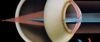
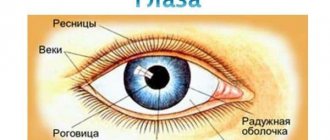
If a person can combine two images from both retinas into one picture, he has developed binocular vision. This unification occurs in the cerebral cortex and is ensured by the fusion reflex. To do this, it is necessary that the brain receives two absolutely identical pictures from both eyes, that is, they must match each other in size and shape. For spatial vision to function, light rays must fall on identical points on the retina. These points are called corresponding points. Each point on one retina has a corresponding point on the other retina. If light falls on them, the pictures come together, as if superimposed on each other. If focusing occurs at different points, then the pictures are different. This leads to diplopia - double image.
Vision will be binocular if:
- there is the ability for fusion (merging images in the brain);
- the eyeballs move in concert and are located symmetrically and parallel in the same frontal and horizontal plane;
- visual acuity is sufficient for normal visual perception of the environment (not lower than 0.3-0.4 diopters);
- there is no aniseikonia, when the eyes see images of different sizes;
- there are no opacities of the cornea, lens or vitreous body, which are accompanied by a decrease in visual acuity in one eye;
- there are no pathologies of the central nervous system.
There are many conditions for the formation of stereoscopic vision. Moreover, the above factors apply not only to the organs of vision, but also to other systems of the body. Lack of binocularity can be a symptom of both eye diseases and non-ophthalmological pathologies.
Four-point color test - an option for home vision testing
Typically, a four-point color test is carried out in a clinical setting using a special apparatus. The operating principle of the device is based on separating the visual fields of the right and left eyes using multi-colored filters. The patient is put on special glasses: a red filter is placed in front of the right eye, and a green filter is placed in front of the left eye. After this, the patient looks into the holes of the device. Depending on the color of the filters, the doctor makes a conclusion about the presence of binocular, monocular or simultaneous vision.
You can conduct a similar test at home if you have red-green glasses. To carry out the test you will need red, green and white items. Place them one to five meters from your eyes and look at them through glasses with colored lenses. If binocular vision is not impaired, colored objects will have the color they originally had. A white object in your perception will become half red and half green. With monocular vision, all objects are “colored” in the color of the lens of the dominant eye.
Binocular vision disorders and eye diseases
Almost all pathologies of the visual organs can be accompanied by impaired binocularity. Binocular vision is absent when:
- anisometropia (different refraction);
- damage to the oculomotor and other eye muscles, which lead to inconsistency in the movements of the eyeballs;
- infectious eye diseases;
- malignant and benign eye tumors;
- mechanical injuries to the visual organs, including burns, which lead to displacement of the eyeballs;
- pathologies of bones and orbital cavity;
- cataracts;
- retinal diseases (hemorrhages, rupture).
Other causes of binocular impairment:
- infectious diseases of the brain;
- poisonings in which the brain stem is damaged;
- tumors in the brain;
- problems with the activity of the nervous system.
This is a list of a few diseases in which binocular vision is absent. Other pathologies of the body can also negatively affect visual functions.
Friendly form of strabismus
Concomitant strabismus is the most common type of binocular vision disorder in children. It occurs due to the following reasons:
- neurological disorders;
- adverse effects on the fetus during the prenatal period;
- chromosomal abnormalities;
- acquired farsightedness or myopia;
- decreased visual acuity in one eye;
- heterophoria (different strength of the muscles of the left and right eyes);
- complications after infectious pathologies.
With the concomitant form of strabismus, the patient has changes in only one of the organs of vision. In this case, the movements of the eye muscles are not impaired, and the angles of deviation from the visual axis are the same. This means that if one eye squints by 5 degrees, then the other one deviates by the same amount.
Concomitant strabismus often looks like a purely external defect and does not cause any particular inconvenience to the patient. This form of strabismus is not accompanied by double vision. However, over time, strabismus can lead to decreased visual acuity. To see any object, a person has to squint and strain his eyes. This leads to visual fatigue and headaches. Therefore, concomitant strabismus must be treated in childhood. Binocular vision disorders in adults are much more difficult to correct.
Treatment of binocular vision disorders
Lack of spatial vision is not an independent pathology. This is a sign of another disease that needs to be treated. After all symptoms of the disease are eliminated, binocular vision is restored. Thus, anisometropia is treated with surgery. You can also correct this visual pathology with glasses or contact lenses.
To restore stereoscopic function, you must first accurately identify the cause of its absence. This can only be done through a thorough examination. In this case, you may have to see not only an ophthalmologist, but also other specialists.
The most common pathology in which normal binocular vision is absent is strabismus (strabismus). With this disease, the movements of the eyeballs are not coordinated. In other words, the eyes look in different directions. One of them may completely disappear from the visual process.
Strabismus can be congenital or acquired. It can be treated surgically, with hardware procedures and with eye exercises. The main direction of treatment for strabismus is the restoration of binocularity.
Diagnostics
There are several home tests that you can use to check your binocular vision yourself:
- Sokolov's method. You need to roll up a tube of paper (like binoculars) and apply it to one of your eyes. You need to place your palm opposite the other eye and hold it at the level of the end of the pipe. If binocular vision is normal, then the person will see a hole in the palm.
- The book method. At a distance of 2 - 3 cm from the tip of the nose, you need to place a pencil and try to read the text of the book. With normal binocular vision, a person can do this without difficulty.
- Kalfa method. You need to hold two pencils in front of you, one in a vertical position and the other in a horizontal position. Then you need to try to connect their ends together. If a person has problems with binocularity, then it will be difficult for him to perform this test.
These tests will only help to preliminary assess the quality of stereoscopic vision. Only a specialist can accurately identify binocular disorders. If the patient has increased visual fatigue, double vision or visible strabismus is noted, then it is necessary to urgently visit an ophthalmologist.
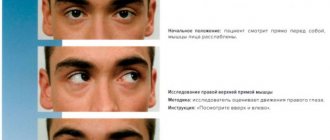
Doctors prescribe the following diagnostic procedures to check binocularity:
- Examination using the “Monobinoscope” and “Synoptophore” devices. These devices not only help to accurately diagnose strabismus and amblyopia, but are also often used for therapeutic purposes.
- Refractometry. Using a special device, the refractive power of both eyes is assessed and compared.
In addition, ophthalmoscopy and biomicroscopy are performed. This allows you to assess the condition of the tissues of the cornea, lens and fundus.
Treatment of binocular vision disorders due to strabismus
There are two main hardware methods for restoring stereoscopic function in strabismus: orthoptics and diploptics. Orthoptic treatment involves training by showing a person two slightly different images that need to be combined. The patient is shown split images through a synoptophore, which he tries to combine into one visual image.
Diploptics is the next stage of treatment. This method involves the doctor using an optical prism, which causes double images. The patient eliminates diplopia through his own efforts. The ophthalmologist changes the prisms, changing the angle of inclination until the eyes become accustomed to looking parallel without the use of lenses.
The final stage of treatment will be eye exercises. There are a lot of them. An ophthalmologist selects a specific technique for each patient.
Binocular vision training
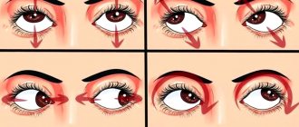
Hardware treatment methods are available only in the clinic, but eye exercises can be done at home. There are two simple exercises that help restore binocular vision. For the first exercise, you will need a sheet of paper on which you need to draw a vertical line approximately 1 cm wide with a marker. The sheet must be fixed on the wall at eye level and moved 1-2 meters away from it. Look at the line and tilt your head down until the line begins to double. Next time, turn your head left and right. The duration of the exercise is 5 minutes. It is advisable to do it three times a day every day. Within two weeks you should notice that your vision has improved.
For the second exercise, you will again need a piece of paper with some kind of drawing on it. Hang it on the wall and move 2-3 meters away from it. Next, you should raise your hand with a clenched fist and a finger extended upward at eye level, but on the same visual axis with the object on the wall. You should look at the tip of your finger and at the drawing alternately. When you look at the drawing, your finger doubles, and when you look at the tip of your finger, the image on the wall blurs. You need to train both eyes for 3-5 minutes daily. After just a few weeks of training, you will notice that your visual acuity has improved.
Therapy methods
Treatment of binocular vision disorders at an early stage is carried out using conservative methods. The following methods of therapy are used:
- Occlusion. The patient wears special glasses, one of which is sealed with adhesive tape. The sticker is applied to the healthy side. This causes the patient to strain the squinting eye. This treatment method prevents the development of amblyopia due to strabismus.
- Hardware techniques. For treatment, “Monobinoscope” or “Synoptophore” devices are used. With their help, they conduct eye exercises to combine several pictures into one. These devices also allow you to stimulate the eye muscles with light signals.
Drug treatment for binocular disorders is auxiliary. Complexes with beta-carotene, vitamins A and C are prescribed. This helps maintain visual acuity. For the paralytic form of strabismus, the use of nootropics, antioxidants and neuroprotectors is indicated.
If there is no effect from conservative therapy for 1.5-2 years, then this is considered an indication for surgery. During surgery, the doctor weakens the eye muscle. This leads to normalization of eye movements and elimination of external signs of strabismus. However, binocular disturbances may persist. Therefore, after the operation, a second course of hardware treatment is carried out using the Synoptophore device.
It is important to remember that treatment of strabismus and amblyopia is best done in childhood. In adults, such vision disorders require long-term and persistent therapy, and sometimes surgical intervention.
Prevention of binocular vision disorders
As for adults, in order to avoid vision problems, you need to follow a work-rest schedule, lead a healthy lifestyle and eat right, regularly visit an ophthalmologist, and do eye exercises, especially if the work involves a heavy load on the visual organs. In addition, you need to consult a doctor promptly if you suspect you have eye disease.
Measures to prevent binocular disorders must be taken starting from the first days after the birth of the child. There are a few simple rules that parents must follow:
- hang toys above the baby’s crib at least 50 cm from his face (otherwise his eyes will squint toward his nose);
- toys should be on both sides of the face;
- Change the location of toys several times a week so that the newborn's gaze does not focus on one point.
At the first alarming symptoms, be sure to show your child to the doctor.