The retina of the eye is the most important structure of the organ of vision with a complex structure that allows it to perceive light impulses. Its immediate task is to receive and transmit visual information, acting as a middle link between the optical system of the eye and the visual parts of the brain. Retinal defects, depending on their severity, become a serious obstacle to the transmission of information or can completely reduce it to zero.
One of the most common retinal defects is dystrophy, which, as a rule, is caused by certain disorders in the vascular system of the eyeball. Suffering from this pathology are mainly elderly people, whose vision is gradually deteriorating. With this defect, damage occurs in the photoreceptor cells responsible for color perception and distance vision. At first, retinal dystrophy may be asymptomatic, so a person is often unaware of the insidious disease present.
Types of retinal dystrophies
Dystrophic defects of the retina are usually divided according to the location and the reasons that caused them. In this regard, the following are highlighted:
Central and peripheral dystrophies. Peripheral retinal dystrophy is very often present in people with myopia. This is due to decreased blood circulation in the eye, which impairs the delivery of nutrients and oxygen to the retina, which is the main cause of all types of peripheral dystrophies.
Congenital and acquired. Such retinal defects can be genetically determined or occur during life due to common diseases and injuries. These include:
- "Senile" dystrophy. Most often, it develops after 60 years. This retinal defect can be combined with senile cataracts caused by the general aging of the human body.
- Pigmentary dystrophy. It is associated with dysfunction of photoreceptor cells responsible for twilight vision. Pigmentary dystrophy is quite rare and is a hereditary disease.
- Dotted white dystrophy. This retinal defect usually occurs in childhood and begins to progress with age. This type of dystrophy is also hereditary.
Project work in biology “Vision defects”
Municipal budgetary educational institution of the Borisoglebsk urban district Borisoglebsk secondary school No. 10
Educational and research work
Vision defects
The work was completed by: Anastasia Shishkina, student 8 “A”
Head: Sudakova Irina Viktorovna, biology teacher 1KK
Introduction
People look at the world each in their own way; some people have clear vision, while others have poor vision. This can be seen through a little experience, for example: looking through a transparent piece of glass and a cloudy one, through the transparent one we see everything clearly and clearly, but if you look through a cloudy piece of glass it will be unpleasant, it will be uninteresting to perceive everything that surrounds you.
Goal of the work:
To find out the factors influencing the deterioration of vision of students of MBOU BGO Secondary School No. 10 in grades 8 and 10.
Tasks:
1) Familiarize yourself with visual defects.
2) Identify the percentage of students with poor and good vision in MBOU BGO Secondary School No. 10, grades 8 and 10.
3) Find out the causes of visual impairment.
4) Learn ways to prevent eye diseases.
Object of study:
human vision
Subject of study:
existing visual defects in humans
Vision
– the ability of a person to perceive information by converting the energy of electromagnetic radiation in the light range, carried out by the visual system.
Due to the large number of stages in the process of visual perception, its individual characteristics are considered from the point of view of different sciences - optics (including biophysics), psychology, physiology, chemistry (biochemistry). At each stage of perception, distortions, errors, and failures occur, but the human brain processes the information received and makes the necessary adjustments. These processes are unconscious in nature and are implemented in multi-level autonomous correction of distortions.
The eye is the organ of vision
The eye is the organ of vision in animals and humans. The human eye is a complex optical system.
The eyeball is located in the orbit of the skull and has three membranes. This:
1) sclera: outer (albuginea) membrane; formed by dense connective tissue, performs a protective function; its transparent front part is called the cornea;
2) choroid: occupies a middle position; contains blood vessels and pigment cells that supply the eye; its visible part is the iris;
3) retina: inner shell; Here are the photoreceptors, as well as the neurons whose axons form the optic nerve.
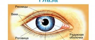
Retina.
The outermost position in the retina is occupied by photoreceptors; deeper there are several layers of neurons that take part in the operational processing of visual information.
Humans have two types of photoreceptors – rods and cones. There are relatively more rods at the periphery of the retina, and cones closer to the middle (opposite the pupil). In the very center of the retina there is an area consisting of the most densely located cones - the macula macula. This is the area of greatest visual acuity. When examining an object in detail, we look directly at it, and the image, refracted in the lens, is projected onto the macula.
The photoreceptor consists of an outer part, a nuclear region and a zone in contact with retinal neurons. The reaction of the photoreceptor to light is possible due to the presence inside its outer part of light-sensitive pigments - substances that are destroyed under the influence of electromagnetic waves of a certain length. The breakdown products of pigments cause a receptor reaction and affect the generation of impulses by retinal neurons.
Cones contain pigments called iodopsins (otherwise known as conopsins). There are three types: red-, green- and blue-sensitive. Each specific cone contains one of the iodopsins, and together they provide color vision.
Types of vision defects:
- astigmatism;
- myopia, or so-called myopia;
- farsightedness, or hypermetropia;
- color blindness, or color blindness;
- Strabismus;
- Cataract;
- Glaucoma.
Astigmatism
Visual defect associated with non-spherical cornea
that is, its different curvature in different planes. (A person clearly sees horizontal lines, but blurredly sees vertical lines, or vice versa)
The reason for the development of this pathological condition is an incorrectly formed cornea of the visual organ. It should also be noted that the development of astigmatism is directly influenced by the displacement of the eye lens relative to the refractive axis. Both of these reasons entail differences in distances, which are essential for focusing the “picture”.
Myopia, or so-called myopia
The eye is either elongated or has a smaller radius of corneal curvature compared to a normal eye. Therefore, a beam of parallel rays is refracted excessively, converging at a point in front of the retina
Myopia can develop for several reasons. The first is to lengthen the eye while maintaining correct refraction. As for the second reason, this is an overly powerful optical refraction, which is more than 60 diopters, with the length of the visual organ within normal limits. Both presented deviations negatively affect obtaining a normal image. In other words, the image is not able to focus on the retina, but is located inside the eyeball. Thus, only a focused image of some objects located at a short distance from the person penetrates the retina.
Farsightedness, or hypermetropia
Shorter length of the farsighted eye or larger radius of curvature of the cornea compared to the normal eye. The beam of rays converges behind the retina.
This defect develops due to excessively weak optical refraction in the visual organs while maintaining the normal length of the eyeball. It should be especially noted that the cause of farsightedness is also the shortening of the eyeball, provided that the refractive optical power is preserved. Due to the fact that the farsighted eye is not able to create a focus on the retina, muscle tension increases significantly. This phenomenon gradually changes the curvature of the lens, which in turn leads to the adaptation of the visual organ to the prevailing conditions. However, this is not enough for normal focusing of the resulting image. When examining objects near the eyes, the muscle tissue of this organ becomes even more tense. In other words, the closer an object is, the further away its image appears on the retina.
Colorblindness, or color blindness
Color vision impairment.
This defect is a congenital disease that is most often observed in men. The essence of this deviation is that patients have impaired correct color perception, which is regulated by photoreceptor cells (cones) in the retina. If a person lacks any type of cones, then he or she has color blindness.
Strabismus
Strabismus is a deviation of the visual axes from the direction of the object in question, in which the coordinated work of the eyes is disrupted and it is difficult for both eyes to fixate on the object of vision. An objective symptom is the asymmetrical position of the corneas in relation to the corners and edges of the eyelids. There are congenital (present at birth or appears in the first 6 months) and acquired strabismus (appears before 3 years).
The causes of strabismus are very diverse. They can be either congenital or acquired:
- the presence of ametropia (farsightedness, myopia, astigmatism) of medium and high degrees;
- injuries;
- paralysis and paresis;
- abnormalities in the development and attachment of the extraocular muscles;
- diseases of the central nervous system;
- stress;
- infectious diseases (measles, scarlet fever, diphtheria, influenza, etc.);
- somatic diseases;
mental trauma (fear);
a sharp decrease in visual acuity in one eye.
Cataract
Cataract is a pathological condition associated with clouding of the lens of the eye and causing varying degrees of visual impairment up to its complete loss. The most common symptom of cataracts is decreased visual acuity. Depending on the location of the lens opacities in the center or on the periphery, vision may decrease or remain high. If cataracts begin to develop on the periphery of the lens, the patient may not experience any changes in vision. Such cataracts are discovered by chance during a routine medical examination. The closer to the center the clouding of the lens is, the more serious the vision problems become. With the development of opacities in the central part of the lens (its nucleus), myopia may appear or worsen.
Glaucoma
Glaucoma is a large group of eye diseases characterized by a constant or periodic increase in intraocular pressure above what is tolerable for a given person. There are two main forms of glaucoma: open-angle and closed-angle. In addition, there are congenital glaucoma.
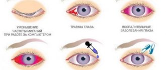
Causes of visual impairment
- Lack of work of the eye muscles.
- Aging of the retina.
- Poor circulation.
- Eye-strain.
- Dryness of the mucous membrane of the eye.
- Lack of vitamins.
- Bad light.
Properties of food products from the point of view of their effect on vision. (benefit, improvement for vision)
Beet.
Contains: phosphorus, sodium, manganese, iodine, vitamins C, B6 B2, PP, E, U, folic acid, carotenoids.
What are the benefits: relieves eye fatigue, cleanses the blood.
Norm per day: 100 g.
How to eat: fresh, boiled, you can make juice.
Rose hip.
Contains: vitamins C, P, B, B2, A, K, E, organic acids, sodium, calcium, manganese, iron.
What is the benefit: ensures the strength and elasticity of the blood vessels of the eyes.
Norm per day: 1 glass.
How to drink: as an infusion.
Chicken eggs.
Contains: protein, lutein.
What is the benefit: the substances prevent the formation of cataracts, protect the optic nerve, and neutralize the harmful effects of the environment.
Norm per day: one egg.
How to eat: boiled and fried.
Carrot.
Contains: beta-carotene, iodine, magnesium, iron, calcium, phosphorus.
What is the benefit: supports the process of cell formation and growth, improves vision functions.
Norm per day: 1 medium carrot.
How to eat: fresh or stewed, adding a little vegetable oil or sour cream. In winter, when carrots are no longer so juicy, prepare freshly squeezed juice with cream from them.
How many people with poor vision are there in the world?
Poor vision 20%
Good vision 80%
Number of people with visual impairments in 8 “A” class
- Astigmatism – 0.27%
- Myopia – 44%
- No problems – 55.73%
Number of people with visual impairments in 10 “A” class
- Astigmatism – 0.25%
- Myopia – 20%
- No problems – 79.75%
Questions that were asked to students
- Since when did your vision begin to deteriorate?
- What do you use your computer for?
- How much time do you spend at the computer?
- At what distance do you watch TV shows?
- How much time do you watch TV shows?
Conclusion
During the work, it turned out that not all students take good care of their eyes. They spend a lot of time on the computer and TV.
Applications
https://ru.wikipedia.org/wiki/Human_vision
https://obuchonok.ru/node/444
https://kopilkaurokov.ru/nachalniyeklassi/prochee/issliedovatiel-skaia-rabota-zrieniie-eto-vazhno
Literature
- Schoolchild's encyclopedia 4000 fascinating facts.
Lyricist John Farndon. Project manager Ian Poulin.
- Children's encyclopedia ROSMEN man
Diagnostics
In order for the diagnosis of retinal dystrophy to be confirmed or refuted, it is necessary to undergo a thorough ophthalmological examination. In modern ophthalmological centers, the diagnosis of retinal defects is performed using special computerized equipment, which makes it possible to create a complete picture of the state of the patient’s visual system.
The examination usually includes:
- Determination of visual acuity;
- Perimetry, for studying visual fields, in order to assess the condition of the retina in the periphery;
- Optical coherence tomography;
- Electrophysiological study to determine the viability of nerve cells of the retina and optic nerve;
- Ultrasound of the internal structures of the eye, including A-B scanning;
- Tonometry with measurement of intraocular pressure;
- Ophthalmoscopy with fundus examination.
Symptoms
| Occurrence (how often a symptom occurs in a given disease) | |
| Color perception disorder | 100% |
| Poor vision at dusk | 90% |
| Rapid eye fatigue when performing visual work (eye fatigue, visual fatigue, eye strain, asthenopia) | 80% |
| Decreased visual acuity (deterioration of vision, poor vision) | 80% |
| Intolerance to bright light (photophobia, light sensitivity) | 60% |
| Double vision (diplopia) | 50% |
general information
In medical practice, a vision defect is often called a refractive error. Such abnormalities are the most common eye problems. The essence of this group of diseases is that the optical system of the eye is unable to focus light rays on the retina, which is our recorder of light stimuli. The main symptom and consequence of this pathological condition is poor vision.
Astigmatism
The reason for the development of this pathological condition is an incorrectly formed cornea of the visual organ. It should also be noted that the development of astigmatism is directly influenced by the displacement of the eye lens relative to the refractive axis. Both of these reasons entail differences in distances, which are essential for focusing the “picture”.
This vision defect in one eye can combine the effects of farsightedness, nearsightedness and normal vision.