Causes of dilated veins in the fundus
Eye diseases and decreased visual acuity associated with dysfunction of the vascular network are increasingly being diagnosed. According to obaglaza.ru, this consequence is caused, first of all, by late seeking medical help and ignoring the symptoms of systemic diseases.
Enlarged veins in the fundus of the eye - causes of pathology
The eyes are a particularly sensitive organ to many pathologies that occur in the body. Their health can be affected by factors such as surges in blood pressure, significant fluctuations in body weight, and a decrease in the degree and quality of the immune system.
The reasons why veins in the fundus may be dilated are as follows:
- hypertension, in which there is a sharp increase in blood pressure, which negatively affects the condition of the veins and blood vessels in the fundus of the eye;
- damage to the kidneys and urinary system, which also increases fluid pressure in the skull and in the veins of the eye fundus;
- deterioration of the condition of the veins, the appearance of cholesterol deposits in them - vascular atherosclerosis causes an increase in blood pressure in the brain and worsens the process of blood movement in the veins of the fundus;
- during pregnancy, especially in the last quarter.
Impaired blood supply to the eyes, hormonal changes, metabolic disorders (primarily diabetes mellitus), and retinal thrombosis can also cause dilation of the veins located in the fundus of the eye. The reason for this negative manifestation lies in changes in the blood supply to the eye tissues and increased pressure. Microcirculation disorders are also often observed.
This lesion is accompanied by a gradual deterioration of vision, the appearance of fog before the eyes, and the causes that occur in adults and children are basically similar. And if adults are characterized by acquired diseases (hormonal imbalances, age-related changes, atherosclerosis of blood vessels and veins), then children are more characterized by congenital anomalies: insufficiency of microcirculation in the tissues of the eye, increased blood pressure due to pathologies of the brain.
Treatment
Obaglaza.ru specialists insist that priority in the treatment of diseases of the vascular system of the eyes is given to the treatment of the disease that caused the damage (hypertension, diabetes mellitus, atherosclerosis, kidney disease, etc.).
To maintain blood vessels and restore visual function of the eyes, the following are effectively used:
- General medications to maintain vision;
- Vitamin therapy;
- Physiotherapeutic procedures, massage;
- Cool baths, walks;
- Oxygen injections under the conjunctiva.
Symptoms and diagnosis of the disease
When the veins of the fundus of the eye are dilated, a number of characteristic symptoms appear, allowing one to identify the initial stage of the pathology, which is more amenable to treatment. Pathological narrowing of blood vessels may indicate a violation of blood circulation and microcirculation in the tissues of the fundus, and the development of various diseases may be noted. For example, atherosclerosis of blood vessels and veins, accompanied by the formation of cholesterol deposits, interferes with the normal movement of blood through them, which causes increased pressure when the veins of the fundus of the eye expand.
Hypertensive angiosclerosis, which causes disturbances in the condition of the fundus veins, causes the following characteristic symptoms:
- swelling of the eyes, which occurs in more advanced stages of the disease;
- feeling of a constant veil before the eyes;
- deterioration in the quality of vision both at close and far distances;
- changes in the tissues of the retina, which is accompanied by a violation of blood microcirculation in it;
- the field of view may decrease, some parts of it seem to fall out of view.
Patients may also complain of headaches that arise due to excessive eye strain; the clinical picture reveals swelling of the optic nerve, an increase in the size of the disc, blood vessels “drown” in the swollen mass, and the veins are excessively tortuous, which impairs blood circulation in them. If the therapeutic effect on the affected part of the fundus is insufficient, the symptoms worsen, which provokes the development of retinal thrombosis.
Symptoms
In the initial stage of diseases or disorders of the vascular system of the eyes, there may be no symptoms at all. Pathology is detected during a thorough medical examination. In more severe and advanced cases, a person may notice a number of symptoms:
- Seeing floaters in the eyes, multi-colored spots, flashes of light.
- Parts of the view may be lost from view.
- Sometimes pathologies are accompanied by blurred vision.
- In more advanced cases, vision decreases to complete blindness.
Obaglaza once again emphasizes that a periodic high-quality ophthalmological examination by a qualified specialist will protect your vision and can diagnose a number of systemic diseases at the very beginning of their development, before symptoms appear.
Treatment of retinopathy and angiopathy
Dilated veins, localized in the tissues of the fundus of the eye, require therapeutic intervention, since if treatment is insufficient, there is a high probability of aggravation of symptoms. In subsequent stages of the disease, more active treatment is required, with the use of medications to restore the elasticity and normal functioning of the veins. Treatment of the underlying disease is based on the impact of the root cause of the pathology of the veins of the fundus, therefore you should promptly pay attention to even minor changes in the quality of vision.
The main direction of treatment is the use of absorbable agents, and their use is carried out in combination with drugs that relieve swelling. Thanks to them, it becomes possible to prevent the possibility of hemorrhage in the tissues of the fundus, which can lead to complete loss of vision.
Thrombosis
Signs of cholesterol deposits forming on the walls of the veins, which can cause the formation of blood clots that interfere with blood circulation, must be promptly eliminated. The transition of the disease to the next stage is fraught with a significant deterioration in the quality of vision, therefore diagnosis of the pathology should be carried out at the first manifestations of changes in vision.
Thrombosis of the central retinal artery is extremely susceptible to disturbances in the blood circulation; its aggravation can be prevented by the combined use of drug decongestant therapy with physical procedures: magnetic therapy, acupuncture.
Inflammation
In some cases, an inflammatory process may occur, and deterioration of vision is accompanied by the possibility of bleeding in the fundus tissues. The therapeutic method is selected depending on the condition of the eye and the presence of edema.
Optic atrophy
Enlarged optic discs, which can cause inflammation of the fundus tissues, can cause gradual atrophy. The following characteristic symptoms are noted:
- blurred vision;
- loss of image clarity;
- flashing “fly” before the eyes.
In difficult cases, in the absence of the necessary therapeutic intervention, retinal detachment is likely to occur, which can lead to complete loss of vision, which cannot be cured in the future.
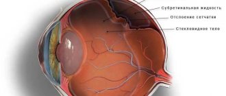
Delaminations
Manifesting itself in the vascular tract of the eye, detachment is accompanied by the destruction of blood vessels, which provokes changes in the flow of blood into the veins. When the retinal layer is destroyed, there is a threat of vision loss, and the treatment method differs in that agents must be used that ensure the germination of blood vessels that ensure normal blood supply to the fundus tissues.
Any damage to the cornea is accompanied by a deterioration in the quality of vision, but it is detachment that is the most serious problem among other eye pathologies.
Retinal tumors
Cancerous lesions can also affect such a delicate and sensitive organ of the human body as the eye. In advanced stages of the pathological process, treatment becomes increasingly difficult; foci of exudation can be localized in different parts of the eye.
The therapeutic method for diagnosing tumor processes is most often based on surgical removal of pathological cells and preventing the worsening of the disease.
Dystrophy
Lesions of the veins of the eye can be accompanied by symptoms of dystrophy, manifested in deterioration of vision, loss of clarity of the picture, and flickering of “spots” before the eyes. Dystrophy may require the use of medication in combination with surgical correction of the retina.
Full-blooded veins of the fundus: causes, treatment
The vessels of the fundus of the eye are a reflection of vascular pathologies occurring in the human body. These are the only blood vessels of the internal organs of the human body that can be seen using non-invasive research methods - various types of ophthalmoscopy.
Vasoconstriction (angiopathy) of the retina is not an independent disease, but almost always indicates the presence of a general somatic pathology in the patient.
I recently read an article that talks about the drug Choledol for cleaning blood vessels and getting rid of CHOLESTEROL. This drug improves the general condition of the body, normalizes the tone of the veins, prevents the deposition of cholesterol plaques, cleanses the blood and lymph, and also protects against hypertension, strokes and heart attacks.
I’m not used to trusting any information, but I decided to check and ordered a package. I noticed changes within a week: the constant pain in my heart, heaviness, and pressure surges that had tormented me before receded, and after 2 weeks they disappeared completely. Try it too, and if anyone is interested, below is the link to the article.
What causes vasoconstriction in the fundus?
Retinal angiopathy, as one of the ophthalmological symptoms, occurs in various diseases and pathological conditions of the human body:
- arterial hypertension;
- neurocirculatory dystonia;
- atherosclerosis;
- hemorrhagic stroke;
- diabetes mellitus;
- kidney diseases (chronic glomerulonephritis, nephropathy of pregnancy);
- traumatic injuries of the cervical spine, neck, head, eyes;
- cervical osteochondrosis;
- hydrocephalic syndrome;
- tumors inside the skull;
- thrombosis of the venous sinuses and cerebral veins;
- infectious diseases of the central nervous system;
- leukemia;
- congenital anomalies in the structure of vascular walls;
- autoimmune systemic diseases (periarteritis nodosa, multiple sclerosis, systemic lupus erythematosus);
- exposure to harmful production factors;
- long-term intoxication of the body (alcohol, drugs, salts of heavy metals, smoking).
These reasons often lead to irreversible changes in the blood vessels of the human body, including the vessels of the fundus.
Narrowing of the retinal arteries can also be reversible. The causes of reversible (reflex) constriction of the fundus vessels include:
- long-term stay in rooms with poor lighting;
- long hours of work at the computer;
- watching TV for a long time, especially with the lights off.
The narrowing of retinal vessels is based on different mechanisms:
- spasms - prolonged contraction of the muscle layer of the vascular walls;
- stenosis - a decrease in the lumen of blood vessels due to plaques present on their inner surface;
- sclerosis – thickening of vascular walls due to the replacement of their muscular elements with connective tissue;
- formation of granulomas or deposition of autoimmune complexes on vascular walls;
- mechanical compression of the arteries supplying the eye from the outside.
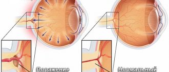
Clinical signs of pathology
Clinically, there are three degrees of retinal angiopathy - first, second (moderate) and third.
The degree of narrowing of the vessels of the fundus can only be determined during ophthalmoscopy.
In the initial stages of retinal vascular narrowing, the patient usually does not have complaints or has complaints that are general.
- headache;
- discomfort in the eyeballs;
- visual disturbances (floaters, fog or spots before the eyes, slight decrease in visual acuity).
Changes in the fundus vessels in the first degree of angiopathy are reversible, as they are functional. Provided that effective treatment of the disease that caused angiopathy is prescribed in a timely manner, these changes can be easily regressed.
As angiopathy progresses, more specific signs appear, by which the clinician may suspect pathology of the fundus vessels:
- throbbing pain in the eyes;
- marked decrease in visual acuity, often myopia;
- narrowing or loss of visual fields;
- impaired light sensitivity.
But given the fact that the narrowing of retinal vessels is not an isolated pathology, the symptoms characteristic of the underlying disease, which caused the onset of angiopathy, come to the fore in the symptoms.
Ophthalmoscopy in the second degree of angiopathy reveals organic vascular lesions that are difficult to regress:
Reader Recommendation:
To clean VESSELS, prevent blood clots and get rid of CHOLESTEROL, our readers use a new natural drug recommended by Elena Malysheva. The preparation contains blueberry juice, clover flowers, native garlic concentrate, rock oil, and wild garlic juice.
Read more…
- significant narrowing of the arteries;
- dilatation and tortuosity of veins;
- retinal hemorrhages;
- retinal vascular thrombosis;
- pallor of the fundus;
- newly formed vessels in the area of the optic nerve.
With adequate treatment of the underlying disease, it is possible to stabilize angiopathy and prevent its further progression.
The most severe (third) degree of angiopathy, which threatens the patient with loss of vision, is clinically manifested by the following symptoms:
a sharp decrease in visual acuity;
ophthalmoscopically:
- straightening the arteries;
- uneven caliber of arteries;
- cotton wool-like lesions;
- massive hemorrhages in the retina;
- swelling of the retina and optic nerve.
Depending on the causes of narrowing of the fundus vessels, the following types of retinal angiopathy are distinguished:
- Hypertensive, which is based on an increase in blood pressure that occurs as a result of persistent spasm of the muscular component of the arterial walls;
- Diabetic, the cause of which is diabetes mellitus. In this case, a high level of blood glucose is observed, which, due to a lack of insulin or its immunity to the body tissues, does not enter the body cells, but settles in the lumen of the capillaries and arterioles;
- Traumatic, which occurs with injuries to the cervical spine, neck or head. As a result of increased intracranial pressure, a reflex narrowing of the arterioles occurs, resulting in retinal hypoxia. Traumatic angiopathy
How to detect pathology?
It is impossible to detect retinal angiopathy with the naked eye. This requires special equipment - ophthalmoscopes, slit lamps, ultrasound machines.
Ophthalmoscopy
An ophthalmoscopy allows the ophthalmologist to examine the arteries and veins of the fundus through the pupil. The most common methods of examining the retina include:
- Indirect ophthalmoscopy (mirror). This procedure is performed using a mirror ophthalmoscope and a magnifying glass. The fundus image is presented as a mirror image and provides only an overview.
- Direct ophthalmoscopy. Examination of the fundus is carried out using an electric ophthalmoscope. This procedure is more informative compared to the mirror procedure.
- Ophthalmochromoscopy. Examination of the fundus with an ophthalmoscope with replaceable filters. The use of colored glasses (red, green and blue) in different combinations during the procedure makes it possible to identify initial changes in the retinal vessels that remain invisible in white.
- Polarizing ophthalmoscopy. Examination of the fundus in polarized light makes it possible to detect retinal edema in the initial stage.
Clarifying research methods
In modern medicine, many other methods of studying retinal vessels are used to clarify the results of ophthalmoscopy:
- Biomicroscopy of the fundus using a slit lamp with fundus lenses. This is a contact method for examining the fundus, in which a three-mirror lens is placed on the cornea, allowing a detailed examination of the entire surface of the retina.
- Doppler ultrasound examination of the eyes. It is carried out through a closed eyelid and has no contraindications. Allows you to study the vessels of the retina and evaluate the speed of blood flow through them. Determines the narrowing of the fundus vessels in the initial stages.
- Laser ophthalmoscopy. With this research method, the retina is illuminated with a laser, and an image of the retina is displayed on a monitor. The advantage of this method is the ability to examine the fundus of the eye while reducing the transparency of the lens and vitreous body.
- Rheoophthalmography. This is a contact method for studying the blood supply to the retina, based on recording pulse waves from the vessels of the fundus. To capture impulses from them, electrode lenses are applied to the cornea.
- Fluorescein angiography of the fundus. This is a highly informative method based on photographing contrasted vessels of the fundus. The contrast agent is administered intravenously. The procedure is performed using a slit lamp with fundus lenses.
- Densitometry. The procedure is performed similarly to fluorescein angiography, but photographs of the retina are taken, which allows one to evaluate the blood flow in it over time.
These methods do not replace each other, but complement each other perfectly. The choice of diagnostic procedure in most cases depends on the doctor’s preferences, territorial and financial accessibility of the diagnostic method, and the transparency of the eye structures.
Narrowing of the fundus vessels can lead to blindness of the patient, so it is very important to identify the pathology in time, establish the causes of its occurrence and prescribe appropriate treatment.
The preservation of the functions of such an important human sensory organ as the eye depends on this.
Source:
Pathologies of the fundus: dilation and narrowing of blood vessels in the eye
In many cases, these diseases signal us about complex systemic diseases of our body as a whole. Thus, narrowing of the fundus vessels will be the first alarming signal for all types of retinopathy, among which there will be secondary manifestations of diabetes mellitus, heart and vascular diseases and many others.
Other pathologies of the fundus will also indicate serious health problems, and their danger is the loss of visual acuity, which most likely will not be restored. These diseases require urgent and immediate treatment.
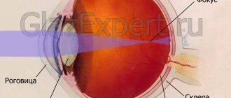
The structure of the fundus and why its diseases can help in diagnosis
By the term “fundus”, doctors mean the inner part of the eyeball, which can be seen during ophthalmoscopy (this is a non-invasive examination, it is carried out by eye doctors using an ophthalmoscope in a dark room). During ophthalmoscopy, without interfering with our body, we can see eye diseases, as well as the first symptoms of many systemic diseases.
So, with the help of an ophthalmoscope, the doctor sees:
- The structure of the vessels of the eye, veins and arteries, their filling, possible narrowing or, conversely, expansion, the presence of hemorrhages.
- Optic nerve and macula, their defects.
- Retina, its thinning (dystrophy), detachments, ruptures.
We ourselves will not be able to see the fundus of the eye, but the doctor will tell a lot about the state of our health simply by looking at the fundus of the eye through an ophthalmoscope.
Fundus pathology is always secondary. Therefore, it is imperative to look for the underlying disease.
Fundus vascular diseases in children
In childhood, as in adults, symptoms of significant visual impairment, damage to the retina and fundus tissues may occur. For children, the most typical manifestation is congenital anomalies in the development of the eyes, in particular the fundus. Since eye lesions in childhood are likely to progress rapidly, an ophthalmological examination of the child should be carried out regularly in order to promptly identify the onset of fundus vein disease.
The blood vessels of a child's eyes are more susceptible to negative effects, especially with a hereditary predisposition. It is the genetic factor that plays a special role in the occurrence and development of any pathology of the fundus veins. As Dr. Malysheva advises in her program about health, children need to undergo a doctor’s examination even at an early age, since childhood eye diseases at an earlier stage of development are more amenable to treatment.
Nature of the vessels
During a medical examination, it is very important to undergo an ophthalmoscopy. Depending on the degree and nature of the disturbances in the venous network of the fundus, the doctor can determine the disease that caused the pathology:
- dilation of venous vessels and narrowing of arterial vessels - evidence of the initial stage of hypertension;
- microaneurysms and hypertrophied branching of venous vessels, which can sometimes be accompanied by retinal edema - develops in diabetes mellitus;
- vascular spasms, thrombosis of the central vein and artery, with a background of the fundus of the eye with a whitish-milky tint (insufficient blood supply) - speak of vascular diseases;
- a bright dot in the area of the macula and swelling of the retina - according to obaglaza ru, indicates a bloodless central artery of the eye;
- retinopathy (insufficient blood supply to the retina) is a complication that is a consequence of hypertension, vascular disease and diabetes;
- swelling of the retina near the projection of the beginning of the optic nerve, strong branching of the veins - indicates a disruption in the functioning of the central vein of the eye or its blockage (with hypertension, renal and vascular pathologies).
Possible complications
In the absence of the necessary treatment, there is a high probability of developing complications that negatively affect vision. The most common complications include:
- deterioration of vision, which with further inattention to the condition of the eyes can develop into complete blindness;
- hemorrhages in the retina - this consequence can occur with improperly selected treatment, significant strain on the eyes, as well as with the development of hypertension, which is expressed in increased blood pressure;
- damage to the cornea, which can provoke the formation of cataracts. These conditions, in turn, can lead to gradual loss of vision and headaches, which significantly reduce the patient’s quality of life.
Retinopathy develops in later stages of the disease, which is likely due to insufficient therapeutic intervention or too late diagnosis of the disease. Therefore, to prevent possible complications and maintain visual acuity, it is recommended to pay attention to any changes in the condition of the eyes in a timely manner.
Diagnostic measures
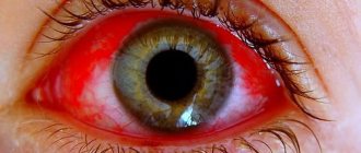
Hyperemia of the conjunctiva of the eye is not considered as an independent pathology. This is just a symptom that can manifest itself with various abnormalities. Therefore, when it is detected, it is necessary to diagnose and prescribe targeted treatment. First of all, the doctor conducts a survey, visual examination of the patient and palpation of the lacrimal sac (the latter to exclude or confirm dacryocystitis). In some cases, this is enough to make a diagnosis. Sometimes additional examinations of the affected eye are required:
- Biomicroscopy is an examination of optical media and organ tissues using a microscope and directed light.
- Ophthalmoscopy is a non-invasive technique for examining the fundus of the eye. It is carried out using an ophthalmoscope. Allows you to identify venous congestion of the fundus, anomalies in the retina, choroid, and optic nerve head.
- Visometry is a standard test of visual acuity using a special table.
- Perimetry - testing the visual field and detecting blind spots (scotoma).
- Gonioscopy is a contact method for studying the structures of the anterior chamber of the eye.
It may also be necessary to conduct laboratory tests and measure intraocular pressure. In some cases, an ultrasound is prescribed.
Disease Prevention
To prevent possible consequences, loss of vision and deterioration of well-being, you should make some changes in your own life. The following points of a preventive approach to problems with fundus veins should be considered important:
- Introducing healthier lifestyle habits: quitting smoking, alcohol.
- Sufficient physical activity to activate blood circulation in the tissues of the brain and fundus of the eye.
- Regular preventive examinations with a doctor, in particular an ophthalmologist.
- Nutrition control - lowering cholesterol levels, introducing more fresh fruits, vegetables, herbs into the daily diet, preventing digestive disorders.
The recommendations listed are easy to apply and will help preserve the vision and health of every person.
Prevention
To prevent the occurrence of hyperemia, you need to adhere to the following recommendations:
- Avoid overstrain of the visual organ.
- At high loads, regularly perform special gymnastics.
- Observe hygiene rules.
- Protect your eyes from contaminants and potential allergens. Use safety glasses or a mask if necessary.
- If the mucous membranes dry out, use moisturizers.
If you have ophthalmological problems, regular visits to your doctor for a preventive examination are required. This is also necessary for some other pathologies, such as diabetes and hypertension.