What is aphakia of the eye
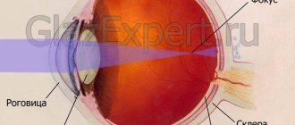
The lens is an elastic, transparent natural lens, due to the constant change in shape of which a person can quickly focus on both nearby and distant objects. It is located inside the capsule and is an integral part of the optical system of the eye. Therefore, any disturbances affecting the lens inevitably affect visual acuity.
The sun's rays, hitting the lens through the cornea and pupil, are refracted and fall on the retina, which is responsible for image formation
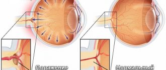
Aphakia is the absence of the lens in the eye.
In this case, refraction changes dramatically, that is, the ability to correctly refract light rays entering through the pupil, resulting in a significant decrease in visual acuity.
Depending on whether both eyes or one are affected, aphakia is distinguished:
- One-sided. This type of pathology is diagnosed most often. It is characterized by the absence of a lens in only one of the eyes.
- Double-sided. It usually occurs after removal of a severe lens that has caused deformation of the lenses of both eyes.
Features of the disease in children
Aphakia is rarely congenital. But in such cases, the lens may be completely absent or stop developing at a certain stage.
More often, the pathology is a consequence of congenital cataracts, the diagnosis of which is not difficult.
In general, the disease progresses and is treated in the same way as in adults. But it is possible to radically solve the problem by installing intraocular lenses only after the child turns 2 years old, since it is at this age that the growth and development of the eye stops.
Structure of the eye - video
First aid
It is quite natural that if such a situation happens for the first time, the animal’s owners may be confused, not knowing what to do in the current circumstances.
In such a case, there is no need to panic.
Pug owners should always ensure that the simplest saline solution is always present in their home medicine cabinet..
It is unlikely that you will be able to do without this remedy if, for example, a speck gets into your pet’s eye.
Fixing the eyeball in the correct position will not be difficult.
To do this, you will need clean gauze, which should be folded several times and thoroughly wetted with the solution . Afterwards, the moistened gauze cloth should be applied to the missing eye and securely secured with a bandage.
If it happens that there is no gauze or solution available, then you can use a clean handkerchief.
A handkerchief should be moistened in salt water and also applied to the prolapsed eyeball. After these steps are completed, you should take the animal to the veterinarian, who will be able to correctly return the eye to its original place.
NOTE!
After such manipulations, the four-legged friend’s eye should be bandaged until complete recovery.
Sometimes the veterinarian is forced to stitch the dog's eyelid shut. After an appropriate period of time, the applied sutures are removed.
In order to speed up the healing process, experts recommend treating the pug's eyes with saline solution, and also using hydrocortisone ointment for this..
It is quite possible to put the eye back in place without the help of a doctor. To do this, you should first wash your hands with soap, then gently press your finger on the apple of your eye, which will allow you to return it to its place.
Then you need to moisten the animal’s eyelids and apply a sterile bandage.
If first aid is provided incorrectly or is absent, the dog’s visual organ may atrophy.
As for veterinarians, they recommend not postponing your pet’s visit to a specialist if such a problem arises.
Expert opinion
Tolkachev Andrey Mikhailovich
veterinarian
“Depending on existing opportunities, you should not put off a visit to the veterinarian for a long time. This is due to the fact that if first aid is not provided in a timely manner, then, as experience shows, the pet may become blind. At home, it is unlikely that anyone will be able to provide adequate first aid. All this is fraught with drying out of the eye and the development of an inflammatory process, which may require longer and deeper treatment.”
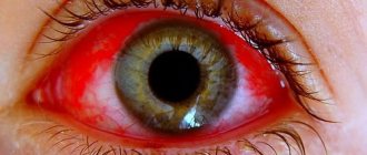
Signs and diagnosis
Patients complain of:
- a sharp decrease in visual acuity;
- obtaining images of different sizes by the healthy eye and the sick one, as a result of which the picture does not merge together;
- difficulty focusing.
It is not difficult to recognize the absence of a lens. Firstly, a person usually knows what preceded the onset of vision problems. Secondly, there are visible changes in the affected eye, which are easy to notice during biomicroscopy, that is, examination of the eye through a slit lamp. So:
Although interviewing the patient is usually sufficient to make a diagnosis, instrumental diagnostics are necessary to exclude other pathologies, since symptoms of aphakia can be observed with dislocation or subluxation of the lens.
Correction methods
The problem of aphakia can be solved using conservative methods or surgical intervention. The latter is more preferable, as it allows you to completely restore binocular vision.
In both cases, the prognosis is favorable, since correctly selected optical correction allows one to maintain high visual acuity and ability to work, but only if there is no damage to the nerve endings.
Conservative therapy
You can increase the visual acuity of an aphakic eye with the help of:
Strong plus glasses can cause annular scotoma, that is, the patient loses an entire area of vision, which leads to painful sensations from the regular sudden appearance of one or another object in front of the eyes. This significantly complicates the lives of patients, as it is difficult for them to cross busy intersections, etc.
Surgery
Surgery to install an intraocular lens (IOL) is used after lens removal.
This type of vision correction is a surgical procedure for installing an artificial plastic lens of a certain strength, the value of which is calculated using specially created computer programs and tables, taking into account:
- lens thickness;
- refractive power of the cornea;
- depth of the anterior chamber;
- length of the eyeball.
The fullness of vision after surgery is planned individually for each patient, guided by his wishes. For example, if the healthy eye is myopic or the patient’s work involves the need to constantly sit at a computer, the IOL is selected in such a way that this can be done without glasses, etc.
Its implementation is possible only after the final formation of the eye structures, which usually occurs by 2 years. Therefore, when treating children with congenital aphakia before reaching this age, it is necessary to use glasses or contact lenses.
There are 4 different types of IOLs, differing in how they are inserted:
- with fixation in the corner of the anterior chamber;
- iris clip lens (pupil);
- extracapillary;
- posterior chamber.
The optimal option is a posterior chamber IOL, which does not interfere with normal pupil dilation and provides better quality of vision, since it occupies the natural place of the lens.
If for one reason or another the optics of the operated eye are incompatible with the optics of the other, another surgical intervention may be performed to implant a second artificial lens of the required strength.
Previously, other operations were used to restore the quality of vision in patients with aphakia, but due to their imperfections and the frequent lack of positive changes, they have not been carried out in recent years.
Consequences and complications
Unilateral aphakia is often accompanied by aniseikonia. This pathology is characterized by obtaining images of different sizes in the healthy eye and the diseased eye, which greatly complicates the life of patients. If left untreated, the disease can cause significant deterioration in vision, loss of ability to work, and disability.
Anyone can experience aphakia. If you do not ignore the problem and consult a doctor in a timely manner, a person has every chance of restoring the affected eye and maintaining normal vision. Otherwise, you should expect a gradual deterioration of the condition and loss of performance.
Most often, the eyeball falls out in brachycephalics (pugs, Pekingese, Shih Tzu, griffins), also in Chihuahuas, Yorkshire terriers. For these groups of breeds, even the slightest physical impact can be dangerous and result in eye loss. In dogs of other breeds and in cats, the eye can also fall out, but this requires a lot of physical impact (fall from a height, car injury), so the prognosis for this group of patients is relatively poor.
Diagnostics
The diagnosis is made based on examination. Additionally, to determine the prognosis regarding vision, an assessment of pupillary-motor reactions, reaction to blinding light, ophthalmoscopy, and ultrasound of the eyeball are used.
Good prognostic signs
The presence of vision, pupillary reflex, reaction to blinding light, normal ophthalmoscopy findings, rupture of less than 2 extraocular muscles, absence of blood in the anterior chamber and vitreous serve as indicators.
Poor prognostic signs
to preserve vision and to preserve the eye as an organ: absent pupillary-motor reactions, absent reaction to blinding light, retinal detachment, rupture of more than 2 extraocular muscles, scleral rupture, vitreous hemorrhage.
Proptosis is often the result of fights and car injuries, so it is necessary to fully examine a patient with proptosis for damage to the bones of the skull, brain, and internal organs.
Actions
The eyeball is kept moist all the time until the moment of reduction; a protective collar is used to prevent self-injury.
After assessing the condition of the eyeball, it is reduced
, even with poor prognostic signs, since some of them (lack of pupillary reactions, reactions to blinding light) may be temporary, and reduction is a quick and low-traumatic procedure, relative to enucleation, which can be performed routinely later.
To realign the eyeball, it is generously lubricated with gel/ointment/viscoelastic, then the eyelids are turned out and lightly pressed on the eyeball until it returns to the orbit. In some cases, a lateral canthotomy is performed first. After reduction, the eyelids are sutured with 1-2 U-shaped sutures, so that the suture material does not come into contact with the cornea.
Video of the operation (technique of suturing the eyelids):
In the postoperative period, local and systemic antibiotics, systemic anti-inflammatory drugs, and a protective collar are used. On days 7-10, the sutures are removed from the eyelids and the eyeball is re-evaluated.
Consequences of proptosis
If the eyeball suffered minor damage due to loss, vision was preserved, such an eye will be able to continue to function well after the sutures are removed.
If the eyeball has lost function due to prolapse, but has retained its anatomical integrity, there is a palpebral and corneal reflex, and there is no dry keratoconjunctivitis, then such an eye can be left under observation (Figure 2). In case of inflammation or ulceration, dryness of the blind eye, it is advisable to remove it.
If the eye is severely damaged during the loss (rupture of the sclera, cornea, hemophthalmos, avulsion of many muscles), then removal of the eye may be recommended a few days after the loss (these several days are used to monitor the general condition of the animal after the injury). This is done in order to rid the animal of a blind, painful eye.
If the condition of the eye allows, then such a patient can undergo eyeball prosthetics instead of removal, this will give a good cosmetic result.
Prevention
To prevent eye loss, it is recommended to avoid holding brachycephalic dogs by the neck, and to prevent small dogs from fighting and playing aggressively with large ones. Dogs with an excessively large palpebral fissure and a shallow orbit can have the palpebral fissure shortened prophylactically (surgically).
Have you ever observed a lack of symmetry in the arrangement of eyelids in friends or yourself? If one eyelid droops too much, or both, this may indicate the presence of the following disease.
Ptosis (from the Greek word - fall) of the upper eyelid means its drooping. Normally, in a healthy person, the upper eyelid overlaps the iris by about 1.5 mm.
With ptosis, the upper eyelid droops by more than 2 mm. If ptosis is one-sided, then the difference between the eyes and eyelids is very noticeable.
Ptosis can occur in any person, regardless of gender and age.
Treatment
If sunken eyes are caused by non-pathological causes, they will not require any specific treatment. It will be enough:
- If you are stressed, sleep more and try to get rid of the source. If this is not possible, then drink special herbs that soothe and alleviate symptoms. Among them are usually mint, lemon balm, chamomile and various herbs that include them.
- If you are overexerted, give up the computer and phone for several days, spend more time in nature, replace paper books with audio. You can also visit an ophthalmologist and ask him to prescribe moisturizing and relaxing drops, as well as gymnastics, to prevent future overexertion.
- If the cause is physical activity, then it needs to be adjusted so that it is no longer unnecessary. Do not train to the point of exhaustion, lift less weights. And to remove the effect of sunken eyes, you should stop exercising altogether for a few days.
- If you have bad habits, it is enough to give them up - or at least indulge in them less.
- If you are dehydrated, you should improve your relationship with water - drink more, eat more soups.
- If you are exhausted, you should allow yourself rest and, if necessary, consult a doctor.
- If you have poor sleep, you should go to bed and get up at the same time, drink soothing herbs at night, ensure complete peace and darkness at night, and also do not read articles on the Internet or play games on your phone at night. If this does not help, consult a doctor, look for the causes of insomnia and ask to prescribe something for it.
- If the reason is the use of certain drugs, you should, again, consult a doctor and make sure that the regimen was prescribed correctly. If everything is in order, all you have to do is wait - when the course is over, your eyes will gradually begin to look healthier.
Types of disease
The types of ptosis include:
- unilateral (appears in one eye) and bilateral (in both eyes);
- complete (the upper eyelid completely covers the eye) or incomplete (closes only partially);
- congenital and acquired (depending on the cause of occurrence).
The severity of ptosis is determined by how much the eyelid droops:
- 1st degree is determined when the upper eyelid covers the pupil from above by 1/3,
- 2nd degree - when the upper eyelid is lowered onto the pupil by 2/3,
- 3rd degree - when the upper eyelid almost completely hides the pupil.
The degree of visual impairment depends on the severity of ptosis: from a slight decrease in vision to its complete loss.
What can it be confused with?
The following pathologies of the visual organs can be mistakenly mistaken for ptosis:
- dermatochalasis, due to which excess skin of the upper eyelids is the cause of pseudoptosis or ordinary ptosis;
- ipsilateral hypotrophy, which is expressed in drooping of the upper eyelid following the eyeball. If a person fixes his gaze with the hypotrophied eye, while covering the healthy eye, pseudoptosis will disappear;
- the eyelids are poorly supported by the eyeball due to a decrease in the volume of the orbital contents, which is typical for patients with false eyes, microphthalmos, phthisis of the eyeball and enophthalmos;
- contralateral eyelid retraction, which can be determined by comparing the levels of the upper eyelids. It should be taken into account that covering the cornea with the upper eyelid by two millimeters is the norm;
- brow ptosis, caused by excess skin in the brow area, which can occur with facial nerve palsy. This pathology can be determined by raising the eyebrow using your fingers.
Causes of the disease
Let us examine in detail the reasons for which ptosis occurs.
Innate
Congenital ptosis occurs in children due to underdevelopment or even absence of the muscle that should be responsible for raising the eyelid. Congenital ptosis sometimes occurs together with strabismus.
When ptosis treatment is not treated for a long time, the child may experience amblyopia (lazy eye syndrome). Congenital ptosis is most often unilateral.
Acquired
Acquired ptosis develops for several reasons and is divided into:
- aponeurotic ptosis
, which is associated with the weakening or stretching of the aponeurosis of the muscle that should raise the upper eyelid. This type includes senile ptosis, which is one of the processes during natural aging of the body, ptosis that appears after eye surgery. - neurogenic ptosis
associated with damage to the nervous system after diseases (stroke, multiple sclerosis, etc.) and injuries. Ptosis can appear with paralysis of the sympathetic cervical nerve, since it is the muscle that innervates the levator pallidum. Along with ptosis, constriction of the pupil (or miosis) and retraction of the eyeball (or enophthalmos) occur. A syndrome that combines these symptoms is called Horner's syndrome. - with mechanical ptosis,
the cause is mechanical damage to the eyelid by foreign bodies. Athletes are at risk because eye injuries are quite common. - false ptosis
(apparent ptosis), which appears with excess skin folds on the upper eyelid, as well as hypotonia of the eyeball.
Determining the cause of ptosis is an important task for the doctor, since the surgical treatment of acquired and congenital ptosis is significantly different.
An interesting fragment from the program “Live Healthy” about ptosis of the upper eyelid
Symptoms of the disease
One of the main manifestations of ptosis is a directly drooping upper eyelid.
The following symptoms of ptosis are distinguished:
- inability to blink or close the eye completely,
- irritation of the eyes due to the fact that there is no way to close them,
- increased eye fatigue for the same reason
- possible double vision due to decreased vision,
- the action becomes habitual when a person sharply throws his head back or tenses his forehead and eyebrow muscles in order to open his eye as much as possible and lift the drooping upper eyelid,
- strabismus and amblyopia may occur if treatment is not started on time.
Chemical burn to the eye
A chemical burn to the eye is also a common hazard that can cause eye leakage. The most harmful substances are alkalis and acids. If they get into your eye, the very first thing to do is rinse it under a strong stream of water. Next, you need to call an ambulance as quickly as possible and wait for the doctor to arrive. The specialist will conduct an examination, prescribe treatment, and also take an image to find out how badly the visual organ is damaged.
Diagnosis of the disease
When identifying a drooping eyelid, which is noticeable even with the naked eye, doctors need to determine the cause of the disease in order to prescribe treatment.
The ophthalmologist measures the height of the eyelid, studies the symmetry of the position of the eyes, eye movements, and the strength of the muscle that should raise the eyelid. When diagnosing, be sure to pay attention to the possible presence of amblyopia and strabismus.
In those patients who have acquired ptosis during life, the muscles that lift the eyelid are quite elastic and elastic, so they can completely close the eye when their gaze is lowered.
With congenital ptosis, the eye cannot close completely even with the gaze lowered to the maximum, and the upper eyelid makes movements of very small amplitude. This often helps diagnose the cause of the disease.
The importance of determining the cause of ptosis is that with congenital and acquired ptosis, different parts of the visual analyzer suffer (with congenital ptosis, the muscle that lifts the eyelid itself, and with acquired ptosis, its aponeurosis). Accordingly, the operation will be performed on different parts of the eyelid.
Treatment of the disease
Neither congenital nor acquired ptosis goes away on its own over time and always requires surgery. It is better to start treatment as early as possible to increase the chances of maintaining vision, because ptosis is not only an aesthetic and cosmetic defect.
The operation is performed by an ophthalmic surgeon under local anesthesia, with the exception of children, sometimes under general anesthesia. The operation takes from half an hour to 2 hours.
Until surgery is scheduled, you can hold the eyelid open during the day with an adhesive tape to prevent strabismus or amblyopia in children.
If acquired ptosis appears due to some disease, then in addition to the ptosis itself, it is necessary to simultaneously treat the provoking disease.
For example, with neurogenic ptosis, the underlying disease is treated, UHF procedures, galvanization are prescribed, and only if there is no result, surgical treatment is prescribed.
The operation to eliminate acquired ptosis is carried out as follows:
- remove a small strip of skin from the upper eyelid,
- then the orbital septum is cut,
- cut the aponeurosis of the muscle that should be responsible for raising the upper eyelid,
- the aponeurosis is shortened by removing part of it and sutured to the cartilage of the eyelid (or tarsal plate) just below,
- The wound is sutured with a cosmetic continuous suture.
During surgery to eliminate congenital ptosis, the surgeon’s actions are as follows:
- also remove a thin strip of skin from the eyelid,
- cut the orbital septum,
- isolate the muscle itself, which should be responsible for raising the eyelid,
- perform muscle plication, i.e. put several stitches on it to shorten it,
- The wound is sutured with a cosmetic continuous suture.
When congenital ptosis of the upper eyelid is severe, the levator palpebral muscle is attached to the frontalis muscle, thereby the eyelid will be controlled by tension of the frontalis muscles.
When the operation is completed, a bandage is applied to the operated eyelid, which can be removed after 2-4 hours.
There is usually no pain during or after surgery. Sutures are removed 4-6 days after surgery.
Bruising, swelling and other effects of surgery usually disappear within a week. The cosmetic effect of the treatment remains unchanged for life.
Surgery to treat ptosis can cause the following side effects:
- pain in the eyelid area and decreased sensitivity;
- incomplete closure of the eyelids;
- dry eyes;
These symptoms in most cases disappear on their own within a few weeks after surgery and do not require any treatment. Some patients may experience subtle asymmetry of the upper eyelids, inflammation and bleeding of the postoperative wound. The cost of surgery to treat ptosis in Russian clinics ranges from 15 to 30 thousand rubles.
Treatment methods
If your eye falls out, you should immediately go to the hospital. Depending on the degree of protrusion of the eyeball, and the collected data based on diagnostic procedures, a treatment plan will be formed. Basically, this pathology is eliminated surgically. It is important to keep the eye moisturized on the way to the hospital. First aid can be provided by the victim himself; you need to apply a clean gauze pad, it must be soaked in “Furacilin” or saline solution. It is important to ensure that the bandage does not dry out and moisten it; this procedure will help preserve vision after repositioning the eyeball.
The surgery is performed under general anesthesia. First, impurities, fibrin membranes and blood are removed. Following this, the prepared eyeball is treated with an antiseptic solution, the external commissure of the eyelids is cut and the damaged eye is carefully set into the bony orbit. After that, stitches are applied, they tighten the eyelids and fix the eye, they are removed after 10 days. During this stage, ophthalmic antiseptics and glucocorticoid injections are used.
Leakage mechanism
The eyeball, due to the fact that it is 2/3 filled with vitreous, is very elastic. This helps him withstand strong impacts, whose kinetic energy will be transferred by the liquid filling deep into the skull. Unfortunately, with such an impact, the thin walls of the orbit (especially the bottom) take the blow and the contents of its cavity, that is, the eyeball, can descend into the maxillary cavity. However, the fatty body of the orbit allows in some cases to protect the eye from damage by bone fragments and keep it from rupture.
If a small damaging object enters the eye, the eye first contracts, causing intraocular pressure to increase. There is a posterior displacement of the iris and lens, as well as their rupture. Instantly the shock wave travels to the posterior pole of the eye and then returns forward. Such vibrations cause damage to internal structures. If the intraocular pressure was very strong (rupture of the sclera from the inside) or a traumatic object violated the integrity of the membranes of the eyeball, then its contents leak out. If the cornea is kept intact, you may notice its swelling and clouding. This intraocular fluid accumulates in the anterior chamber of the eye. In case of serious damage, a hyphema forms here - hemorrhage.
Traumatic damage to the retina leads to its ruptures, and the blood entering the vitreous saturates it, and hemophthalmos occurs.
With timely and proper medical care, the hemorrhage can resolve, but the eye remains intact. However, when injuries cause optic nerve atrophy or other disorders leading to atrophy of the eyeball in humans, there is a need for surgical removal of the organ from the orbit. This operation (evisceration) eliminates a serious source of possible infection for the entire body. The eye socket is also cleaned if the eye has leaked and cannot be restored.
Endophthalmitis caused by clostridia. Arrow points to opaque perforated cornea
With endophthalmitis, infection occurs in the internal structures of the eyeball - the membranes and the vitreous body. As a rule, infectious agents are introduced during penetrating trauma, failure to observe surgical asepsis, or a perforated corneal ulcer. An active inflammatory process covers the entire eyeball, pus is formed in large quantities, permeating its tissues, resulting in their complete melting.
Exudate leaking outward may be incorrectly interpreted as eye leakage. However, the disease has serious consequences - tractional retinal detachment and subatrophy of the eyeball. As a result, such an eye must be surgically removed to prevent the development of an abscess and sepsis of the brain. That is, people lose an eye in the same way as if it leaked.
Eye injury
If a foreign object (for example, shavings or wire) penetrates the visual organ, it begins to shrink and intraocular pressure increases. Moreover, if the integrity of the eye membranes has been compromised, the contents of the eye leak out. If the cornea remains intact, it begins to swell and become cloudy.
If you receive any eye injury, you must seek help as quickly as possible. This must be done in order to prevent loss of the eye, and with it vision. In addition to carrying out therapy to preserve the visual organ, measures are often taken to disinfect the body. For open injuries, wash the injury site with an antiseptic and apply a compress with medicinal ointment.
It is also worth noting that if a foreign object gets into your eye, do not try to remove it yourself; it is best to also consult an ophthalmologist. The specialist has the necessary equipment with which it can be removed quickly and painlessly.
How to proceed
In case of an eye injury, especially if it was suffered by a child, the victim must be taken to a doctor as soon as possible. Often, a bruise can mask a tear in the sclera, posing a threat of leakage of the eye in the future. In addition, one should always be wary of the development of an infectious process, therefore, if possible, the injured organ should be covered with a sterile napkin and, in this form, wait for an ambulance.
All infectious and inflammatory diseases of the eyes, including “harmless” conjunctivitis, dacryocystitis, barley, and the like, must be treated under the supervision of a doctor in order to prevent the generalized development of infection of the visual organ.
Cover the eye with a sterile bandage and refer the victim to a doctor
Other cases
There are other conditions under which the eyeball can leave the orbit and still remain intact. Some people may also call this condition eye leakage.
Why this might happen.
Tumor
A growth in the orbit or along the optic nerve physically increases pressure in the orbit and pushes the eyeball outward. The danger is posed by both the tumor itself and the fact that the blood vessels and optic nerve in this case experience tension and tension, which can gradually lead to their atrophy, and the extraocular muscles are pinched and cannot perform their function. Without the ability to lubricate it with tear fluid, the covering of the eyeball will become inflamed and feel dry.
Treatment is carried out surgically, removing the tumor. If it is malignant, the operation is aimed at cleansing the entire contents of the orbit (removal of the eye) down to the bone walls.
Prevention measures and prognosis
The final conclusions about the preservation of visual functions are made after the sutures are removed. The prognosis for recovery depends on the speed of medical care and the severity of eyeball loss. If you quickly go to the hospital, according to statistics, in 90% of cases, visual functions are fully preserved. In extremely severe cases with rupture of the optic nerve or ophthalmic muscles, the prognosis for recovery is less favorable.
As a preventative measure, doctors advise taking very careful care of the affected organ of vision. Put as little physical stress on him as possible, exercise carefully, and do not lift heavy weights. If possible, completely abstract yourself from traumatic activities. Undergo mandatory examinations with an ophthalmologist. Preventive surgeries are prescribed to reduce the possibility of recurrent prolapse of the orbit of the eyeball.