Causes
The problem may be a consequence of various factors. The eye vessels narrow due to the development of other pathologies. If you do not cope with the provoking factor, there is a risk of progression of the pathology and the appearance of another disease. This will cause damage to the walls of the eye vessels.
The key factors that cause the anomaly include the following:
- Arterial hypertension. A constant increase in pressure negatively affects the condition of the walls of blood vessels. This leads to the destruction of their inner layer. In addition, high pressure causes capillaries to rupture. Third degree hypertension is always accompanied by vasoconstriction.
- Lesions of the head, spine, eyes. Such injuries immediately increase intracranial pressure. They can provoke a violation of the integrity of blood vessels and hemorrhage.
- Diabetes. In such a situation, the walls of the blood vessels of the eyes and the whole body suffer. This is due to an increase in glucose levels. As a result of such processes, the lumen of blood vessels is reduced, which leads to problems with blood flow in the organ of vision. If treatment is not started immediately, a person may become completely blind.
- Hypotension. This disease causes noticeable pulsation in the eyes and provokes the formation of blood clots.
In addition, the provoking factors of angiopathy include the following:
- Smoking;
- Drinking excessive amounts of alcohol;
- Chemical intoxication;
- Osteochondrosis;
- Age over 30 years - but in some situations children are also susceptible to the disease;
- Food poisoning;
- Congenital vascular anomalies.
What you need to know about pathology
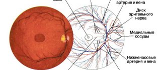
Angiopathy is an abnormal transformation of the vascular tone of vessels of various sizes in the fundus. They become abnormally convoluted, narrow or widen. As a result, the speed and volume of blood flow, as well as innervation, changes. Violations can be seen by an ophthalmologist. They indicate the presence of more serious diseases that caused this symptom.
It's important to understand. If the blood vessels in the eyes are narrowed or the capillaries are dilated, then this is a consequence of some disease, and not an independent disease.
This clinic indicates the presence of a pathology that interferes with normal hemocirculation, changing the tone of the veins and arteries. If the situation is not corrected in time, necrotic changes in the retina, deterioration in the quality of vision or its loss (complete or partial) are possible.
As a rule, a similar situation is diagnosed in patients over 35 years of age who have certain chronic diseases. In children, pathology is rarely recorded.
Symptoms
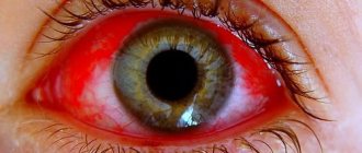
If the vessels of the fundus of the eye are narrowed, floaters appear before the eyes. Patients often experience darkness in their eyes, sometimes dark spots, pain and stinging occur. The pathology is often accompanied by headaches.
After work that involves high concentration of attention, pulsation occurs.
After some time, a person experiences visual disturbances. They develop, becoming more persistent.
It is worth considering that the symptoms of the disease depend on its type.
Hypertensive angiopathy
A chronic increase in pressure destroys the walls of blood vessels, leading to damage to their inner layer - the endothelium. As a result, compaction of these areas is observed. After some time, they undergo fibrotic processes.
In the area where the vessels intersect, compression of the veins is observed, which leads to impaired blood flow. As a result, conditions are created for the formation of blood clots and hemorrhages.
A person has high blood pressure, rupture of individual vessels. As a result, angiopathy transforms into retinopathy.
A typical symptom of hypertension is vascular tortuosity. According to statistics, at the early stage of hypertension this symptom is diagnosed in 25-30% of people, while at the third stage all people experience abnormal processes.
In difficult situations, hemorrhages occur, the retina becomes cloudy, and destructive processes are observed in its tissues.
Diabetic angiopathy
When diabetes appears in a patient, the structure of the small vessels of the retina and larger vessels of the internal organs is disrupted. As a result of these processes, the patient becomes disabled.
A complex consequence of disorders is retinopathy. It occurs in 90% of people with diabetes. This deviation can be detected already in the initial stages of the disease. This is carried out during an ophthalmological examination, when there are still no symptoms from the organ of vision.
Deterioration of vision is a late sign that indicates the irreversibility of changes. With prolonged diabetes, vision is impaired so severely that the person becomes unable to work.
Complete blindness with this disorder is 25 times more common in patients with diabetes than in those who do not have this disease.
Traumatic angiopathy
This form of pathology is caused by compression of the skull, chest, and abdominal cavity. It can also be a consequence of a neck injury. Damage to the blood vessels of the organ of vision is caused by a sharp increase in pressure and compression of the blood vessels in the neck area.
Characteristic symptoms of such an anomaly include severe vasoconstriction and hemorrhage in the retina. This process is characterized by a sudden deterioration in vision. Moreover, it is not always possible to normalize it.
Hypotonic angiopathy
Weakening of vascular tone and a decrease in the rate of blood flow during hypotension form the prerequisites for the appearance of blood clots.
This type of illness is accompanied by a noticeable expansion and branching of the arteries, a sensation of pulsation in the veins, which a person can feel. Many people also experience headaches and dizziness.
Patients often experience weather dependence.
Juvenile angiopathy
The second name for this little-studied anomaly is “Eales disease.” This condition is extremely rare. It manifests itself in the form of inflammation of the retinal vessels, which has an unclear etiology.
With this disease, hemorrhages occur. They can be localized in the area of the retina or vitreous body. There is also a risk of connective tissue overgrowth. This leads to dangerous consequences such as retinal detachment or the development of cataracts.
In addition, the anomaly can cause glaucoma.
How to detect pathology?
It is impossible to detect retinal angiopathy with the naked eye. This requires special equipment - ophthalmoscopes, slit lamps, ultrasound machines.
Ophthalmoscopy
An ophthalmoscopy allows the ophthalmologist to examine the arteries and veins of the fundus through the pupil. The most common methods of examining the retina include:
- Indirect ophthalmoscopy (mirror). This procedure is performed using a mirror ophthalmoscope and a magnifying glass. The fundus image is presented as a mirror image and provides only an overview.
Direct ophthalmoscopy. Examination of the fundus is carried out using an electric ophthalmoscope. This procedure is more informative compared to the mirror procedure.- Ophthalmochromoscopy. Examination of the fundus with an ophthalmoscope with replaceable filters. The use of colored glasses (red, green and blue) in different combinations during the procedure makes it possible to identify initial changes in the retinal vessels that remain invisible in white.
- Polarizing ophthalmoscopy. Examination of the fundus in polarized light makes it possible to detect retinal edema in the initial stage.
Treatment
In order for the treatment of narrowing of the vessels of the fundus to produce results, the underlying pathology should be treated.
To slow down or completely stop the progression of changes, it is necessary to use drugs to normalize blood sugar levels and antihypertensive drugs. It is also recommended to follow a special diet.
The rate of abnormal changes in blood vessels depends on the effectiveness of therapy for the underlying pathology. Treatment should be combined.
It is not only the ophthalmologist who should monitor therapy. In such a situation, supervision of a therapist or endocrinologist is required.
In addition to medications, physiotherapy methods are used. There may also be a need for local therapy and nutritional correction.
If you have diabetes, following a diet is as important as taking medications. You should remove foods that contain a lot of carbohydrates from your diet. Instead of animal fats, you should eat plant foods.
The daily menu should include vegetables and fruits. Eating fish is also beneficial. The menu should also include dairy products. Be sure to control your weight and monitor your sugar content.
Medications
Vascular damage requires the use of a whole range of medications:
- Drugs to normalize blood flow. These include Trental, Actovegin. It is worth considering that this category of products should not be used by women during pregnancy and lactation. They are also contraindicated for children. If it is necessary to treat these categories of patients, the decision on the appropriateness of a particular drug must be made by the doctor.
- Means for reducing the permeability of vascular walls. This group includes calcium dobesilate and parmidine.
- Substances to reduce platelet aggregation. These include dipyridamole and ticlodipine.
- Vitamins. It is important to take vitamins B, C, P, E.
Courses of therapy usually last 2-3 weeks. They must be repeated 2 times a year. All substances can be used only after consulting a doctor.
If you have diabetes, it is important to use medications to lower your blood sugar and stick to your prescribed insulin dosage. In case of atherosclerosis and arterial hypertension, means are needed to normalize blood pressure parameters and cholesterol levels.
In addition to systemic medications, the ophthalmologist may prescribe eye drops. The most effective means include the following:
- Vitaminized substances - these include lutein complex and anthocyanin forte;
- Vascular agents – emoxipin, taufon.
Medicines ensure normal blood flow in the eyes. Thanks to this, the patient's condition improves significantly.
Physiotherapy methods
For treatment to be effective, it must be comprehensive. Most often, the following methods are used to treat angiopathy:
- Magnetic influence;
- Acupuncture;
- Laser exposure.
Folk remedies
In addition to standard therapy, you can use home recipes:
- Mix 100 g of chamomile, immortelle, St. John's wort. Take the same volume of birch and yarrow buds. Add 500 ml of boiling water to 1 large spoon of the mixture and leave for 20 minutes. Bring the strained product to the original amount. Take 1 glass in the morning and before bed. In the evening after using the product, it is forbidden to drink or eat.
- Take 1 small spoon of mistletoe powder, add 1 cup of boiling water and leave to steep overnight. Take 2 large spoons twice a day. Treatment should last 3-4 months.
- Mix 15 g of lemon balm and valerian rhizomes, add 50 g of yarrow. Mix 2 small spoons of the mixture with a glass of water and leave for 3 hours. Infuse the composition in a cool place. Place in a steam bath for a quarter of an hour, cool and strain. Add water to make 250 ml. Take throughout the day in small portions. This treatment should be continued for 3 weeks.
Narrowing of the eye vessels indicates various pathologies and can provoke negative health consequences. To minimize the likelihood of illness, you should consult a doctor in a timely manner and strictly follow his instructions.
Types and causes of rare angiopathy
There are some types of pathologies that are diagnosed relatively infrequently. For example, juvenile angiopathy.
It is difficult to determine why the blood vessels in a child’s eyes are narrowed and often this question remains unresolved, but the situation is quite dangerous and requires an immediate solution. In premature infants, pathogenesis may be associated with birth trauma or as a result of intrauterine developmental anomalies.
Angiopathy during pregnancy in the early stages of pathogenesis is not something very dangerous, but if the process is neglected, tissue detachment may occur. More often the disease appears at the end of the second and third trimester. As a rule, the cause is hypertension or other pathologies that weaken or narrow small blood vessels.