Types of eye cysts
The disease is classified according to causes and clinical symptoms:
- Congenital cavity - occurs due to dissection of the iris. It can develop within several years after birth, but appears at a young age before 7 years.
- Spontaneous cyst on the eyelid - serous (in terms of fluid content) and pearly (resembles a white dense ball). The causes are unknown, cavities appear at any age. It is known as a lower eyelid cyst because it most often appears on the lower eyelid.
Serous tumor of the upper eyelid. - The traumatic type (or corneal cyst) is formed due to the impact of epithelial cells on the eye organ. For example, an eyelash can get into the mucous membrane of the eye and gradually grow.
- Exudative - develops due to eye diseases (for example, glaucoma).
- Cyst on the conjunctiva of the eye - there are two types: implantation (after surgical treatment) and retention (due to stagnation of lymph and purulent fluid).
Tumor on the conjunctiva of the eye organ. - Stromal - the cavity appears unexpectedly, can move throughout the eye, and then disappear. Causes significant discomfort.
- Dermoid cyst of the eye - is formed due to fragments of epithelial cells of the hair, skin and nail plates.
- A Moll cyst develops due to blockage of the sweat glands of the eye.
Most often, an eye cyst is conjunctival, as it occurs on the conjunctiva.
Diagnosis of the disease
To make an accurate diagnosis of a cyst that has formed, it is worth going through a number of procedures:
- External examination of the skin of the eyelids and face.
- Visiometry - this study reveals a person’s visual acuity, which may be reduced due to certain diseases.
- Perimetry.
- Analgizemetry - allows you to determine the sensitivity of the cornea.
- Biomicroscopy of the eye.
- Examination of the eyes for the presence of transparency of their environment.
- Ophthalmoscopy.
Chalazion treatment
If we talk about Moll cysts, then you will have to take scrapings from the affected skin to determine antibodies to the virus. A general blood test is also taken to determine whether inflammation is present in this disease. When diagnosing a dermoid cyst, computed tomography or MRI is considered an effective method; they visualize in detail the required area with the cyst.
Symptoms
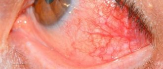
The neoplasm at the initial stage causes virtually no discomfort and grows asymptomatically. You can detect a cyst by external examination or palpation of the eyelids - the ball will be dense and painless, felt like a small nodule under the skin.
Most cysts are not detected after a couple of weeks because they resolve on their own. However, they often increase in size, causing significant discomfort and affecting vision. The neoplasm enters the second and third stages of development imperceptibly, showing clinical signs:
The tissues of the eyelid do not close tightly due to the tubercle that has arisen.
- narrowing of the field of vision - depending on the location of the eye, it ceases to cover most of the space. Most often it develops when the cyst is on the outside of the conjunctiva;
- clouding - vision becomes cloudy, the picture is not fully perceived, since the cyst actually blocks the view, being on the eyeball;
Swelling of the eyelids.
- flickering before the eyes;
- bright flashes on the back of the eyelids;
- dull pain that bursts the eye organ from the inside;
- the tissues of the eyelid do not close tightly due to the tubercle that has arisen, and deformation develops;
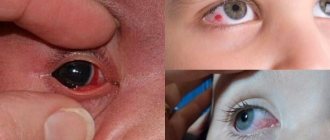
- redness of the eyeball;
- swelling of the eyelids (often affects the upper ones, less often the lower ones).
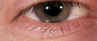
Externally, the cyst resembles a ball with yellow contents in the center. Many people mistake the tumor for a purulent pimple if it is located on the eyelids. The significant difference is that the cavity ball is dense and moves under pressure rather than being squeezed out like a pimple. Also, around the cyst on the eyelid there is no characteristic redness like inflammation.
Symptoms of the disease
|
|
The neoplasm may have a red-brown, transparent, whitish-blue tint. Once these symptoms appear, you should consult a doctor. A timely diagnosed cyst is a guarantee of quick treatment.
Return to contents
Causes
The development of a cyst on the eye is provoked by a number of factors, mainly eye diseases:
- Congenital abnormalities (for example, delamination of iris pigment).
- Eye infections (conjunctivitis).
- Diseases of the eyelids (stye).
- Worm infestation.
- Inflammation of the sclera.
- Long-term course of treatment, including surgery to restore vision.
- Mechanical damage.
- Constant friction due to contact lenses.
In rare cases, decorative cosmetics and false eyelashes can provoke the unwanted development of a benign cavity.
The tumor also appears as a postoperative complication. Some causes of eye cysts are due to external influences (for example, dust or sand).
Causes of the disease
Heredity is one of the main factors of the disease. A cyst can appear in the womb, early or teenage years. Chronic illnesses, abuse of tobacco products, and various alcoholic beverages carry the greatest risk to the fetus. And they lead to the appearance of dermoids in the eyes.
Another cause of the neoplasm can be trauma, wounds, mechanical damage, surgery, or removal of the pterygium. According to ophthalmologists, a parasitic and inflammatory process can lead to the disease. There are cases when the disease occurs without any prerequisites.
Impaired fluid outflow from the glands and inflammatory processes can be caused by low-quality decorative cosmetics. It contains a lot of oils that cause clogged pores.
Hypothermia also causes cyst formation. It is often found on the lower eyelid, but can also appear on the upper eyelid. The pathology is not related to the skin. If you press on it, it will be able to move freely. The eyelid turns red. And the sizes can vary from a few millimeters to 1 cm.
Diagnostics
To complete the picture, an ultrasound of the eyeball is performed.
Diagnostic procedures for eye tumors vary depending on the factors of formation. The tumor is detected using special equipment, including mirror and lens systems. To complete the picture, an ultrasound of the eyeball is performed (allows us to find out how deep the cyst is in the tissues of the eye). But, if the disease is caused by helminthic infestation or inflammation of the sclera, additional clinical blood tests are performed.
Surgery
It happens that it is not possible to cure a Moll cyst using general methods; in this case, you need to resort to surgery. The cyst must be removed; if this is not done, there is a huge risk to health.
This operation is not classified as complex. During this procedure, the doctor simply squeezes the site of the cyst formation and removes all its contents using a sharp object. The duration of the operation usually does not exceed half an hour, and almost immediately after its completion the patient is sent home. Any operation to remove a cyst from the eye is performed under local anesthesia. To avoid the inflammatory process after surgery, doctors apply an ointment that has a disinfecting effect to the place where the cyst was. The patient's eye must be bandaged, and the bandage can be removed after three days, but only after examination by a specialist.
Treatment
After detection of a cystic lesion of the eyeball, treatment is determined according to the type of tumor, its location and the size of the tumor. Growth rates are also taken into account. The main treatment is surgery. The operation takes less than half an hour, there are practically no complications. Timely treatment allows you to avoid such radical measures. Sometimes physical therapy and regular eye rinsing are sufficient.
Physiotherapeutic procedures:
Gentle eyelid massage.
- UHF exposure;
- soft eyelid massage;
- temporary heating using lasers;
- warm compress based on medicinal herbs;
- washing with infusions of medicinal herbs;
- electrophoresis.
Physiotherapy is completely contraindicated if the cyst has formed after an infection. When heated, the inflammatory process may develop into an abscess, or the neoplasm will rupture and fluid will flow into the eye area.
Drug treatment
Medicines are prescribed if the disease occurs after an infection, or as a complication of vision pathology.
The medication course consists of anti-inflammatory drugs in the form of drops.
The drug course consists of anti-inflammatory drugs in the form of drops (drops are instilled directly if a tumor of the conjunctiva of the eye is detected) and ointments for lesions of the eyelids. Additionally, antihistamine emulsions are prescribed to relieve secondary symptoms.
Causes
There are many reasons why the disease develops.
Quite often, a cyst occurs due to poor hygiene. Bacteria that enter the eyes through the palms cause inflammation.
A cyst can occur after an injury to the eyelid, since during this period the integrity of the skin is compromised.
Contact lens wearers who also do not follow hygiene rules often develop this disease.
Decreased immunity and excessive use of antibiotics cause changes in the normal composition of tear secretions.
The use of low-quality and cheap cosmetics can provoke inflammatory processes. For their manufacture, harmful materials are usually used, which lead to clogging of pores.
Various allergic reactions can lead to this disease.
Experts come to the conclusion that constant hypothermia is the main factor for the development of cysts.
As a rule, a Moll cyst does not require surgical treatment; it goes away on its own over time. It is recommended to use antiseptic drugs. However, if a cyst bothers a person for several days, it is necessary to consult a doctor. Don't put off visiting a specialist.
Removing a cyst on the eye
A tumor on the eye must be removed using various methods. Intensive growth of a tumor is an indicator for surgical intervention.
Laser removal
The cyst is removed in a gentle manner, leaving healthy tissue unaffected. The laser acts in a targeted manner, producing a bactericidal effect. Characterized by rapid rehabilitation and a short postoperative period. There are no defects in vision or appearance. It is practiced only for small formations that do not show a growth trend.
Dissolution of tissue occurs when a laser beam is applied.
The whole process consists of three important stages:
- preparing the eye for laser exposure (the tissue around it is treated with a solution, a micro-hole is made);
- dissolution of neoplasm tissue;
- aspiration (pulling out contents from the eye).
Tissue dissolution occurs when a laser beam is passed through a thin tube whose walls protect healthy tissue. The laser burns the neoplasm tissue, and at the same time the damaged vessels are fused. The liquefied contents are removed using a vacuum tool - it is pulled out of the cavity.
Surgical removal
Indicators for surgery are intensive growth, or an advanced state, and if drug therapy has proven to be ineffective. Surgery is often the only solution, since the eye area is dangerous to be affected by long-term drug treatment. This will also lead to relapse or the development of cysts. Surgery is required if a dermoid cyst of the eye appears.
The site for excision of the tumor is marked.
Excision of the cyst does not take more than an hour; the operation is performed under anesthesia. Afterwards, a bandage with antibiotic ointment is applied, and the eye is completely covered with a protective bandage. The patient is observed for a week, or comes for examination on his own within 5 days. Dressings are only done in the hospital to avoid infection. The bandages are removed after the operated area has completely healed. Medicines are prescribed to support the immune system, weakened due to the intervention.
After the operation, a bandage with antibiotic ointment is applied, and the eye is completely covered with a protective bandage.
After complete removal of the cyst, the eye organ quickly recovers. There are no complications. It is recommended to avoid physical and psychological stress for six months. It is also necessary to avoid stressful situations. Intense sports, swimming and baths or saunas are completely contraindicated. Exposure to high temperatures and steam combined with humidity can cause the cyst to recur.
The only noticeable drawback is that after the operation, vision is fully restored within 1-2 months. A complication may also arise in the form of decreased vision, after which maintenance therapy or even prophylactic wearing of glasses will be required.
Classification
The tumor that appears can be localized on the upper or lower eyelid. It's individual. Often, hollow formations simultaneously affect both eyelids. On palpation, a dense knot is heard under the fingers. Externally, the eyelid protrudes slightly, this depends on the size of the formation.
The disease is classified according to its causes:
- Congenital. It can be diagnosed in infants. Dissection of the iris occurs.
- Traumatic – occurs after surgery, injury, or eye trauma.
- The consequence of glaucoma is the formation of an exudative cyst. The cause may also be the use of medications that cause the smooth muscles of the eye to contract.
- Dermoid. Occurs when the development of epithelial cells is disrupted. After opening, hairs and small particles of skin can be found inside the hollow formation. A cyst may not bother a person for a long time or cause pain. With its active growth, a decrease in vision is observed. There is a risk of malignancy formation.
- A Moll cyst on the eyelid provokes the development of lacrimation and photophobia. There is swelling. Inside the eyelid, rashes begin to form; they are transparent and bubbly. Several bubbles merge with each other, this increases the size of the formation. The cyst may begin to fester. In this case, the person feels a general weakening of the body, and the body temperature may rise. In this area, sensitivity to stimuli of any nature increases.
- Another type of pathology is a spontaneous cyst. It can occur on the upper or lower eyelid, or affect both. The size reaches a small bead. It is impossible to find out the cause of its occurrence. These types of formations include pearly and serous cysts.
In most cases, diagnosis of formation occurs in late stages. Previously, it does not manifest itself and does not cause discomfort or pain. Using an ophthalmic lens and mirror, the doctor examines the patient and then makes a final diagnosis.
Folk remedies
Most folk methods involve the use of decoctions based on medicinal herbs. It is important to remember that such treatment can only be used after examination in a hospital. It provides support and can provoke resorption of the cyst, but will not cure if the tumor occupies a large area.
The most commonly used means:
- Algae tincture - dry seaweed called Fucus is used. The collection is purchased at the pharmacy. Three tablespoons of the dry mixture are poured with boiling water into a thermos and allowed to brew overnight. Then pour into an ice tray and freeze. Use the resulting cubes to wipe the eye area and eyelids.
Fucus is poured with boiling water, after which it is frozen into cubes and ground at the site of the tumor. - Herbal infusion - you should take cornflower petals, plantain leaves and cumin. All herbs are well crushed and then poured with boiling water. The cooled solution is filtered. You should drop 3 drops into the eyeball at least five times a day.
- Guava decoction - only leaves are used. The affected eye is washed three times a day.
The following decoctions are used as compresses and for rinsing: acacia leaf based, chamomile tea, rose water with honey.
Treatment of a cyst in the eye with folk remedies can aggravate the course of the disease, so it is recommended to consult a doctor. The main thing to remember is that traditional methods are only an auxiliary means of treatment.
Treatment of cysts on the eyelid (upper and lower): surgery, laser, drugs
A cyst on the eyelid is a benign formation that occurs under the influence of many factors, for example, mechanical damage to the organ of vision, the development of infection or congenital defects.
When choosing a treatment method for a Moll cyst on the eyelid (also called the disease), the doctor must take into account the severity of the symptoms, the course and form of the disease.
Features of therapy for cysts
When diagnosing a cyst, doctors, as a rule, are in no hurry to prescribe a special course of therapy, preferring to wait. But if the tumor does not resolve on its own within 2-3 weeks, then the patient is prescribed a complex of treatment.
It will depend on how long ago the pathology developed, the size of the formation, as well as the presence of concomitant pathological processes.
Ignoring the symptoms of the disease can lead to serious complications, including: deformation of the lens of the eye, decreased visual acuity, and deterioration in the patient’s quality of life.
Various methods can be used to treat cysts, from the use of medications to surgical treatment. For maximum therapeutic effect, an integrated approach can be used.
Prevention
Any prevention involves maintaining visual hygiene. It is necessary to wash off makeup thoroughly, use only clean contact lenses and for a short period of time. Massage your eyelids for at least a minute during the day. Wash your face daily with clean water, rinsing your eyes and avoid getting soap suds.
The prognosis for treatment of any neoplasm in the eye is always favorable. It is necessary to follow all doctor's recommendations to avoid recurrence of the disease or its complications.
Reasons for appearance
Risk factors that increase the likelihood of occurrence:
- Intoxications of various types (medicines, radiation exposure) during the period of intrauterine development of the fetus, genetic disorders, mutations.
- Inflammatory processes: infectious, parasitic, allergic. Mechanical damage (injuries, burns).
- Age-related changes (dystrophic involutive processes).
- Postoperative processes in tissues.
- Long-term use of medications (ointments, drops).
- Wearing contact lenses.
- Poor quality cosmetics.
Cyst treatment
There is a false opinion - “it doesn’t hurt, so it doesn’t need to be treated.” Or “it will resolve on its own.” However, cases of self-healing are so rare that you should not hope for them. You should also forget about the complications described above. Therefore, when a cyst is detected, treatment must be started immediately.
If the size of the cyst is small and there is no inflammation, you can limit yourself to conservative treatment methods.
These methods include:
- Medication. A number of medications are used to treat the underlying causes of the disease. Hormonal medications eliminate imbalances in the endocrine system. General strengthening and vitamin complexes consolidate the result. In the presence of background stress, sedatives are used.
- Aspiration. Using a thin needle, liquid is drawn out of the cavity. The cavity can be filled with a medicinal composition; this is one of the methods of treatment.
If the disease does not respond to conservative treatment methods, has progressed to dangerous forms, or complications have arisen, surgical methods are used:
- Laparoscopic surgery. Minimally invasive intervention using a laparoscope, which makes a puncture in the mammary glands. It is used if the size of the cyst is small.
- Surgical removal of the formation. This is done by cutting fabric. It is used if the size of the tumor is large. During the operation, the portion of the gland affected by the cyst can be removed.
- Surgical removal of the mammary gland. It is used if almost the entire gland is affected by polycystic disease. After this, reconstructive breast surgery is performed.
Since the organ being operated on is external, the operation is not complicated. Does not require a long recovery period after it. Modern surgical technology makes it possible to minimize postoperative scars. After the punctures and incisions heal, they become barely visible or even invisible.
The following treatment methods used in conjunction with conservative and surgical methods should be added here:
- Diet therapy. It will help get rid of the risk of cyst re-formation. By normalizing nutrition, they eliminate the risk of obesity and hyperexcretion of estrogen.
- Physical training. Moderate physical activity will restore your metabolism. This significantly reduces the risk of relapse.
- Psychotherapy. If there is constant stress in a woman’s life, there is a possibility that the disease will return. It is necessary to eliminate stress and restore the patient’s psycho-emotional state.
By diagnosing problems in the early stages, surgery can be avoided. Following simple rules of prevention will prevent the recurrence of the disease.
Causes of pathology
An eye cyst can vary in structure and mechanism of formation. But there are a number of prerequisites that create favorable conditions for its development.
These include:
- developmental anomalies during pregnancy. This includes anomalies in the structure of the visual organs and the layering of non-specific tissues;
- inflammatory processes on the eyelids;
- viral and bacterial eye infections and their complications;
- injuries, scratches, bruises, ingress of foreign objects;
- eye surgeries (lens replacement, strabismus correction, etc.);
- complications, side effects after long-term use of drops and eye ointments;
- prolonged allergic reaction;
- glaucoma;
- constant irritation from wearing lenses or incorrectly attached false eyelashes. The mucous membrane and skin of the eyelids rub, resulting in infection and epithelial particles;
- diabetic retinopathy. The integrity of the retina is damaged. Its surface becomes uneven. As a result, not only vision deteriorates, but edema forms and cysts grow;
- parasitic infection (helminths make their way from the intestines and are brought in with dirty hands).
These reasons do not guarantee the development of neoplasms. But they create favorable conditions for their formation. Why, under the same conditions, some patients develop pathology and others do not, science does not know.

