Why is blepharitis dangerous?
In ophthalmological practice, blepharitis represents a large group of polyetiological disorders that often recur. Their treatment is a complex, time-consuming process. The advanced course of the disease leads to complications such as keratitis, uveitis,
In this article
- Why is blepharitis dangerous?
- Causes and complications
- Features of pathogenesis
- Classification
- Common symptoms of eye blepharitis in adults
- Symptoms of various forms
- Complications
- Diagnosis of blepharitis
- How to treat eyelid blepharitis in adults?
- Treatment of seborrheic blepharitis
- Treatment of allergic blepharitis
- Actions in case of relapse of the disease
- Prognosis for blepharitis
- Prevention
chalazion, as well as a decrease in the optical ability of the eye. The prevalence of the disease worldwide is quite high and reaches 35%. The risk group includes women and men in the age group from 40 to 65 years. The disease is rare among children.
Causes and complications
A large number of causes are involved in the occurrence and development of the disease. Depending on the etiological factor, the following are distinguished:
- Infectious blepharitis, which is caused by pathogenic staphylococcal microflora, mycotic lesions (fungi), and mites. The lesion involves the superficial epidermal part of the eyelid and the hair follicles of the eyelashes.
- Non-infectious blepharitis caused by other damaging factors (traumatic, allergic).
Inflammatory diseases of other anatomical structures may also be the cause. The chronic nature of eyelid damage is determined by the presence of an infectious agent in the foci of the pharyngeal tonsils with tonsillitis, maxillary and frontal sinusitis (sinusitis, frontal sinusitis, pansinusitis), hard tissues of the tooth (with cariogenic processes, purulent pulpitis), as well as in the soft tissues of the maxillofacial area (abscesses) , phlegmon).
In rare cases, the cause is herpes simplex viruses (I, II, III), intestinal gram-negative bacteria, yeast fungi, and molluscum contagiosum. Blepharitis can be accompanied by complications that complicate the course of the disease and clinical symptoms. These include:
- Conjunctivitis.
- Uveitis.
- Keratitis.
- Chalazion.
- Stopping eyelash growth.
- Corneal injury.
- Deterioration of vision (astigmatic manifestations, myopia, farsightedness).
- Spread of the inflammatory process to the optic nerve.
Inflammatory diseases of the eyelids
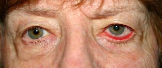
Blepharitis is a bilateral inflammation of the eyelid margins. Depending on the location, blepharitis is divided into anterior marginal and posterior marginal. Anterior marginal blepharitis is a local manifestation of an infectious lesion of the skin of the eyelids. Depending on the symptoms, it is divided into simple, scaly, ulcerative and angular. Posterior marginal blepharitis is a consequence of dysfunction of the meibomian glands. Depending on the cause of blepharitis, a distinction is made between meibomian and demodectic.
Blepharitis is accompanied by itching and pain, a feeling of heaviness of the eyelids and a foreign body, increased visual fatigue, and general discomfort. With scaly (seborrheic blepharitis), many small scales appear on the skin of the edges and eyelashes. With ulcerative blepharitis, purulent crusts and ulcerations form along the edges, and the eyelashes stick together. Posterior marginal blepharitis (dysfunction of the meibomian glands) is accompanied by redness and thickening of the edges of the eyelids, hypersecretion of the meibomian glands, accumulation of yellowish-gray foamy secretion in the outer corners of the eye, hyperemia of the conjunctiva of the eyelids. With demodectic blepharitis, thickening and redness of the edges of the scales, crusts, and white muffs on the eyelashes are noted.
If the cause of blepharitis is identified early, treatment is short and the outcome is favorable.
Treatment of blepharitis is etiotropic, general and local. Local treatment consists primarily of careful hygienic care of the eyelids. In case of ulcerative blepharitis, the crusts and discharge are removed with a damp cotton swab. Rough crusts are first softened with a damp lotion or by lubricating the edges of the eyelids with ointment.
An ointment consisting of a corticosteroid and an antibiotic is applied to the edges of the eyelids. In cases of conjunctivitis or marginal keratitis, additional eye drops are instilled.
If there are no ulcers or after they have healed, massage the edges of the eyelids on a glass rod daily. The edges of the eyelids are dried and degreased with alcohol or ether on a cotton swab, the eyelids are massaged, and then the edges of the eyelids are lubricated with an alcohol solution of brilliant green. At night, the edges of the eyelids are treated with ointment containing corticosteroids and antibiotics.
For demodectic blepharitis, ointments are used that act on the causative agent of the disease. Before going to bed, the edges of the eyelids should also be generously covered with ointment, this disrupts the life cycle of mites.
Complications of blepharitis - entropion and trichiasis - are eliminated with the help of plastic surgery.
Treatment of blepharitis should be systematic and long-term. For successful treatment it is necessary to establish the etiology of the disease. The best preventive measure is the correction of those disorders that contribute to its occurrence.
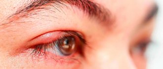
Barley is an acute purulent inflammation of the sebaceous gland of the skin of the eyelid. The causative agents are pyogenic microorganisms, most often staphylococcus. Limited redness and swelling appear in the affected area. After 2-3 days, a purulent core begins to form in the center of the lesion. Then a purulent pustule forms, with reactive hyperemia and edema noted around it. After three days, the pustule usually opens and pus is released from it. A small wound forms at the site of the stye and a scar forms.
Sometimes barley occurs as a boil with abscess formation. In this case, very pronounced infiltration with thickening of the skin of the eyelids predominates. After opening the boil, a crater-shaped depression is formed, at the bottom of which there is a necrotic purulent plug. The necrotic tissue is rejected, and a rough scar forms in its place.
Acute meibomitis or internal stye is an inflammatory process on the inside of the eyelids, and not on the outside, as with stye. The disease is caused by inflammation of the glands of the cartilage of the eyelids.
Treatment of barley and meibomitis is most often local. In the area of the affected skin of the eyelids, cauterization is done with 70% alcohol or an alcohol solution of brilliant green. The edges of the eyelids are lubricated with ointment, and antibiotic drops are instilled. In severe cases and general malaise, oral antibiotics and multivitamins are prescribed.
Chalazion or hailstone is a sluggish and almost painless chronic inflammation of the gland of the cartilage of the eyelids. In some cases, it develops after acute meibomitis or stye. In the thickness of the cartilage of the eyelid, a dense, round, easily palpable compaction of various sizes is formed.
For small sizes, a long-acting corticosteroid is injected into the chalazion cavity. For large chalazions, treatment is surgical on an outpatient basis.
Abscess, or phlegmon of the eyelids , is a limited or diffuse infiltrative purulent inflammation of the tissues of the eyelid. It can occur due to direct infection during damage to the eyelid, spread from surrounding structures, or become a consequence of metastatic spread of infection from other foci (pneumonia, sepsis, etc.). The most common pathogens are gram-positive cocci or anaerobes.
When the disease occurs, a feeling of “fullness” and pain in the eyelid area appear; the skin of the eyelid is hyperemic, tense, shiny, the eyelid is sharply painful on palpation; fluctuation is possible; due to dense swelling of the eyelid, the palpebral fissure is sharply narrowed or closed; lacrimation and mucous discharge from the conjunctival cavity appear; regional lymph nodes enlarge; have symptoms of general intoxication.
Features of pathogenesis
The pathogenesis of blepharitis consists of a number of sequential processes:
- Disturbance in the process of washing away triggers and infections from the surface of the eye, development of xerophthalmia as a result of decreased functioning of the lacrimal glands;
- Endogenous allergization of the body, possible with general somatic pathologies that contribute to changes in the cellular and chemical composition of the lacrimal gland secretion;
- Provoking factors (hypovitaminosis, dust particles, prolonged exposure to the sun) that trigger the inflammatory process.
As a result, triggers and microbial flora affect the eyelid, and a symptom complex characteristic of this disease develops.
Classification
The disease has different localization and is divided into the following forms according to location:
- Marginal anterior (the process involves the ciliary edge);
- Posterior marginal (with damage to the lacrimal glands and lacrimal sacs);
- Angular (inflammation is localized in the corners of the eyes).
The following classification of blepharitis depending on clinical manifestations is generally accepted:
- Simple form;
- Demodectic mange (caused by a tick-borne agent);
- Osteofolliculitis (ulcerative-necrotic manifestations);
- Seborrheic (caused by dermatic lesions of the eyelid epithelium);
- Allergological;
- Rosacea-blepharitis;
- Mixed form.
Common symptoms of eye blepharitis in adults
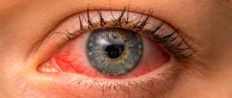
All forms of blepharitis, as for any inflammatory disease, are characterized by a single symptomatic complex: hyperemia (redness), swelling (edema), pain, dysfunction.
The classic variant of the clinical course is accompanied by itching, overstrain of the visual analyzer, hypersensitivity to light stimuli and physical factors. Depending on the damage to the ducts of the lacrimal gland, either xerophthalmia (dryness of the eye shell) or increased lacrimation develops. This contributes to infection of soft tissues and the organ of vision, and the spread of microbial agents. The inflamed epithelium becomes covered with a whitish or yellow coating, the eyelashes stick together or fall out. Eye discharge leads to the appearance of plaque and sticking of eyelashes.
Symptoms of various forms
Depending on the form of the disease, the following symptoms of eyelid blepharitis in adults are distinguished:
- The simple form of the disease is characterized by an inflammatory lesion of the upper eyelid: it thickens, becomes swollen, and painful. A whitish mucous secretion accumulates in the corners of the orbit. The conjunctival membrane turns red, its permeability increases. The tear ducts expand, and due to an increase in their diameter, the infectious agent spreads to other membranes of the eye.
- The main manifestation of seborrheic blepharitis of the eyelid in an adult is scaly cells of the keratinized epidermis. They are located between the eyelashes on the inflamed eyelid, compress the follicles, block the mouths of the sebaceous glands on the thickened and hyperemic edge of the eyelid. When the scaly form is advanced, depigmentation (loss of pigment) and loss of eyelashes, partially or completely, are possible. The scales can also be located on the eyebrows, skin around the orbit, and hair.
- The ulcerative-necrotic form is characterized by the appearance of crusts, upon removal of which an ulcerated epithelium is revealed. Depending on the stage of the process, the lesion may take the form of erosion, ulceration, or a necrotic area with a white fibrinous coating. After the wounds heal, scars or scars remain in their place, which slow down or stop eyelash growth. In severe cases, poliosis (depigmentation of the eyelash shaft) and madarosis (loss of the eyelash hair follicle from the epidermis of the eyelid) are observed. The aesthetics of appearance are disrupted, the skin wrinkles and increases in size.
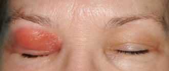
- The allergic form is characterized by a sudden onset and progressive course. Most often, exogenous agents are causative factors. Patients complain of severe swelling, profuse lacrimation, pain and hypersensitivity to physical irritants. With this form of the disease, an “allergic bruise” (dark purple skin color) is observed.
- Blepharitis caused by tick-borne demodicosis is accompanied by incessant itching. It is felt more clearly in the morning and subsides in the evening. The outer edge of the eyelid takes on a roll-like shape, swells and becomes hyperemic. A viscous secretion from the lacrimal gland is released from the corner of the eye, which collects and dries. The appearance of the eyes deteriorates, the circulation of tears may be disrupted, as a result of which toxic substances are retained in the epidermis.
- Blepharitis-rosacea is accompanied by the development of red nodules (nodus) on the thickened eyelid, which can merge. The presence of small diameter ulcers is noted, from which yellow contents are released.
Diseases of the human eyelids: treatment, prevention, symptoms, signs, causes
• redness of the eyelids;
• burning and severe itching in the eyes;
• gluing eyelashes;
• fear of bright light;
• headache;
• the appearance of excessive eye sensitivity;
• the formation of dry scales between the eyelashes, which are tightly glued together (during treatment, such scales will begin to fall off, without damaging the skin);
• loss of eyelashes;
• eyelid inversion is observed in advanced cases.
• formation of purulent ulcers in the eyelash growth area, which will bleed and become covered with a yellow coating (after the ulcers heal, visible scars remain in their place);
• the patient has severe weakness and fear of bright light;
• excessive loss of eyelashes;
• pain when blinking;
• temperature increase.
3. The demodectic form of blepharitis is observed when the eyes are damaged by mites.
• the appearance of very severe itching in the eyes;
• sleep disturbance;
• the appearance of sticky fluid released from the eyelids;
• visible redness of the eyelids;
• gluing of eyelashes.
In addition, when examined under a microscope, mites can be clearly seen in the eyelash growth area.
4. Allergic blepharitis in most cases occurs together with conjunctivitis.
• constant tearing;
• blurred vision;
• sore throat;
• the appearance of cutting pain in the eyes;
• damage to both eyes at once;
• rapid eye fatigue;
• severe sensitivity of the eyes to cold, wind and light;
• formation of a thin film on the eyes;
• burning in the eyes;
• changing the direction of eyelash growth;
• feeling of heaviness of the eyelids.
Complications
If treatment is unsatisfactory or advanced, blepharitis can be complicated by other diseases. These include:
- inflammatory lesions of the eye membranes (uveitis, keratitis, blepharoconjunctivitis, conjunctivitis);
- dry eye syndrome (xerophthalmia);
- acute and chronic varieties of meibomitis, chalazion;
- orbital abscesses;
- phlegmon of the orbital region;
- ulcerative necrotic lesions of the sclera and cornea.
One of the most dangerous complications is a decrease in the optical ability of the eye, as a result of which vision is partially or completely lost. Most often, inflammatory diseases tend to become chronic and recur over many years.
Diagnosis of blepharitis
The disease is determined by an ophthalmologist based on a thorough diagnosis. He interviews the patient, clarifies complaints, and collects the necessary data (allergic status, concomitant diseases, information about genetics). During the examination, visible symptoms characteristic of the inflammatory process are noted (redness of the eyelids, their swelling, the presence of accumulated secretions). The diagnosis is verified using laboratory and instrumental studies.
Visual acuity is determined using Sivtsev or Orlova tables (for children) from a distance of 5 meters. This method is subjective and does not provide an accurate assessment.
Objective methods of assessing vision include refractometry (detection of myopia, presbyopia, astigmatic disorders) and skiascopy.
To examine the eyelids and adjacent tissues, as well as the membranes of the eye in an adult, biomicroscopy of the eye and fundus is used. Additionally, the state of the accommodative capabilities of the lens and lens fibers, the optic nerve (its color, shape, diameter) is assessed.
In the case of demodectic blepharitis, a microbiological examination of eyelashes is prescribed to identify mites; in the case of an allergic form, allergological skin tests are performed to determine the irritant. Histological examination methods make it possible to exclude oncological diseases (basal, squamous cell carcinoma). Tissue collection is performed using a biopsy.
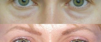
Swelling of the eyelids
Allergy
Diabetes
Pyelonephritis
11719 November 16
IMPORTANT!
The information in this section cannot be used for self-diagnosis and self-treatment.
In case of pain or other exacerbation of the disease, diagnostic tests should be prescribed only by the attending physician. To make a diagnosis and properly prescribe treatment, you should contact your doctor. Swelling of the eyelids: causes of occurrence, what diseases it occurs with, diagnosis and treatment methods.
Definition
Swelling of the eyelids is a pathological condition caused by the accumulation of fluid in the loose subcutaneous fatty tissue of the periorbital zone. It can be sporadic or rare, caused, for example, by insufficient sleep. Normally, swelling of the eyelids goes away on its own within 24 hours. If an unpleasant symptom takes on a permanent form, a full diagnosis of the body is required.
Swelling of the eyelids in most cases signals the development of serious pathological processes in the body.
Types of swelling of the eyelids
Swelling of the eyelids can be classified according to several parameters.
Firstly, edema can be of inflammatory (infectious, allergic) and non-inflammatory origin. In the first case, swelling of the eyelids is accompanied by redness, pain, increased temperature of the tissues of the periorbital zone, and lacrimation. The above signs are not typical for non-inflammatory edema.
Secondly, swelling of the eyelids can be unilateral or bilateral. Thus, unilateral edema is more often infectious, while bilateral edema has a wider range of causes.
Thirdly, edematous tissue can be soft and dense, which plays an important role in determining the cause of the development of edema.
Finally, swelling of the eyelids may be the only manifestation of edema syndrome, or may be combined with edema of other localizations, incl.
with swelling distributed throughout the body. Possible causes of eyelid swelling
Edema is the accumulation of excess fluid in tissues outside the blood and lymphatic vessels. There are several factors that contribute to the release and accumulation of fluid in the tissue. These include:
- high pressure created by the flow of blood inside the vessels;
- increasing the permeability of the vessel wall;
- a decrease in the concentration of blood plasma proteins, which, due to osmotic forces, retain the liquid part of the blood inside the blood vessel;
- an increase in the concentration of proteins and inorganic ions in tissues that attract water like a magnet.
The listed mechanisms play a dominant role in the development of eyelid swelling.
Among the more rare causes are a violation of the outflow of venous blood and a violation of the outflow of lymph due to narrowing of the lumen of blood vessels (for example, a blood clot, compression of a vessel from the outside, trauma). Symmetrical swelling of the eyelids that appears in the morning may be a consequence of consuming excessive amounts of liquid the night before bed.
Such swelling usually disappears on its own with proper drinking regimen. What diseases cause swelling of the eyelids?
First of all, swelling of the eyelids may indicate kidney disease:
- glomerulonephritis
- inflammatory damage to the glomeruli of the kidneys after a streptococcal or other infection; - secondary damage to the renal glomeruli in diabetes mellitus
,
chronic hypertension
; - pyelonephritis
is a disease (usually of infectious-inflammatory origin) characterized by damage to the renal collecting system and interstitium; - tubulointerstitial nephritis
- a group of diseases that primarily affect the kidney tubules, for example, toxic damage to the kidneys from salts of heavy metals; - other pathological processes in the kidneys, leading to the development of acute kidney injury
or
chronic kidney disease
.
Other pathologies that are characterized by the appearance of swelling of the eyelids include infectious diseases
of bacterial and viral origin.
Swelling of the eyelids is also a characteristic sign of allergic reactions, in particular angioedema
(Quincke's edema), manifested by swelling of the soft tissues of the head and neck: eyelids, mucous membranes in the pharynx and larynx.
This condition is life-threatening and occurs in response to the body’s contact with allergens coming from food, inhaled air, etc.
Dense persistent swelling of the eyelids is typical of
myxedema
- acquired insufficiency of the thyroid gland, which is characterized by a state of hypothyroidism - a decrease in the concentration of the hormones thyroxine and triiodothyronine.
Widespread swelling, incl. swelling of the eyelids, develops with severe heart failure
, however, “cardiac” edema is characterized by its spread from bottom to top: from the feet and legs to the upper half of the body.
Edema also develops when the synthesis of plasma proteins by the liver is impaired, which occurs, for example, in liver cirrhosis
.
Which doctors should I contact if swelling of the eyelids occurs?
Since there are many causes for the development of edema, the first doctor you should contact is a general practitioner or. After a comprehensive clinical, laboratory and instrumental examination, the patient can be referred to specialized specialists - a nephrologist (a doctor specializing in kidney diseases), an allergist-immunologist, an ophthalmologist, a hepatologist.
Diagnosis and examination of eyelid edema
The main goal of diagnosis is to establish the cause of eyelid edema. A thorough clinical examination gives the doctor a lot of information. However, kidney disease, one of the main causes of eyelid swelling, may have mild clinical manifestations. In this case, laboratory and instrumental diagnostics are required, which usually includes the following studies.
- clinical blood test with a detailed leukocyte formula to determine the presence of an inflammatory process in the body;
How to treat eyelid blepharitis in adults?
Treatment of eyelid blepharitis in adults is a long and labor-intensive process that requires an integrated approach. Depending on the etiology and pathogenesis, the therapeutic tactics chosen by the doctor may vary.
The non-drug approach is based on compliance with sanitary and hygienic standards, washing, removing mucous secretions from the corners of the eye and Blepharogel applications.
Drug treatment for a simple form of blepharitis is reduced to the use of the following groups of pharmacological drugs:
- Antiseptics (Demoten, Okomistin) in eye drops for a course of no more than 7-10 days;
- Non-steroidal anti-inflammatory drugs to destroy pathogenic microflora (Metronidazole) in the form of ointments for a course of 30 days;
- Antibacterial drugs (if bacterial flora is present) with a broad spectrum of action;
- Anti-allergy medications (“Allergodil”, “Ocumend”, “Gilan Comfort”) in the form of drops;
- Drops that moisturize the membranes of the eye and replace tears (“Systane Ultra”, “Vizin”, “Visomitin”, “Inoxa”, “Cationorm”).
Ophthalmologists advise following a diet rich in vitamins and minerals. The use of vitamin complexes “Blueberry Forte”, “Vitrum Vision”, “Glazoroz” and “Okuwait Lutein Forte” will help restore imbalance in the body.
- 1. Ankyloblepharon
- 2. Coloboma of the eyelid (coloboma palpebrae)
- 3. Epicanthus
- 4. Entropion of the eyelids (entropium palpebrarum)
- 5. Eversion of the eyelids (ectropium palpebrarum)
- 6. Lagophthalmos, or “rabbit’s eye” (lagophthalmus paralyticus)
- 7. Dermoid cyst
- 8. Hemangioma of the eyelids
- 9. Neurofibromatosis (Recklinghausen's disease)
LECTURE No. 7. Congenital pathologies and neoplasms of the eyelids
1. Ankyloblepharon
When examining newborns, congenital fusion of the eyelids in the form of dense scars or thin bridges is extremely rare. This congenital pathology is called ankyloblepharon and occurs as a result of a violation of the reverse development of eyelid fusion in the fetus in the seventh month of pregnancy.
Treatment. Immediate dissection of adhesions and plastics.
2. Coloboma of the eyelid (coloboma palpebrae)
Occasionally, along the edge of the eyelid, usually the upper one, a congenital defect of various sizes is found in the form of a triangle with the base downwards (this is the so-called coloboma).
Treatment. Plastic replacement of the defect with a free graft or pedicle flap.
3. Epicanthus
Some children have a wide bridge of the nose due to the presence of semilunar skin folds located on both sides of the bridge of the nose and covering the inner canthus. This congenital anomaly is called epicanthus. Epicanthus is often combined with ptosis and strabismus.
Treatment is surgical.
4. Entropion of the eyelids (entropium palpebrarum)
Entropion (entropion) is an abnormal position of the eyelid, in which its edge with growing eyelashes is partially or completely turned towards the eyeball. There are cicatricial, spastic and senile entropion.
Etiology and pathogenesis. With cicatricial entropion, the etiological factors are trachoma and other conjunctival processes leading to the development of scars in the conjunctiva, as well as chemical and thermal burns of the conjunctiva. Cicatricial shortening of the conjunctiva and curvature of the cartilage tighten the edge of the eyelid and deflect it towards the eye.
The occurrence of spastic entropion occurs due to spastic contraction of the age-old part of the orbicularis oculi muscle. Chronic inflammatory processes in the conjunctiva, causing irritation of the orbicularis oculi muscle, lead to its convulsive contraction, as a result of which the cartilage of the eyelid can rotate around its length towards the eye.
Senile entropion occurs in old age due to age-related changes. Age-related accumulation of orbital fat and loss of skin elasticity lead to the fact that the eyelid (usually the lower one), not meeting support from the eye, easily turns inward not only during convulsive, but also during simple closure of the palpebral fissure.
Clinical picture.
1. Inversion of the eyelid is cicatricial. The eyelid is trough-shaped, convex forward. The posterior edge of the eyelid edge is rounded, the entire edge of the eyelid and its skin surface are turned towards the eye, the eyelashes injure the eyeball, causing severe irritation. The disease develops slowly, gradually progressing. Friction of the eyelashes and thickening of the eyelid margin contribute to the formation of corneal infiltrates and ulcers. The same long-term course is observed with cicatricial entropions caused by burns of the conjunctiva, since entropion of the eyelid leads to chronic irritation of the mucous membrane, and then to its inflammatory infiltration. A similar picture is given by trichiasis (a congenital abnormality in the location of the eyelashes). With trachoma, inversions are often observed in combination with trichiasis (eyelash shoots are directed in different directions).
2. Spastic inversion of the eyelid. The edge of the eyelid with eyelashes and the skin surface of the eyelid are turned towards the eye and irritate it. The absence of scars on the conjunctiva distinguishes this form from cicatricial volvulus, and the presence of spasm distinguishes it from senile volvulus.
3. Entropion of the eyelid due to senility. The edge of the eyelid with eyelashes and the skin surface of the eyelid are turned towards the eye and cause irritation. The course is long-term, chronic, with a tendency to constant progression.
Treatment. For cicatricial volvulus, treatment is surgical. Prevention comes down to treating the underlying disease.
With spastic volvulus, therapy comes down to treating the underlying disease and placing disinfectant ointments and drops into the conjunctival sac. Persistent entropions are often eliminated by simply cutting the external commissure of the eyelids.
In case of severe irritation that cannot be treated, surgical intervention is performed.
In case of accidental entropion, sometimes it is enough to put the eyelid in the correct position and fix it in this position with narrow strips of adhesive plaster. Astringents and zinc drops must be injected into the conjunctival sac.
In case of persistent volvulus, surgery is indicated.
5. Eversion of the eyelids (ectropium palpebrarum)
This is a condition opposite to inversion, i.e., characterized by the distance of the eyelid from the eye and accompanied by lacrimation due to inversion of the lower lacrimal punctum. It is often observed due to scar changes after burns and other injuries to the eyelids.
Treatment. Plastic surgery.
6. Lagophthalmos, or “rabbit’s eye” (lagophthalmus paralyticus)
If, when the eyelids close, they partially cover the eyeball, then in the absence of changes in the eyelids, this may indicate the presence of paresis or paralysis of the facial nerve innervating the orbicularis eyelid muscle. Lagophthalmos appears as a consequence of otitis, purulent mastoiditis and some infectious diseases. The danger of this lesion is that the eye, especially at night, remains open. The cornea in the lower segment dries out and becomes cloudy.
Treatment. Ointment dressings, temporary stitching of eyelids when keratitis appears, plastic surgery.
7. Dermoid cyst
A small round-shaped neoplasm is found at the internal or external commissure of the eyelids in children. It has an elastic consistency, sometimes dense, not fused to the skin, but often connected to the periosteum, does not decrease when compressed, and is painless. This is a dermoid cyst of the orbit. It arises from the detached parts of the ectoderm and is located mainly in the area of bone sutures. Increases in size slowly. Treatment. Surgical removal of the cyst along with the capsule.
8. Hemangioma of the eyelids
Often in children in the first days after birth, a tumor with a reddish or bluish tint is discovered on the eyelids. This is a hemangioma. Its shape can be different: capillary, cavernous, recemotic, etc. With capillary hemangioma, a flat dark red spot is determined on the eyelid, consisting of dilated superficial vessels. Cavernous hemangioma can be more massive, often grows into the thickness of the eyelid, and leads to more severe external changes. Hemangiomas have a tendency to rapidly expansive growth.
Treatment. Rapid surgical removal of hemangioma often involves simultaneous skin grafting. Depending on the shape and size of the hemangioma, cryotherapy, sclerosing therapy (injection of alcohol, quinine-urethane, etc.) and radiotherapy are also indicated.
9. Neurofibromatosis (Recklinghausen's disease)
With this systemic disease, plexiform neurofibroma of the upper eyelid often occurs in the form of a diffuse tumor. In the thickness of the eyelid, dense cords along the cutaneous nerves are felt. Persistent symptoms of neurofibromatosis are café-au-lait spots on the skin. If, along with the eyelids, the ciliary nerves are affected by neurofibromatosis, then various changes in the eyes are possible; hydrophthalmos is often observed in children.
Treatment of plexiform neurofibroma of the eyelids involves excision of tumor-like tissue, which, however, does not prevent relapses and progression of the process.
Table of contents
Treatment of seborrheic blepharitis
For seborrheic blepharitis, adults are prescribed syntomycin emulsion (1%). With its help, the eyelid is cleared of crusts. After the procedure, it can be lubricated with fish oil or Blepharogel. Ulcerative areas are treated with methylene blue, brilliant green solution or colloidal silver emulsion. Antiseptic treatment is carried out throughout the entire treatment (sodium sulfacyl in the form of drops, tetracycline, levomikolev ointments). It is allowed to use keratoplasty agents to heal the ulcerated epidermis and restore its cellular composition (Solcoseryl, Methyluracil).
NSAIDs are prescribed in the form of ointments (Hydrocorizon, Prednisolone, Dexa-Gentamicin), as well as eye drops (Dexamethasone, Albucid, Tsipromed, Vizin, Inoxa). In the absence of tears and the development of xerophthalmia, it is advisable to use artificial tears or Oftagel.
Turn of the century
Specific treatment for entropion of the eyelid comes down to blepharoplasty. In the preoperative period, the use of special adhesive bandages is indicated to prevent intraoperative complications. In the early stages of the disease, it is recommended to apply U-shaped sutures from the area of the skin under the ciliated edge to the lower parts of the tarsal plate. These measures ensure the correct anatomical position of the eyelid, the effect lasts for 3-4 months. The surgical technique for spastic entropion involves making a small incision in the orbicularis oculi muscle in the pretarsal region or complete excision of the Riolan muscle bundle. In the atonic form, it is necessary to remove the triangular section of the tarsal plate along with the adjacent layers of muscle and skin. This technique secures the eyelid in a horizontal position.
In case of cicatricial entropion, a small part of the mucous membrane should be transplanted to the area of the posterior edge of the eyelid or the intercostal zone (Sapezhko method). The Wis method of surgical intervention is ineffective, since in most cases it leads to the re-formation of a keloid scar. To eliminate entropion of the eyelid after an injury or burn, it is necessary to reconstruct the posterior plate of the eyelid, followed by transplantation of the own mucous membrane from the lips to the damaged areas. This technique is also recommended for Lyell and Stevens-Johnson syndromes.
Conservative treatment is based on the daily use of artificial tears, gels containing dexpanthenol, and eye ointments at night (alternating thiamine ointment with antibacterial agents). In case of spastic entropion in the early stages, it is recommended to instill topical anesthetics. Drug therapy is unable to eliminate organic pathology of the eyelids and is used only in the early stages to reduce the clinical manifestations of the pathology.
Forecast and prevention of entropion
The prognosis for entropion of the eyelid in case of timely treatment for life and ability to work is favorable. Only irreversible damage to the cornea (formation of a cataract, ingrowth of newly formed vessels) leads to a decrease in visual acuity. With entropion formed as a result of toxic epidermal necrolysis or malignant exudative erythema, the prognosis is questionable.
Specific measures to prevent entropion have not been developed. To prevent further progression of the pathology, it is necessary to maintain eyelid hygiene, moisten them in a timely manner, together with moisturizing the conjunctiva and cornea. In the early stages of the disease, an examination by an ophthalmologist is indicated twice a year. A mandatory stage of the examination is eye biomicroscopy.
Treatment of allergic blepharitis
In this case, treatment is prescribed by an allergist according to the following scheme:
- Antihistamines (Allergodil, Systane Ultra, Okumend, Gilan Comfort) in the form of eye drops for up to 10 days;
- Broad-spectrum antibacterial drugs;
- Non-steroidal anti-inflammatory ointments (“Prednisolone”, “Hydrocortisone”);
- Ointment "Tavegil";
- Lecrolin drops, etc.
In the chronic form of the disease, as well as in the presence of itching and profuse tears, it is recommended to eliminate contact with the allergen forever as soon as possible. It should be remembered that it is completely impossible to cure chronicity; the doctor can only stop the process.
Blepharitis: causes
Diseases of the eyelids include blepharitis, styes (internal and external), meibomitis, MGD, abscess and cellulitis of the eyelids, chalazion, eyelid lesions due to rosacea, erysipelas and herpetic inflammation of the skin of the eyelids.
Due to their close proximity to the eyes, and their cosmetic effect, eyelid problems can cause disproportionate anxiety to the patient. The differential diagnosis is wide, and the temptation to look at this symptom “superficially” with a cursory examination should not distract from the need to carefully collect anamnesis.
Causes of human eyelid diseases
Common reasons:
- barley;
- blepharitis;
- meibomian gland cyst (chalazion);
- xanthelasma;
- obstruction of the lacrimal duct.
Possible reasons:
- periorbital edema, eg orbital cellulitis, herpes zoster, angiogenic edema, nephrotic syndrome, insect bite;
- ectropion (inversion of the eyelid);
- entrogschon - entropion of the eyelid (can be caused by trichiasis);
- eczema (seborrheic, allergic);
- ptosis (congenital, with oculomotor nerve palsy, Horner's syndrome, myasthenia gravis, senile ptosis, myotonic dystrophy);
- muscle problems (myokymia, blepharospasm).
Rare reasons:
- malignant tumor, such as basal cell carcinoma;
- benign neoplasms, for example papilloma, hemangioma;
- dacryocystitis;
- alopecia;
- molluscum contagiosum;
- lice.
comparison table
Stye Blepharitis Stye Blepharitis Chalazion Xanthelasma Tear duct obstruction
| Soreness | Yes | No | Available | No | No |
| Long history | No | Available | Available | Yes | Available |
| Visible swelling | Yes | No | Yes | Yes | No |
| No tearing | No | No | No | No | Yes |
| Itchy eyelids | No | Yes | No | No | No |
Diagnosis of human eyelid diseases
Examination methods
Basic: lipid profile.
Additional: OAM, OAK, assessment of liver function.
Auxiliary: tests performed for ptosis; biopsy.
- Lipid profile: If xanthelasmas are present, this may indicate hypercholesterolemia.
- Urinalysis: proteinuria in nephrotic syndrome.
- OAK: an increase in white blood cells occurs during an infectious process, such as cellulitis.
- Liver function assessment: hypoproteinemia in nephrotic syndrome.
- Tests to further diagnose the cause of ptosis (usually performed by specialists), such as chest x-ray (for Horner's syndrome), edrophonium test (for myasthenia gravis), blood sugar levels and CT/MRI of the brain (for oculomotor palsy).
- Biopsy - if a malignant process is suspected.
A Meibomian cyst is often misdiagnosed—by patient and physician—as a stye, especially if it becomes infected.
Entropion with secondary trichiasis may be missed as a cause of recurring eye pain and tearing, especially in the elderly.
Myokymia, a recurrent focal twitching of the orbicularis oculi muscle, is a harmless symptom, but may irritate or alarm the patient.
Orbital cellulitis requires urgent treatment in a hospital.
Bilateral eyelid ptosis that worsens during the day may indicate myasthenia gravis.
Newly diagnosed unilateral ptosis requires investigation—possible diagnoses range from diabetes mellitus to malignancy.
Loss of eyelashes is a poor prognostic sign for alopecia.
Unilateral loss of eyelashes with or without visible blepharitis may be a sign of an eyelid tumor.
The most common diseases of the eyelids are blepharitis - inflammation of their eyelid margins. Typically, the inflammation involves the leading edge of the eyelid - anterior blepharitis. Inflammation of the posterior margin of the eyelids is called “posterior blepharitis” or “meibomitis.” Blepharitis can be infectious (S.
aureus) or non-infectious (seborrheic, with uncorrected refractive errors, with gastrointestinal diseases), clinically occurs in two forms - scaly and ulcerative. With blepharitis, the patient is bothered by itching, burning discharge on the eyelids and sticking of the eyelids, especially after sleep.
Biomicroscopy reveals scales and crusts on the eyelids and eyelashes, redness and thickening of the edges of the eyelids, possible loss or whitening (poliosis) of eyelashes, ulcers along the edge of the eyelid.
Treatment includes careful hygiene of the eyelid margin.
- Warm compresses on eyelids daily. The temperature of the towel used to make the compress should be 45 °C, changed every 2-3 minutes, the duration of the procedure is 10 minutes.
- Eyelid scrubs (Systane, Alcon eyelid wipes; or using baby shampoo applied to a cotton pad).
- Antibiotic ointment on the edge of the eyelids before bed (ofloxacin or erythromycin eye ointment).
- Eyelid expression (massage) using a cotton swab (3-10 min).
- Special glasses Blephasteam, which warm and moisturize the air near the eyelids.
For meibomitis, it is rational to add to the treatment regimen:
- oral doxycycline or erythromycin;
- Okomistin for treating eyelids 2-4 times a day.
Meibomian gland dysfunction (MGD) is observed in many common diseases and disorders (seborrhea, menopausal syndrome, rosacea, Sjogren's syndrome), which lead to the development of one of the forms of MGD: seborrheic or obstructive. MGD almost always accompanies dry eye disease, although there may not be concomitant blepharitis. This must be taken into account when prescribing therapy.
Treatment for MGD includes:
- therapy for the general disease that caused MGD;
- eyelid hygiene;
- eyelid expression (massage) using a cotton swab (3-10 min);
- Blephasteam glasses;
- lubricant that improves the lipid film;
- doxycycline orally or erythromycin.
External stye - inflammation of the sebaceous gland of Zeiss or eyelash bursa; internal stye develops when part (lobule) of the meibomian gland becomes inflamed. Treatment includes the prescription of dry heat, antibacterial ointments (ofloxacin or erythromycin eye ointment), after opening the purulent focus - careful hygiene of the eyelids using damp cotton pads, instillation of Okomistin or Vitabact.
Chalazion can be multiple, especially in the presence of meibomitis. Self-resorption and opening are possible. Treatment is most often surgical; in the early stages, Kenalog is injected into the thickness of the chalazion.
Ocular manifestations of rosacea
An idiopathic skin disease, the onset of which usually occurs in middle age. Damage to the eyelids in this disease is usually bilateral, characterized by the development of blepharitis, more often meibomyitis with recurrent chalazions. Treatment of eyelids for rosacea includes oral antibiotics tetracycline or erythromycin or doxycycline; skin gel with metronidazole for eyelids.
Allergic lesions of the eyelids
In order to answer the question of what eye blepharitis is, it is usually enough to use only two words, one of which denotes the organ, and the other the name of the pathological process.
There are a huge number of eye diseases in cats.
There are a lot of eye diseases in cats; let’s look at the symptoms and causes of the most common diseases.
Wounds and bruises
Inflammation of the eyelids
- Hypothermia, colds, viral or inflammatory diseases.
- Frequent inflammatory diseases of the eye, in particular chronic blepharitis, conjunctivitis and others.
- Barley. Inflammation of the sebaceous gland can also cause blockage of the canal, and hence the onset of chalazion.
The immediate cause of the appearance of a chalazion, according to ophthalmologists, is a blockage of the sebaceous gland duct of the eyelid. This gland is also called the meibomian gland. As a result of this process, the lipid (in other words, protein) secretion formed in the gland is not able to come out, and therefore accumulates in the lumen of its duct.
Before talking about what a chalazion looks like, it’s worth talking about the features of its formation.
It should be noted that in its development, “hailstone” goes through several stages or stages. This is a rather important point, because the stage of chalazion determines the symptoms with which this disease manifests itself, and the treatment method that should be chosen in order to effectively cope with the disease.
First of all, it is worth noting that there are a huge number of reasons why eyelid inflammation occurs, but most often such a defect is provoked by the following factors:
Micromite bite. These insects are mainly located in human hair or skin. And this can happen due to a decrease in human immunity. Human sensitivity to various environmental factors. The overwhelming majority of irritants are: low-quality decorative cosmetics, animal hair and even house dust. If a person has a defect such as farsightedness and does not use glasses, then in such a situation the eye muscles will be constantly in a tense state. As a result, the person will begin to suffer from constant itching, which will make him want to rub his tired eyes. And when you rub your eyes, there is a high probability of getting an infection, which provokes an acute form of blepharitis.
In these cases, the eyelids also suffer. Eyelid diseases can be caused by other factors.
Defects in the digestive system provoke metabolic failure. Therefore, in the vast majority of cases, blepharitis is detected against the background of diabetes mellitus, colitis and gastritis. And the whole point is that due to these ailments, a person experiences disruptions in the normal functioning of the sebaceous glands located between the eyelashes, which increases the risk of developing inflammation of the eyelids.
1. The bite of micromites, which can live in the hair and skin of people. This happens when a person’s immunity sharply decreases. Immunity itself may decrease due to stress, overwork, the presence of chronic diseases or a recent viral illness.
Actions in case of relapse of the disease
During relapses, antiseptics and anti-inflammatory eye drops are prescribed. If blepharitis is the result of acute respiratory viral infections, the doctor usually prescribes antiviral drugs and antibacterial ointments. It is recommended to maintain good hygiene (rinse your eyes).
Folk remedies that are safe due to herbal origin and the absence of chemical additives can help stop inflammatory processes:
- To increase the body's immune abilities, it is recommended to consume a decoction of ginseng, radiola, ginger or rowan every day.
- Eye hygiene can be carried out using infusions of tea, pine needles, lungwort (diluted one teaspoon per glass of hot water). Before use, the decoction must be cooled to room temperature.
- You can lubricate the affected areas with natural oils of sea buckthorn and rosehip.
- To increase the body's resistance to bacterial flora and fungi, it is recommended to drink herbal teas and vitamin mixtures daily.
Prevention
To prevent inflammatory diseases of the eyelids, you must adhere to the following recommendations:
- Maintain good hygiene.
- Avoid getting dust particles into your eyes.
- When using glasses or lenses, pre-treat them with antiseptic treatment.
- When working for long periods of time, take breaks to avoid increased eye strain.
- Stick to a fortified diet using special complexes (Strix Forte, Vitalux Plus, Mirticam).
- Do not use someone else’s decorative cosmetics or hygiene items to avoid infection.
- Take a course of immunomodulators to increase the resistance of the immune system to pathogenic microorganisms.
- Hardening and contrast showers are the key to long health.
Blepharitis of the eyelids in adults is a rather unpleasant disease, but with a timely and correct approach, it can be treated. To strengthen the immune system and speed up recovery, you can resort to folk remedies, but it is worth remembering that self-medication is unacceptable.
Treatment of eye diseases
Very often, cats are affected by eye diseases. The animal begins to get nervous, scratch its eyelids, and profuse lacrimation appears. How to help your pet? Let's take a look at the diseases that affect cats' eyes.
How to treat chalazion on the eye? This disease is easily diagnosed based on external examination and palpation. One of the characteristic signs is difficulty blinking.
Once the disease is diagnosed, it is necessary to choose a treatment regimen. There are three most popular schemes: classic, corticosteroid injections and surgical removal.
The classic treatment regimen for chalazion of the lower and upper eyelid is used for small and developing formations.
If this regimen does not help, corticosteroid injections may be prescribed. The drugs are injected with a thin needle directly into the chalazion cavity through the inner surface of the eyelids. As a rule, after this, after a few days, it completely resolves.
If this method does not help, then surgical removal of the chalazion capsule and all its contents is used. The operation is performed in a hospital setting and under local anesthesia. It takes 20-30 minutes, including preparation.
After the introduction of anesthesia, the eye quickly loses sensitivity. The affected area is grabbed with a special window clamp. A small incision is made from the inner surface of the eyelid with a scalpel, through which the capsule and all its contents are removed. After this, the incision is sutured and a sterile dressing is applied.
The operation to remove a chalazion is one of the simplest and safest, but it can also have a number of consequences: swelling of the eye, hematomas, inflammatory processes, and so on. To reduce the risk, it is necessary to follow the rules of postoperative hygiene and follow medical recommendations.
In general, most cases of chalazion can be successfully treated, but relapses can sometimes occur. In this case, it is necessary to strengthen the immune system, normalize lifestyle and get rid of pockets of chronic infections in the body.
Watch the video for the operation to remove a chalazion using a laser from the lower eyelid.
Considering the fact that the described disease affects the organ of vision and manifests itself very noticeably, it, as a rule, does not go unnoticed. After all, the eye is a rather important and delicate organ, and therefore anyone suffering from this disease is interested in one simple question: if a chalazion appears on the eyelid, what to do?
The most correct answer to this question will probably be the following: in order to understand how to remove a chalazion, you should consult a doctor, because only he can choose the right, effective way to get rid of this disease.
Like many other eye diseases, hailstones can be treated in several ways. There are usually two of them: conservative (i.e., medication or non-surgical) and surgical (i.e., involving surgery). However, often a third is added to them - folk remedies.
All these methods are designed to make the chalazion resolve or, which is much better, break through. But, unfortunately, not all of them guarantee that the disease will not reappear.
1. Increase your immunity, especially after a recent illness. To do this, you need to have healthy sleep, eat well, exercise and harden yourself. It is also mandatory to take vitamin complexes.
2. Lead a healthy lifestyle and give up bad habits.
3. Do not expose yourself to stress, as it lowers immunity.
4. Be sure to wear glasses if you have eye problems.
5. You cannot use other people’s hygiene products and towels.
6. You need to carefully observe eye hygiene.
7. Do not use low-quality cosmetics if your eyes are sensitive.