The main function of vision is to transmit visual information about the world around us in the form of electrical signals to the brain. The eyes are connected to the human central nervous system by optical nerves, thanks to which a person reacts quickly to the picture he sees.
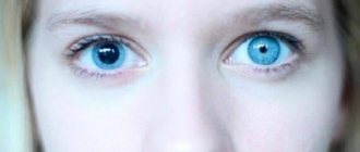
And any deviation in the work or structure of the organ of vision can lead to dire consequences. One of them is anisocoria - a condition when there is a noticeable difference in the size of the pupil. In this case, one eye works normally, in the other the pupil does not react to light, remaining at a fixed size.
Pupillary reflex, its importance for full vision
The visual image perceived by the eye passes through the cornea, pupil, lens, and vitreous body before reaching the retina. Depending on the brightness of the light flux penetrating the eyeball, the size of the pupil in the iris changes.
It is the responsibility of the muscle fibers to narrow or widen this dark hole in the iris. The sphincter muscle, to narrow it, surrounds the opening with circular fibers, and the dilator is formed by radial muscle fibers extending out like the spokes of a wheel. Both muscles move under the influence of parasympathetic and sympathetic nerves.
Bright light causes the iris to shrink, and the light flux entering the eyeball becomes less. When the lighting level decreases, the activity of nerve fibers is inhibited, the sphincter relaxes, and the pupil becomes dilated.
With a high degree of physical activity or a surge of emotions, the dilator fibers dilate the pupil. Nerves and muscles set it in motion and help form a clear image when a person examines nearby objects or tries to see something in the distance.
Symptoms of anisocoria
Anisocoria can be asymptomatic, but can bring many unpleasant minutes, causing:
- dizziness, headaches;
- decreased visual acuity, appearance of spots before the eyes, diplopia (double vision);
- nausea leading to vomiting;
- motor dysfunctions: paresis and partial paralysis, hand tremors;
- impaired coordination of movements.
In addition, anisocoria leads to:
- increased eye fatigue, especially during exercise;
- drooping, drooping of the upper eyelid – so-called ptosis;
- corneal edema, pain;
- deterioration of eyeball mobility;
- protrusion of the eyeball forward. This phenomenon is called “proptosis”.
Forms of violation, their features
The origin of anisocoria can be different, therefore, both congenital and acquired forms of the disease are distinguished.
The abnormal structure of the iris is associated with abnormalities in the functioning of the muscles and nerves of the eye. If the difference between the pupils is small, no more than one millimeter, then this is considered normal, especially since it does not affect visual acuity. Statistics say that physiological deviation occurs in every fifth inhabitant of the planet.
If nerve or muscle conduction is impaired, when the pupil does not respond to the brightness of the light flux, you should consult a doctor. After all, this can be a manifestation of various types of diseases, both ophthalmological and neurological, traumatic or infectious.
One pupil is dilated
This symptom indicates high blood pressure. This is possible if the corresponding eye has been injured. Consequently, a state of high blood pressure develops only within it.
This is due to excess secretion of intraocular fluid, which begins to put pressure on the eye from the inside. This manifests itself in a large pupil. This condition leads to glaucoma within a few years. The disease is expressed in the destruction of cells of the optic nerve. There is a gradual narrowing of the visual fields. The final stage is blindness. Return to contents
Unilateral mydriasis with normal reaction to light
This just indicates glaucoma. Normally, one pupil is larger than the other. But if you point a bright light source at the apple of your eye, it will shrink in size. Perhaps it will even become the same as the second pupil. But when the light source is removed, the pupil will enlarge again. This is due to the application of internal pressure. Because of him, the apple of his eye simply cannot be normal.
Such a reaction indicates glaucoma and the need to measure intraocular pressure.
Unilateral mydriasis with no response to light
This condition is usually associated with exposure to drops of atropine or its analogues. However, if an infection gets into the eye, the pupil may also not respond to light. But the infection must get into the specified eye. After all, the eyes are interconnected. And if processes common to the entire body occur, then mydriasis will be observed on both organs of vision.
Provoking reasons
Pathological changes in the iris lead to a lack of pupillary response to light, accommodation, or the ability to clearly see objects at any distance. There are many reasons and diseases in which the pupils become different sizes and anisocoria develops:
- An overly dilated pupil is associated with damage to the oculomotor nerve . A disorder occurs due to an aneurysm, acute cerebrovascular accident, or brain tumor.
- Parasympathetic denervation is often infectious in nature . Herpes zoster damages the eye socket, and the pupil reacts poorly to light or reacts at a slow pace.
- Different reactions to light in the pupils occur in patients with meningitis, tick-borne encephalitis .
- The patient's Eydie syndrome is associated with degenerative processes occurring in the parasympathetic neurons of the ciliary ganglion. The pupil's tendon reflexes decrease, the pupils become different sizes and blurred vision appears.
- Sympathetic innervation of the eye is diagnosed with enlarged lymph nodes in the neck, a tumor located at the base of the skull, or thrombosis of the carotid artery . An oncological tumor in the upper part of the lung also leads to damage to the neuron. In this case, the reaction to light of the pupils is not impaired, but, looking at the patient, there is a feeling that one eyeball is located deeper in the orbit than the other. This condition is called Horner's syndrome or simple anisocoria. It may be accompanied by pain in the face or radiating to the arm, and circulatory disorders in the vessels of the brain.
- A sharp dilation of the pupil followed by a reduction in all visual reactions occurs when glaucoma leads to an ischemic state of the iris.
- The pupil acquires an abnormal shape as a result of mechanical damage to the organ of vision or traumatic brain injury . In such cases, inflammation of the iris and choroid occurs.
- Damage to the centers of vision located in the cerebral cortex also causes a sharp dilation of the pupil. After a stroke, such deviations are also possible.
- The pupils may dilate or narrow unevenly after taking medications used in medicine - Atropine, Pilocarpine, Physostigmine. The size of the pupils changes unilaterally after using drugs: Cocaine, Amphetamine.
Anisocoria is not an independent pathology, it is only a symptom of abnormalities in the functioning of the brain and the structure of neural connections.
For what reasons does a child's pupils dilate?
The bases of the indicated condition can be divided into two large groups. They should be described in more detail:
Physiological reasons
Associated with lack of light. Because the organ of perception gets used to new conditions of visibility and adjusts to them. Physiological reasons include a number of other factors. For example, during a stressful situation, strong fear, excitement, physiological expansion occurs.
| This is due to the release of adrenaline into the blood. In case of poisoning, enlargement of the pupils also occurs. Thus, the organ of perception reacts to the toxic substance that affects it. |
Pathological causes
It differs from the physiological one in that it does not go away over time. After all, in case of poisoning or fright, the pupil will return to normal as soon as the influence of external factors stops. For example, a person will calm down or toxins will be removed from the body.
And for pathological reasons, it is necessary to take medications and treatment. In general, in such conditions, an enlarged pupil is an indicator of health problems. It results from injury or illness. Return to contents
Preeclampsia in pregnancy
This is internal poisoning. It occurs during pregnancy. In fact, this is toxicosis, which is often diagnosed during pregnancy. The disease is a dangerous condition that gradually develops. In its most severe stage, it is called preeclampsia.
During this period, toxicosis reaches its maximum level. This is combined with the presence of protein in urinary secretions and weight gain. This condition threatens the woman herself and the fetus. Along with the above reasons, pupil dilation is also noted. But it must necessarily exist against the background of an increase in body weight, pressure changes and other signs listed above.
Various chronic encephalopathies
| They can lead to catastrophic consequences. Such conditions are expressed in severe poisoning and metabolic disorders. In any case, the brain cells gradually die. |
As a consequence, a malfunction of the brain occurs, which is always expressed in a violation of behavior, the function of perceiving the surrounding reality, and motor abilities.
At the same time, chronic encephalopathies are often congenital in nature. The child receives this diagnosis due to poisoning in the womb. As a rule, this is associated with taking drugs during pregnancy, poisoning and other severe negative effects. Consequently, the baby is born with encephalopathy. Externally, this condition manifests itself in enlarged pupils.
Return to contents
Schizophrenia
This concept does not have an exact formulation. It cannot be clearly classified, since it is associated with severe mental disorders.
| However, schizophrenia is a severe psychiatric illness that cannot be fully cured. A person experiences a variety of symptoms. |
This could be associating oneself with another being, various manias, obsessions. In general, the patient is not able to relate correctly to his judgments and actions.
This condition can only be maintained at a relatively harmless level with the help of intensive treatment in specialized clinics. Against the background of schizophrenia, in most cases there is dilation of the pupils.
A brain tumor
The clinical picture and course of the pathology are very similar to the signs of encephalopathy. After all, with a tumor there is death of brain cells.
| The neoplasm is rapidly growing. It will inevitably put pressure on the healthy part of the brain. The consequence of this condition is an increase in pressure. |
The result will be an increase in pressure inside the eye. Because the capillaries are directly connected to the brain. Therefore, the pressure in the cranium affects the state of intraocular pressure. And a manifestation of this will be enlarged pupils.
Hyperthyroidism
Hyperthyroidism is the excessive production of hormones by the thyroid gland. It is expressed in changes in the composition of the blood, a general disruption of the body’s functioning, and leads to enlarged pupils. However, this sign is not observed in all cases. The danger lies in the prospect of infertility. If the development of hyperthyroidism is not stopped, the disease will lead to the inability to conceive. One of its manifestations is dilated pupils.
| However, this disease does not occur in childhood. It is typical only for adults. |
If both pupils are dilated and do not react to light
This means there are serious effects on the body and on the eye in particular. After all, it is the reaction to lighting that is the natural cause of pupil enlargement. If it is absent, this indicates illness or poisoning. The main reasons should be indicated:
- Severe fear, rage and other emotional states;
- Poisoning;
- Injuries.
All of these factors can lead to loss of pupillary response to bright light.
Return to contents
Inflammation of the brain (encephalitis) or its membranes (meningitis)
These conditions are dangerous because there is inflammation. During this process, brain cells are damaged, as a result of which the organ stops working normally.
In fact, there is death of a certain part of the brain affected by the disease, and it is able to develop, occupying more and more areas. This difficult and life-threatening process is accompanied by dilation of the pupils. But it always occurs against the background of behavioral disorders and severe headaches.
| Therefore, patients turn to doctors with other symptoms, since dilated pupils are the least of their concerns. |
Botulism
This pathology is infectious in nature. When pathogens enter, severe poisoning of the body occurs. As a rule, the infection enters the body through food and consumed liquids. In this regard, the development of the disease occurs. It is quite fast and takes only a few days.
Botulism is manifested by a feeling of dry mouth, tachycardia. A mandatory symptom will be large pupils combined with loss of reaction to directed bright light.
| To ensure you avoid botulism, you should not eat raw or poorly processed meat products. In this case, the disease cannot be transmitted. It is food and water that pose the danger. |
Brain swelling
This condition develops gradually. As a rule, it is infectious or traumatic in nature. Regardless of the cause, cerebral edema is the final stage of the development of a number of diseases. Strong pressure is generated. As a result, it affects the eyeball. As a result, the pressure inside the eye also increases, which is expressed in an increase in the diameter of the pupils.
Return to contents
Shock states
They are often observed during accidents, after injuries and similar critically dangerous situations. Shock states are always accompanied by nervous tension and the release of adrenaline into the blood. The tension reaches such a degree of expression that reality ceases to be perceived. A person gets lost in time, may not recognize loved ones, and answers simple questions inappropriately.
| The expression of such conditions is dilated pupils. Therefore, after injuries, doctors always shine a light in a person’s eyes. |
If the pupil reacts to light, then there is no state of shock. If there is no reaction, then sedatives are used, which are administered by injection.
Traumatic brain injuries
Often, they lead to a state of shock. Especially if associated with severe pain. Then the pupil dilates from the impact of painful shock and remains in this form for a long time, not reacting to light.
| It should be noted that the injury itself does not have immediate consequences for the pupil. Its expansion is the result of shock. |
Increased intraocular pressure may also occur. It is accompanied by an enlargement of the pupil. This occurs due to injuries to the organ of perception or impacts to the head area.
Epilepsy and increased intracranial pressure
The pathology reaches vivid expressions in the form of seizures. In general, the course of the disease is associated with the presence of high intracranial pressure. Therefore the pupil dilates. After all, there is a strong influence on him from within. Therefore, the pupil will not react to light, remaining enlarged.
Return to contents
Poisons and toxins
| The toxic effect leads to damage to internal organs. It covers the stomach, excretory system, liver and other organs. The action of poisons and toxins can paralyze or disrupt the functioning of the nervous system. |
Toxic substances enter the circulatory system and are delivered to all organs without exception, including the eye. This leads to an enlarged pupil. And it will remain dilated with a lost reaction to light until the toxic effect of toxins or poison is eliminated.
Acute alcohol or nicotine intoxication
In these cases, the development mechanism is the same as when ingested by poisons. After all, alcohol-containing products and nicotine are the same toxins. Therefore, the body perceives them the same way as any other poison. Accordingly, there will be a loss of the pupil's reaction to light. It will go away after eliminating the effects of alcohol and nicotine.
Pupil dilation when taking drugs
The toxic effect of drugs causes the effect of a wide pupil that does not respond to light. The body perceives drugs as alcohol, nicotine, poisons. In any case, these are toxins poisoning it.
Medicines
When conducting a fundus examination, the patient is instilled with atropine, which dilates the pupil. The effect lasts for several hours. But then the apple returns to normal. Analogues of atropine are mezatron and irifrin drops. They also lead to this condition for some time.
Anisocoria in children and adults: features
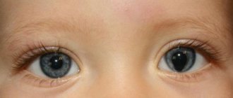
A congenital disorder in the functioning of the pupil can be noticed in infants, but this may be a physiological phenomenon that takes place over several years.
Dysfunction of the pupil of one eye can develop as a result of birth trauma or genetic predisposition. If the child’s parents discover that his pupils are unevenly located or of different sizes, then you should consult a doctor and check for concomitant diseases, such as drooping upper eyelid, strabismus, or restriction in the movement of the eyeball.
In children older than one year, the symptom of different pupils may appear as a result of a brain tumor or increased intracranial pressure.
In such cases, there is a decrease in the diameter of the pupil in a dark room, although the child’s image clarity does not suffer, he sees well what is far or close. The abnormality of the pupil is manifested by blurred vision, double vision, and fear of light. This should alert the child’s parents, and a visit to the doctor is mandatory.
The causes and diseases that provoke pupils of different sizes can manifest themselves both in young people and in adults after fifty years.
The appearance of malignant or benign brain tumors is a phenomenon that often occurs in young women. The response of the pupil to irritation by light in such cases is delayed, but when looking into the distance it slowly begins to expand. The pathological phenomenon is combined with blurred vision.
In older people, the difference in pupils is especially noticeable after a stroke or increased intracranial pressure.
Causes of aniridia
The cause of congenital absence of the iris is a defect in the human genome, in particular this concerns chromosome 11 and the PAX6 gene. The chromosome has two sections (p and q), one of which is shorter than the other. A gene is part of DNA and is responsible for the synthesis of important compounds, without which a number of the body’s capabilities are lost.
Aniridia was described back in the 19th century, at which time scientists agreed that this pathology is the result of a genetic defect that is transmitted through an autosomal dominant pathway. The PAX6 gene encodes information about the structure and function of the embryonic eyeball. In addition, this region of DNA contains information about part of the central nervous system.
In this regard, when the PAX6 gene breaks down, damage occurs not only to the eyeball, but also to some parts of the central nervous system. In some cases, a complete absence of the organs of vision occurs.
The PAX6 DNA region is inherited in a dominant manner, which means that the presence of at least one abnormal copy in the parents leads to a defect in the child. If both parents had a damaged copy of the gene, the disease is more severe. In this case, the fetus may die during intrauterine development or shortly after birth.
When a child is born with an abnormality of genetic material, he has characteristic signs that include underdevelopment of the brain, skull, nose, absence of eyes and large ears.
In absolute aniridia, a genetic mutation of a region of chromosome 11 (PAX6 gene) is involved. A person has two copies of this region, one of which is inherited from the father, and the other from the mother. If one of the copies is broken and the other is preserved, and the person has no signs of the disease, then the gene is recessive. If, in the same situation, the patient has symptoms of the disease, then the gene is dominant. Aniridia refers to the latter case.
Complex of therapeutic measures
Since anisocoria is only a symptom, treatment is aimed at getting rid of the cause of its occurrence.
Hematomas and brain tumors obtained as a result of birth injuries or after an accident lead to the fact that the patient often exhibits disturbances in the conduction of the optic nerve and damage to the muscle fibers of the iris.
In this case, the influence of physiotherapy on the restoration of tissue cells is great due to increased nutrition and stimulation of metabolic processes.
Magnetic waves help improve cerebral circulation and normalize blood pressure. Infrared radiation helps relieve muscle spasms. Electrical stimulation sessions activate processes inside cells and tissues aimed at regeneration.
Prescription of medications occurs after a complete examination and identification of the cause of the deviation. The main efforts are directed at treating the underlying disease, a sign of which is a narrowing or dilatation of the pupil in one eye.
Among the prescribed drugs: corticosteroids to relieve inflammation, antibacterial agents that actively affect pathogenic microorganisms.
Anisocoria caused by eye trauma is treated with medications that relax the muscles of the iris. These include drops Irifrin, Atropine. The ophthalmic drug Cyclomed and Midriacil, which belongs to the group of anticholinergics, is used to dilate the pupil.
Among folk remedies, inflammation of the membranes of the eye is relieved by liquid aloe extract used for lotions. An infusion of a mixture of carrots and dry stinging nettle, taken in the amount of two tablespoons, is prepared from half a liter of boiling water. After two hours, drink the drink; such daily treatment will strengthen your eyesight.
Correctly selected treatment will help you get rid of the disease. In some cases, surgical intervention may be necessary.
Causes of anisocoria
There are two types of anisocoria:
- physiological. Normally, every fifth person has a slight difference in the size of the pupils.
- pathological Eye diseases that can lead to anisocria: glaucoma, inflammatory diseases of the eye (iritis, uveitis), eye tumors
- pathological in general human diseases: viral infection, syphilis, brain tumors, cranial nerve palsy, Horner's syndrome, migraine, cerebral aneurysm.
Consequences of violation
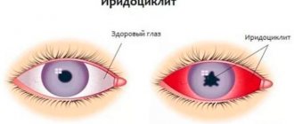
Disturbances in the functioning of the eye muscles and nerve fibers can lead to the patient developing inflammatory processes in the iris, iritis. They usually occur in people under forty years of age, less often in children and the elderly.
During the course of the pathological process, the pattern of the membrane changes, becoming blurred, and visual acuity decreases. The patient feels constant pain in the head, radiating to the temporal region. The chronic form of inflammation can result in eye atrophy.
With diplopia, or double vision, the image is blurry. This greatly tires a person, he begins to see objects poorly, feels discomfort, and dizziness. A neurologist and ophthalmologist will help identify the cause and prescribe treatment.
Different pupil sizes often lead to strabismus, which develops in children due to uncoordinated activity of the eye muscles. The organ of vision that squints does not participate in the visual process and becomes lazy. It is possible to cure this form of pathology in children with the help of medications and wearing special glasses.
Avoiding the unpleasant consequences of acquired anisocoria is the task of specialists, to whom a patient with damage to the nerve fibers of the eye should contact in a timely manner.
Symptoms of aniridia
Among the ophthalmological symptoms of aniridia are the following:
- High photosensitivity;
- Strabismus;
- Nystagmus that is uncontrollable;
- Decreased overall visual acuity;
- Development of glaucoma, cataracts;
- Decreased corneal transparency;
- Anomalies in the structure of the optic nerve, retina, lens;
- Amblyopia.
In some cases, patients experience specific external signs: narrowing of the palpebral fissure, wrinkles on the skin of the forehead. This is the result of increased photosensitivity of the eyes. Often, patients with aniridia have a specific sullen look.
In the area of the cornea in patients, vessels are completely absent or their number is significantly reduced. Most vessels form growths on the cornea, which are called pannus.
The anterior chamber angle in people without anomaly is responsible for the circulation of intraocular fluid and stable intraocular pressure. In the absence of the iris, and against the background of the proliferation of the vascular network from altered arteries, problems arise with fluid circulation, which leads to the development of intraocular hypertension and glaucoma.
In addition, with aniridia, the lens of the lens can also be damaged. This biconvex lens is normally located behind the iris and is responsible for concentrating light rays on the surface of the retina. Next, the information received is transformed and transmitted to the central structures of the brain. With aniridia, dislocation of the lens occurs, most often its subluxation. As a result, the patient gradually experiences a decrease in the permeability of the eye media to light rays, which leads to cataracts.
In addition to the listed deviations, with aniridia there is underdevelopment of the retina, in particular its fossa. The resulting foveal hypoplasia is accompanied by a serious decrease in the quality of vision. When the optic nerve is involved in the pathological process, the situation becomes even worse.
All these defects and developmental anomalies affect the functioning of the body as a whole.
Prevention of polycoria
There are no specific methods for preventing polycoria. Nonspecific measures involve eliminating the influence of harmful factors during pregnancy and preventing infection of the fetus by pathogenic agents that can cross the hematoplacental barrier. Polycoria is a serious pathology that is predominantly congenital. The only way to cope with the disease is surgery. If surgical intervention is not possible, symptomatic therapy is performed.
What function does the pupil perform?
The element has two main tasks: expansion (mydriasis) and contraction (miosis). Thanks to this, the hole regulates the flow of light rays penetrating into the visual apparatus.
Low light intensity increases the diameter of the pupil and allows objects to be seen clearly. If the light stream is too bright, the hole at the reflex level narrows. This minimizes light entering the retina and ensures high visual acuity. Also, such a protective mechanism prevents the harmful effects of too intense light on the retina and helps avoid burns.
Many people have a logical question: why is the pupil black? The reason lies in the fact that a minimum of light penetrates into the hole. Accordingly, the eyeball is dark, so the pupil appears black.
Another important function of the element is the ability to filter out flows penetrating the peripheral area of the lens. This makes it possible to compensate for spherical distortions. In other words, such a disadvantage as a glow around objects is eliminated.
If the pupil increases in size at dusk, then color perception decreases and visual acuity deteriorates. However, this increases the retina's ability to perceive light at low intensity. A person gets the opportunity to navigate in space and distinguish the silhouettes of large objects.
Functions
It is no coincidence that the pupil of the eye is located in the center of the iris; this location helps the rational distribution of solar and artificial lighting over the entire surface of the retina.
This hole in the eye ensures sharpness and clarity of human perception of the world. Its dysfunctions lead to deterioration of vision, “blurring of the picture,” and “blurring” of surrounding objects.
Thanks to the correct functioning of the pupil, people can perceive reality both during daylight hours and in the dark. This is why a person's pupils dilate at night and he can navigate quite well in low light conditions. During the day, by narrowing, the pupil reduces the intensity of sunlight, allowing you to see the surrounding space and objects in it without irritation.
Not only the muscles of the eye, but also retinal cells (cones and rods), as well as the nerve plexuses of the autonomic and parasympathetic systems, and neurons of the brain are actively involved in the work of healthy pupils. In addition, the pupil carries out the processes of accommodation (a natural mechanism that ensures stability of vision when moving the gaze from a distant object to a near one, and vice versa)
Therefore, a person easily and smoothly switches his attention to any near or distant objects.
The internal tension of the pupillary muscles (convergence reaction), which is necessary for the normal functioning of the lens and retina when fixing gaze at a close distance, is also provided by the pupil.
Diagnosis and treatment of anisocoria
An ophthalmologist will be able to make a diagnosis of “anisocoria” based on examination, studying the reaction of the pupils in the dark, in the light, studying the speed of their reaction, and symmetry under different conditions.
In addition, a biomicroscopic examination, diascleral transillumination using a diaphanoscope, and, if necessary, angiography, ultrasound, MRI and CT will be performed. The doctor can also use mydriatics - special drops that can cause artificial dilation of the pupil.
There are also special tests - tropicamide and phenylephrine and pilocarpine - to help make the diagnosis.
Physiological anisocoria, which does not cause any problems to the patient, does not require treatment. Only a cosmetic defect can cause inconvenience.
For anisocoria associated with diseases of the body, treatment consists of eliminating the underlying disease.