A nevus is a mole that can degenerate into a malignant neoplasm.
Sometimes it is located in unusual places - the conjunctiva, iris, retina.
It occurs in people of any age – middle-aged, elderly, young children.
If a pigmented nevus appears, it is necessary to identify the cause and consult a specialist. Proper treatment will reduce unpleasant consequences. If a mole is left untreated or removed at home, there is a high risk of complications. The treatment for nevus is laser removal. The procedure is safe, bloodless, and does not require a hospital stay or long rehabilitation. Suitable for people of any age.
When a mole appears or for prevention, you must regularly use sunglasses.
Causes
According to its origin, nevus can be congenital or acquired. The first type appears in a newborn as a small spot on the eyeball, increasing in size over time. It does not have a negative effect on vision, since there is no effect of the mole on the capillaries and visual impairment.
The reasons why nevus appears in the conjunctiva of the eye or other parts of the organ of vision:
- hormonal imbalances (menopause, pregnancy, puberty, sexual dysfunction);
- severe stress;
- heredity;
- age-related changes;
- taking hormonal contraceptives;
- UV radiation;
- infectious processes;
- damage to mucous membranes and skin.
A mole can be located on the conjunctiva, iris, retina, choroid (choroid of the eye), and vary in size and color.
Symptoms and signs of moles under the eye in women
A mole under the eye becomes noticeable as soon as it appears there. If a small red spot appears on the eyelid, which does not cause pain, then such a nevus does not pose any threat. It usually appears due to vascular disorders or at the time of hormonal imbalance. But if several such spots form, then you should visit a doctor.
Black growths can be large
In this case, it is extremely important to monitor their shade. If the growth suddenly begins to darken even more or change color around the perimeter, then you should be alarmed
Such a change in the appearance of a mole means serious disruptions occurring inside the body.
Brown nevi that appear on the lower or upper eyelid may be itchy. They can be light brown in color, which can make them difficult to notice right away.
Why is a nevus on the eye dangerous?
The first thing that is dangerous about a nevus is its possible degeneration into a malignant formation - melanoma. Determining a tumor is quite difficult: any mole on the eye changes color, shape, and outline.
Other complications due to untimely removal and treatment of a mole on the eye, depending on the type:
- the spot increases in size, takes on a different shape, compresses the vessels of the organ of vision;
- tissue detachment;
- dystrophic changes in the retina and epithelium of the eye;
- nevus of the iris leads to decreased vision and the sensation of a foreign body;
- deterioration in the quality of vision and subsequent loss of it.
In some cases, moles do not lead to complications other than causing psychological discomfort to a person. They are visible to the patient and others.
Iris mole
This is a spot formed on the iris itself. Iris nevus is divided into the following types:
- Typical. The most common pigmented nevus. A flat configuration is more common, but you can also find a slightly convex shape; they rise slightly above the upper eye shell. This type is dangerous because it leads to deformation of the pupils and inversion of the pigment border.
- Diffuse. It appears as small nodules on a small stalk.
- Lisch nodules. This type occurs in all people with neurofibromatosis type 1. Small dots appear in large numbers on the iris.
- Freckles. They are also a type of mole.
What to do with a nevus on the eye
If a mole appears on a child’s eye, no action is required.
In adults, laser treatment is used. Medicines and physical therapy do not affect the nevus. Home methods are not suitable for removing a mole - they can harm the patient’s health and cause irreparable harm.
If a nevus appears on the eye, you need to visit a specialist and register. Regularly monitor the state of education. There is a risk of a mole degenerating into a malignant formation; it is very difficult to conduct a histological examination of eye tissue. Therefore, it is recommended to remove the nevus.
Nevus removal
The procedure is carried out in a clinic without placing the patient in a hospital. Previously, to remove a nevus, they resorted to surgery using a radio or microscalpel.
Currently, local anesthesia and laser technologies are used. The procedure is safe, does not harm the patient’s health, and does not lead to the formation of a deep postoperative scar. Another advantage of using a laser is that there is no blood.
Material and methods
From 2009 to 2013, we examined 52 patients with pigmented tumors of the conjunctiva. The age of the patients ranged from 2 to 65 years. Of these, 48 patients had nevus of the conjunctiva and lacrimal caruncle, 1 patient had melanoma of the lacrimal caruncle, 3 patients had melanoma of the conjunctiva.
Of the 48 patients, 16 had a progressive nevus of the conjunctiva, 5 had a nevus of the lacrimal caruncle, and 27 had a stationary nevus (Fig. 1).
Rice. 1. The ratio of the occurrence of conjunctival nevi and the lacrimal caruncle.
All patients underwent a general ophthalmological examination, including visometry (Huvits sign projector, CCP-3100, Korea), tonometry (TOMEY FT-1000 pneumotonometer, Japan), biomicroscopy (TOMEY TSL-5000 slit lamp, Japan), ophthalmoscopy (fundus lens 78 diopters OCULYAR, USA), as well as B-scan, optical coherence tomography (OCT) of the anterior part of the eye. During the study, we carefully studied the structure of the formation itself, paying attention to the color, contours, nature of the tumor boundaries, surface characteristics, the presence of pigment and its dispersion, the state of the surrounding tissues and limbus when the formation is localized in the immediate vicinity of it, the presence of spread to the cornea (Fig. 2 ). The formations were measured using a regular ruler. Traditionally, the minimum and maximum diameters of the base of the formation were assessed. Ultrasound biomicroscopy was used in patients with conjunctival tumors to determine the thickness of the tumor, obtain a complete description of its structure, and information about the condition of the underlying tissues. The study was carried out using an ultrasound machine Nemo XG SSA-580A. A sensor with a frequency of 8 MHz was used. OCT was used in patients with conjunctival tumors extending to the cornea to determine the depth of corneal lesions (Fig. 3). The study was carried out using a Visante OCT device (Carl Zeiss, Germany).
Rice. 2. Progressive nevus of the conjunctiva.
Rice. 3. Optical coherence tomogram of the anterior segment of the eye with conjunctival melanoma spreading to the cornea.
Cytological examination was performed in all patients with suspected malignant tumors of the conjunctiva. Material for cytological examination was obtained by making an impression smear from different areas of the tumor surface onto a fat-free glass slide. Hematoxylin and eosin were used as dyes.
Histological studies of the surgical material were performed in all patients who underwent surgical excision of adnexal tumors. The number of mitoses, the degree of lymphocytic infiltration, mitotic activity, pigment content were studied, and also determined which structures were affected by the tumor (macroscopically and histologically), the degree of therapeutic pathomorphosis and the distance of tumor cells from the resection margins.
In case of suspicion of conjunctival melanoma giving distant and distant metastases, the patient underwent a special oncological examination, which made it possible to identify the stage of the oncological process and choose the right treatment tactics.
For this purpose, radiography, computed tomography, nuclear magnetic tomography, and ultrasound research methods were used. Chest radiography was performed to detect metastases in the lungs.
Ultrasound (US) has been used to screen for distant abdominal metastases. This method does not involve radiation exposure, is more accessible, and therefore has been successfully used.
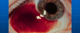
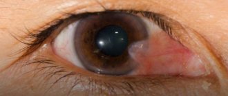
Excision of the tumor within healthy tissue using microsurgical techniques with conjunctival plastic surgery was performed in 12 patients with progressive nevus. One 6-year-old patient had invasion into the upper layers of the sclera, and therefore a superficial sclerectomy was performed; 4 people refused surgery, 2 of them are under observation. 4 patients with nevus of the lacrimal caruncle were operated on; 1 patient also refused surgery. Surgical treatment was performed in 3 patients with conjunctival melanoma. When removing it, a 0.04% mitomycin solution was used intraoperatively. During the operation, delimiting coagulation of the formation was carried out, retreating 3-4 mm from its visible boundaries, followed by its excision along the outer border of the coagulation shaft. After this, the bed was subject to additional coagulation. Plastic surgery of the conjunctival defect was performed by moving local tissues and applying the required number of sutures (Vicryl 8−0). Interferon α-2β was used as adjuvant therapy (Fig. 4). One patient with melanoma of the lacrimal caruncle was sent abroad for treatment (Fig. 5).
Rice. 4. Photograph of a patient with melanoma. a - before surgery; b - after surgery.
Rice. 5. Photograph of a patient with melanoma of the lacrimal caruncle.
Prevention
A mole on the retina and other parts of the organ of vision appears due to the accumulation of melanin. The main preventive measure is to wear sunglasses or special contact lenses with UV filters to protect the eyes from exposure to sunlight.
Additional measures are to eliminate the causes that cause retinal nevus and its other varieties:
- timely treatment of infectious diseases;
- combating stress (taking medications if necessary);
- leveling hormonal imbalances, if possible;
- healing of damaged mucous membranes and skin in the early stages:
- regular visits to the ophthalmologist.
If you have a nevus, you need to monitor the size and color of the formation.
Diagnostics
Detection of a nevus usually occurs by chance during ophthalmoscopy - an instrumental examination of the fundus of the eye. To establish a final diagnosis, dynamic observation of the formation and regular examinations of the fundus using color filters are necessary. Choroidal nevus is well defined in red light, and when green light is used, the stationary nevus “disappears”. With a green filter, changes in retinal tissue are determined, which are characteristic of a formation with a progressive course. In some cases, ultrasound makes it possible to determine the distance of the formation. Conducting fluorescein angiography with a contrast agent when diagnosing stationary nevi confirms that there are no changes in the adjacent vessels. With progressive formation, an effusion of fluorescein is detected from the vessels adjacent to the nevus through their defective walls.
Useful video
Causes of birthmarks on the choroid of the eye. Effective treatment of this type of nevus.
Author's rating
Author of the article
Alexandrova O.M.
Articles written
2031
about the author
Was the article helpful?
Rate the material on a five-point scale!
( 2 ratings, average: 3.00 out of 5)
If you have any questions or want to share your opinion or experience, write a comment below.
Is treatment necessary?
Typical stationary types do not require treatment. In contrast, progressive, suspicious, or atypical choroidal nevi should be detected as early as possible. They require constant monitoring and ultrasound examinations of the eye several times a year.
The tendency to change should be alarming; progression serves as an indication for its removal by laser photocoagulation (cauterization).
Most cases of choroidal nevi of the eye have a favorable prognosis; undergo medical examinations on time, and everything will be fine with you.
Leave comments below if you think moles are dangerous, and don’t forget to share the article with your friends.