Causes
Paralysis of the eye muscles, both complete and incomplete (paresis), is either congenital, due to insufficient development or complete absence of the muscle, or due to aplasia of the corresponding central nucleus, or acquired.
Paralysis involves either one muscle (single or isolated paralysis) or several muscles of one eye, supplied by one or different cranial nerves, or several muscles of both eyes. In both of the latter cases, they speak of ophthalmoplegia, and a distinction is made between ophthalmoplegia, when there is paralysis of the muscles that move the eyeball, and ophthalmoplegia, when the intraocular muscles, the oculomotor nerve, namely the sphincter of the pupil, and the accommodative muscle are paralyzed.
Acquired ophthalmoplegia in a relatively small number of cases is caused by the fact that the muscle itself or its vagina is affected. In this case, injuries and inflammatory processes play the main role; less often, gumma or trichinosis may be the direct cause of paralysis.
Most often, there are cases in which dysfunction of the muscle is caused by damage to the motor cranial nerve. Such lesions can be localized along the entire length of the nerve, from its central origin to its entrance into the muscle.
Accordingly, a distinction is made between central and peripheral ophthalmoplegia. The first is of cortical, nuclear or radicular origin, the second is of basal or orbital origin.
The main diseases or processes that cause pathological changes and disorders of the function of nerves or their nuclei are of a very diverse nature; This includes especially syphilis, which affects almost half of all paralysis of the eye muscles, further, tuberculosis, rheumatism, trauma, poisoning (lead, alcohol, mushroom, meat, sausage and fish poisons, carbon monoxide), infectious diseases, such as, for example, diphtheria, influenza, less commonly measles, scarlet fever and malaria, then Gerlier's disease and beriberi, herpes zoster, diabetes, gout, parasites, and finally, diseases of the stomach and lungs.
Local processes in the central nervous system and its membranes caused by primary diseases may consist of meningitis, tumor formation, also gumma or solitary tubercle, endarteritis, aneurysm, embolism, sinus thrombosis, circulatory disorder, hemorrhage, foci of softening, atrophy, sclerosis, abscess, dropsy ventricles, periostitis and periosteal tumors (exostosis), empyema and, finally, osteomyelitis.
Secondary changes in the motor cranial nerves are of an inflammatory nature (neuritis and perineuritis), or are caused by the pressure of a neoplasm or effusion; in the latter case, the consequence may be severe atrophy and even complete destruction of the affected nerve.
Functions of the optic nerves
The oculomotor nerve, which is located inside the muscular ophthalmic funnel, performs several important functions - it ensures all movements of the eyeball, response to the light of the pupils, raising and lowering the upper eyelid. The nerve gives signals to the muscles, which with its help are innervated, that is, controlled. The trochlear and abducens nerves are involved in this process. All of them are paired and belong to the group of cranial nerves.
The coherence of this system is simply amazing. The impulses are given so quickly that a person cannot even comprehend most of the involuntary movements of the eyelids and eyeball. It is the oculomotor nerve that ensures the safety of the visual organs. at the slightest threat of foreign objects entering the eyeball, the nerve gives a signal to close the upper eyelid. It provokes blinking, in which the surface of the eye is washed with tear fluid, so that it does not dry out.
Complete or partial damage to the oculomotor nerve disrupts the functioning of all eye muscles and provokes the development of serious, often irreversible diseases.
Symptoms and clinical picture of ophthalmoplegia
The clinical picture of ophthalmoplegia consists of the following, partly determined objectively, partly subjective disorders:
• Limitation of eye mobility.
• More significant secondary ocular deviation.
• Diplopia.
• Visual dizziness and unsteady gait.
• Incorrect head position.
Limitation of eye mobility
It can be caused primarily mechanically, due to a tumor in the orbit, tenonitis, symblepharon, muscle disease, and so on; Of course, these causes must first be excluded so that a diagnosis of ophthalmoplegia due to loss or limitation of motor innervation can be made with complete certainty.
The most striking sign of ophthalmoplegia is a lack of eye mobility in one direction. Mobility can be completely lost (paralysis) or only weakened (paresis). Lack of mobility can, however, only be imaginary, due to the fact that the size and configuration of the palpebral fissure, the position of the lacrimal caruncle, and finally, the structure of the eye itself simulate it.
The (absolute) mobility of the eye is examined in such a way that one eye is closed and the other is forced to follow the movements of an object, for example a finger, in all directions to the most extreme physiological limits. The normal border is considered to be reached: outwardly, if the outer edge of the cornea touches the outer commissure of the eyelids, inwardly, if the inner edge of the cornea slightly extends beyond the lacrimal caruncle or if the lacrimal punctum approximately corresponds to the center of the pupil; up and down there are no definite boundaries, mobility in these two directions is individually very different, but with some skill here too you can soon find reference points for assessing mobility. With minor restrictions on mobility, clear results cannot be obtained in this way and therefore it is better to examine the (relative) mobility of both eyes together using the method just indicated. If at the same time one eye lags behind the other, then one can quite reasonably assume a disorder in the movement of this eye. Often, paralysis of the eye muscles is indicated by a peculiar twitching movement of the eye and even clear nystagmus, especially with extreme deviation of the eye to the side. Sometimes the mobility of both eyes may also be limited symmetrically in one or more directions; This especially happens when looking up, while such a restriction of friendly or associated movements to the right and left is rarely observed.
Finally, in all cases where ophthalmoplegia can be suspected, it is necessary to examine the ability of convergence, forcing the patient to fix the tip of the finger, which is increasingly brought closer to the tip of his nose; the lag of one eye, although it does not necessarily indicate paralysis, is still important for the assessment of other established symptoms.
More significant secondary eye deviation
If, when studying the relative mobility of both eyes, you move the fixed finger in the direction of the paralyzed muscle, it turns out that the paralyzed eye, due to the limitation of its mobility, cannot follow the finger beyond a certain limit, so only the healthy eye fixes. The visual lines of both eyes in this case are no longer set to the same fixation point; the visual line of the squinting eye is deviated, it converges or diverges with the visual line of the healthy eye, that is, the paralyzed eye squints. The degree of deviation, called primary, is measured by angle. If you now cover the healthy eye with frosted glass, then the paralyzed eye takes over the fixation, as far as possible. The paralyzed muscle now receives a stronger impulse, but is unable to follow it or only partially follows it. Half of this impulse is transmitted to the other, normally mobile eye, which therefore represents a very strong deviation, much more significant than the deviation of the other eye, limited by the limits of mobility of the affected muscle. This deviation, which can be accurately determined behind ground glass, is called secondary and, as follows from the above, in ophthalmoplegia it is greater than the primary deviation.
Diplopia
This is the most important symptom of ophthalmoplegia. At first it is often noticed only occasionally, but subsequently it always occurs when the eyes deviate in a certain direction. Diplopia, of course, does not occur when one eye is blind; Further, it should be noted that some patients very quickly learn to suppress the imaginary image.
Visual dizziness and unsteady gait
Vertigo is called visual dizziness because it is of peripheral origin, that is, caused by paralysis of the eye muscle, as opposed to cerebral dizziness. The named phenomena interfere especially with paralysis of the muscles that lower the eyes, and immediately disappear if the affected eye is closed. Usually the patient does this himself. In such cases, you can also place a prism with the base down in front of the diseased eye, and a prism with the base up in front of the healthy eye.
Incorrect head position
To relieve the affected muscle from a stronger contraction and prevent diplopia, the patient, every time he wants to look in a direction that requires a special action from the paralyzed muscle, completely impossible or only partially possible, turns his head so that his eyes are directed to the side, the opposite of diplopia. This head rotation is called “vicarious” because the neck muscles act instead of the paralyzed eye muscle.
The position of the head is pathognomonic for almost every paralysis and often this symptom alone can make a probable diagnosis. When turning the head to the left, the muscle that turns the eye to the left is paralyzed, for example, the left external rectus muscle; when turning and lowering to the left and slightly tilting the head forward, the superior oblique muscle is paralyzed.
Symptoms and diagnosis of individual muscle paralysis
Having outlined the most important data concerning ophthalmoplegia, we should now turn to a consideration of paralysis of individual muscles.
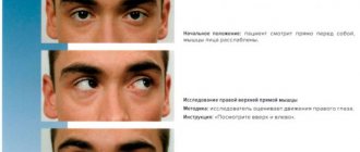
Right external rectus paralysis
If there is complete paralysis of the right external rectus muscle, then the eye cannot be displaced from the average position outward (towards the temple); with extreme effort on the part of the patient, it is noticed that the eye can turn outward several degrees with jerky movements up and down, thanks to the action of both oblique muscles, which slightly turn the eye outward. The patient keeps his head turned around a vertical axis to the right, that is, towards the painful side.
During the experience of palpation, the hand passes by the object outwards, both images are of the same name, located parallel to each other and at a certain distance from each other in the horizontal direction, and the distance between them increases, the further the object moves outwards; the boundary line between the areas of single vision and diplopia is approximately vertical and therefore corresponds to the midline. Visual dizziness almost always occurs in fresh cases.
Incomplete paralysis (paresis) of the right external rectus muscle
The function of the external rectus muscle is not completely lost, the eye can move to the right beyond the midline, but this movement is more or less limited, often so small that it is barely noticeable and is detected only by jerky movements at the extreme limit of contraction. The feeling of dizziness is easily eliminated by slightly turning the head to the right; Diplopia appears, other things being equal, more to the right, the lower the degree of paralysis; often it is noticed only at the very outer periphery.
Right external rectus paralysis with antagonist contracture
After a more or less long-standing paralysis of the abducens nerve, a contracture of the internal rectus muscle may develop, which is expressed in the fact that, despite the unchanged deviation of the eye, the field of view for single vision is narrowed due to the fact that the border of diplopia is more or less shifted to the left; Moreover, diplopia may exist throughout the entire visual field. The nature of diplopia is the same as with simple paralysis.
Paralysis or paresis of the right internal rectus muscle
Turning the eye inwards, that is, to the left, is completely impossible or is more or less limited. In case of complete paralysis, the second pair of muscles, namely the superior and inferior rectus muscles, which also turn the eyes somewhat inward, can act in a compensatory manner to the extent that the contraction of their eyes brings them to the middle position. When turning to the left, the right eye deviates outward; When fixed with the affected eye, the deviation of the healthy eye becomes even stronger. Crossed diplopia, as the internal rectus muscle turns the eye inward; therefore, the imaginary image is located inward (toward the nose) and moves away more and more towards the healthy, in this case the left, eye; Both images are located side by side and in the horizontal plane show neither inclination nor difference in height. The boundary line between the areas of single vision and diplopia is almost vertical, but at the top it is inclined more towards the healthy eye than at the bottom. Therefore, if diplopia does not yet occur when looking in the horizontal direction, then it may appear when looking up. Likewise, diplopia, as is clear, occurs faster when the fixed object approaches the eye. The sometimes developing secondary contracture of the external rectus muscle in some way enhances the paresis, reduces the area of single vision and increases the distance between both images. A compensatory rotation of the head therefore occurs around the vertical axis to the left, towards the virtual image; in the same way, during the experience of palpation, the hand misses the object inwards.
Paralysis or paresis of the right superior rectus muscle
The superior rectus muscle strongly lifts (rotates) the eye upward and weakly deflects it inward. With complete paralysis, her right eye, during binocular fixation, deviates downward and somewhat outward, so the compensatory position of the head is expressed in the fact that the head around the horizontal axis is tilted back, but also slightly to the side.
Since the superior rectus muscles are relatively rarely activated, dizziness is little noticed, mainly when climbing stairs, and generally only when looking up. During the experience of palpation, the hand passes by the object above it, since both images stand one above the other and the imaginary image is located above. Due to the fact that the superior rectus muscle also turns the eye inwards and slightly tilts the vertical meridian, the images are crossed and slightly tilted. The vertical distance between them is greatest when the superior rectus muscle acts with the eye in the abducted position, the lateral distance is most significant in the middle position, and finally, the oblique one - with the eye in the abducted position. The border of diplopia with paralysis of the right superior rectus muscle goes from top left to bottom right.
If the right superior rectus muscle is only in a state of paresis, then the line of sight, depending on the degree of paresis, may be more or less elevated, and accordingly the deviation is expressed. If the paresis is very minor, then it often manifests itself only in the fact that diplopia appears when looking outward and upward.
If a secondary contracture of the antagonist, that is, the inferior rectus muscle, has occurred, then the area of diplopia extends to the lower part of the visual field, and it should only be noted that, due to the stronger convergence caused by the contraction of the inferior rectus muscle, both images in the lower part of the visual field are the same. Further, it should also be mentioned that with paralysis of the superior rectus muscle, due to a stronger nerve impulse to it, the associated muscle that lifts the upper eyelid causes a wider opening of the palpebral fissure.
Most important for diagnosis is that the vertical distance between images increases with the abducted position of the eye.
Paralysis or paresis of the right inferior rectus muscle
With complete paralysis of the right inferior rectus muscle, the right visual line can be lowered down not directly, but only with the help of the superior oblique muscle. Here, the isolated rotating action of the latter also clearly appears, and therefore the eye also lags behind when looking down, up, and slightly outward, and the vertical meridian is slightly inclined. Since the inferior rectus muscle strongly lowers the eye downward, slightly deflects it inward and rotates, both images are located above each other, with the imaginary one being lower, in addition, they are crossed and located not parallel, but somewhat obliquely to each other. Dizziness and disorders with binocular vision are much stronger than with paralysis of the superior rectus muscle, since the gaze is directed downward in most cases. Therefore, the patient keeps his head tilted downwards (towards the chin) and slightly towards the painful side, in this case the right. The field of diplopia is located below the horizontal line and on the diseased (right) side it goes slightly upward.
If the muscle is only in a state of paresis, then the deviation, secondary and primary, etc., is less significant, as is diplopia, but the feeling of dizziness and disorientation are expressed to almost the same extent as with complete paralysis, because, as As already mentioned above, the gaze is mostly directed downwards.
With contracture of the antagonist, that is, the superior rectus muscle, the area of diplopia also extends to the upper part of the visual field; you just need to remember that both images are clearly crossed downwards, but upwards, due to the preponderance of inward rotation caused by the superior rectus muscle, they can cross to a lesser extent or even be of the same name.
Paralysis or paresis of the right superior oblique muscle
The superior oblique muscle, together with the inferior rectus muscle, lowers the eye downward. If only the oblique muscle acts, the eye turns outward and undergoes a rotational movement, so that the vertical meridian tilts inward. The superior oblique muscle causes the strongest lowering of the eye in the adducted position of the eye, and the strongest inclination of the meridian - in the abducted position. Therefore, if the superior oblique muscle is completely paralyzed, then downward movement of the eye, although possible, is limited, especially when the eye is in the adducted position; due to the fact that lateral deviation of the eye under the influence of the oblique muscle is impossible, slight convergence occurs, and the vertical meridian tilts outward, especially when the eye is in an abducted position. Insufficient lowering of the eye is most clearly noticed when the gaze should be directed inward and downward.
The primary deviation occurs upward and inward. When the object being fixed is located inward from the eye, the vertical deviation occurs, of course, earlier and to a greater extent than in the case when the object being fixed is located to the right (laterally).
The superior oblique muscle weakly lowers the eye downward, so both images are only slightly apart in height and the image of the paralyzed eye is located lower; since the superior oblique muscle also turns the eye outward, the images are of the same name, but they are clearly inclined, namely, they converge at the upper ends, since this muscle strongly rotates the eye. The distance between the images in height increases with the abducted position of the eye, and the inclination increases with the abducted position, which is quite understandable based on the physiological action of the muscle. It should be noted that the virtual image in most cases seems to the patient to be located closer and often also has a smaller size.
The patient holds his head turned forward around the transverse axis and around the diagonal axis towards the healthy eye. Diplopia is observed mainly in the lower half of the visual field, the border line of diplopia goes from the upper and healthy side to the lower and diseased side. The feeling of dizziness and misorientation are quite significant because, as already noted, the gaze is mostly directed downwards.
If ophthalmoplegia is incomplete, then eye movement disorders are barely noticeable and diplopia is of decisive importance, on the basis of which only a diagnosis can be made.
If contracture of the antagonist, that is, the inferior oblique muscle, has occurred, then the deviations towards the healthy eye increase, as a result of which the boundary line of diplopia becomes steeper and diplopia also exists in the upper part of the visual field. The vertical distance between the images, as well as their deviation, become larger, but (and this is very important) at the same time, as in the lower part of the visual field both images were the same, in the upper part they can be crossed, due to the fact that the inferior oblique muscle turns the eye outward more than the upper straight line turns inward.
Paralysis or paresis of the right inferior oblique muscle
Under the influence of contraction of the inferior oblique muscle, the eye turns upward and outward and, since it at the same time rotates the eye, the vertical meridian tilts to the lateral side. If there is complete ophthalmoplegia of the inferior oblique muscle, then its action, of course, is impossible and the eye therefore lags behind downwards and inwardly. If the eye is adducted, then the deviation is more pronounced downward. If the eye rises upward in an abducted position, the vertical meridian is strongly inclined to the medial side.
The inferior oblique muscle lifts the eye upward, so both images stand on top of each other.
Diplopia is observed mainly in the upper part of the visual field and the line limiting the area of diplopia goes from below and from the healthy eye upward and towards the diseased eye.
Compensatory rotation of the head occurs around the horizontal axis posteriorly and around the oblique axis towards the painful side.
If there is only paresis, then deviations can hardly help in making a diagnosis; the latter is placed on the basis of diplopia.
If secondary contracture of the superior oblique muscle occurs, then diplopia is also observed in the lower left, and subsequently in the lower right part of the visual field. The distance between the images is greater, but it should be noted that the images of the same name in the upper part of the field of view are crossed in the lower part.
The affected eye should be considered the eye whose image, when examined for diplopia and the movement of the fixed object in the direction of action of the affected muscle, moves faster than the image of the other eye. If it is determined that rapid movement occurs not in one direction, but in several, then there is ophthalmoplegia of several muscles. But such a diagnosis can often be more easily made by an objective examination of eye mobility. Complex paralysis can occur in one eye or both; its existence in both eyes can be easily recognized both by the mobility disorder and by diplopia.
Paralysis of muscles innervated by the oculomotor nerve
The upper eyelid is drooping due to paralysis of the muscle that lifts the upper eyelid; Only with extreme effort does the patient manage to slightly open the palpebral fissure by contracting the frontal muscle. If you lift the upper eyelid up with your hand, the eye turns out to be deviated outward, the pupil is wider than normal. If we examine the movements of the eye, it turns out that they are only possible outward, in addition, the eye, when the patient wants to look down, turns downward and outward. All external and internal eye muscles are paralyzed, with the exception of the muscles innervated by the abducens and trochlear nerves, as well as the dilator muscle, innervated by the sympathetic nerve. As for diplopia, in the primary position of the eyes both images are: crossed; spaced apart from each other in the horizontal direction; inclined and slightly spaced apart in height. The crossover is explained by the lack of action of the internal rectus muscle, which is also joined by the superior and inferior rectus muscles, which also turn the eye inward. These same muscles explain the distance of images in the horizontal direction, as they turn the eye to the side. The difference in image height depends on the inferior oblique muscle. The superior and inferior rectus muscles cancel out their action. In addition, the inferior oblique muscle, as a rotator of the eye, determines the oblique position of the images.
If the oculomotor nerve palsy is not complete, then the relationship is more complicated, and often a diagnosis cannot be made on the basis of movement studies alone, but it can be made on the basis of diplopia according to certain rules.
There may be paralysis or paresis of all the muscles innervated by the oculomotor nerve, or only some of them; often even for a long time, sometimes throughout life, only one muscle is paralyzed, namely, usually the levator upper eyelid, along with which the superior rectus muscle is often paralyzed.
A frequent combination is the paralysis of the following muscles: the levator superior eyelid, the superior, inferior and internal rectus and inferior oblique muscles (the so-called external ophthalmoplegia), therefore, the paralysis of all muscles that move the eyeball with the exception of those innervated by the abducens nerve and trochlear nerve; further isolated paralysis of the sphincter and accommodation (internal ophthalmoplegia). In most cases, both of these forms of paralysis are observed in both eyes, but not at the same time, but at some intervals one after the other.
Paralysis of both external rectus muscles
If both external rectus muscles are paralyzed, then, of course, outward movement of both eyes is impossible or limited, the eyes are in a position of pathological convergence. It is interesting, however, that in this case there may be no diplopia along the midline, and it appears only on both sides of this line. Of course, diplopia has the same name and when moving to the right, the right image moves faster, and when moving to the left, the left one.
Paralysis of the external rectus muscle of one eye and the internal rectus muscle of the other eye
If the external rectus muscle of the right eye and the internal rectus muscle of the left eye are paralyzed, then movements to the right are limited or impossible, but in all other directions eye movements occur normally. Since mobility restrictions cancel each other out (compensate), then in this case there is no diplopia if the fixed object is moved in all directions. On the contrary, it can occur with convergent movements. Since both of these muscles are associated with each other, these and similar paralysis, such as paralysis of both superior rectus muscles, are called associated paralysis. It is interesting that in some paralysis of both superior rectus muscles there is no diplopia, and then it must be assumed that the associated oblique muscles are also involved in the paralysis (functionally).
Differential diagnosis of ophthalmoplegia
The most important symptom of ophthalmoplegia, diplopia, can also be caused by other disease processes that should be excluded through careful examination; for example, diplopia can be caused by a tumor, abscess, or aneurysm of the orbital cavity, which displaces the eyeball and often also limits its mobility. With concomitant strabismus, diplopia is also sometimes observed, especially in the initial stages, or it can be induced artificially using colored glass, a prism, etc.
In differential diagnostic terms, it is important that the distance between both images with concomitant strabismus is constant, while with paralysis it increases in the direction of the action of the affected muscle. A further important difference between ophthalmoplegia and concomitant strabismus is that the secondary deviation of the eye in paralysis is greater than the primary one, while both angles of deviation in strabismus are approximately the same and remain the same when the position of the fixing object changes. Finally, the examination should pay attention to whether diplopia continues when one eye is closed, that is, whether there is monocular diplopia, as occurs with lens luxation, cortical cataracts and corneal facets.
Symptoms indicating the development of diseases
There are three main symptoms to look out for:
- Constant or frequently recurring double vision due to a violation of the coordinated functioning of the muscles;
- Involuntary movements of the eyeball, which makes it impossible to focus on an object, this phenomenon is called nystagmus;
- Severe headache, discomfort in the eye sockets due to constant muscle spasm.
If you contact
doctor with similar manifestations, he will definitely send you for additional examination. Symptoms can occur singly or all at once. They cannot be ignored.
Eye exercises can alleviate the patient's condition somewhat. circular movements of the eyeball, alternating turns to the sides, up and down, focusing vision on points located at different distances - all these are useful exercises.
Outcomes
Congenital ophthalmoplegia exists throughout life and rarely, and even then only to a moderate extent, leads to secondary antagonist contracture.
With acquired ophthalmoplegia, the outcome may be:
• Complete cure.
• Incomplete cure, that is, limitation of mobility may disappear, but periodic or less often concomitant strabismus remains with independent or artificially induced diplopia and unchanged primary and secondary deviation of the eye.
• Persistent existence of paralysis or paresis with a secondary contracture of the antagonist, which in the further course becomes more and more intensified, so that as a result the eyeball remains motionless or almost motionless in the extreme position that it assumed as a result of the action of the antagonist. Often there is also a permanent abnormal position of the head.
Treatment
Treatment of ophthalmoplegia should be cause-and-effect. The course of eye paralysis may vary; regeneration of innervation may take up to 6-12 months.
From the very beginning of ophthalmoplegia, exercises for the extraocular muscles are recommended, which are aimed at reducing the hyperactivity of the antagonist muscles of the affected muscle. Botulinum toxin can be injected into the muscles of the affected muscle antagonist to reduce the angle of strabismus and eliminate double vision. It is advisable to use prismatic glasses or surgical treatment of the eye muscles of the affected eye or both eyes.
Diagnostic measures
To identify diseases caused by disorders of the eye muscles, diagnostic tests are performed in the ophthalmologist's office. The specialist monitors the motor activity of the muscles. Sometimes ultrasound, CT, and MRI give excellent results. Only a doctor can determine what tests are necessary in a particular case with a particular patient. You should not refuse diagnostics; this is the only way to make a final diagnosis and begin timely treatment.
Therapy for such diseases is complex. It includes various exercises, physical. procedures, taking medications. Sometimes patients require surgery to preserve their vision.
source
Forecast
The prognosis of ophthalmoplegia, of course, depends primarily on the etiological factors, the localization of the process and the nature of the underlying disease. The most favorable prognosis is given by ophthalmoplegia caused by diseases of the orbit (periostitis, inflammation of Tenon's bursa, phlegmon, trauma), then paralysis, depending on intoxication; Ophthalmoplegia with tabes dorsalis, multiple sclerosis and progressive paralysis also sometimes quickly disappears.
The prognosis for so-called rheumatic peripheral paralysis is less favorable, but still in many cases not bad. The prognosis for nuclear localization of the lesion is unfavorable, even if it is of syphilitic origin.
The information presented in this article is intended for informational purposes only and cannot replace professional advice and qualified medical care. If you have the slightest suspicion that you have this disease, be sure to consult your doctor!
Facial nerve: treatment in children
Damage to the facial nerve in children is more common than damage to other cranial nerves, which is due to its anatomical features. The facial nerve is supplied with blood from the external carotid artery system, therefore, when the head is hypothermic, spasm of the external carotid artery leads to nerve ischemia, swelling and compression of the facial nerve. Compression of the facial nerve develops especially easily when the process is localized in the narrow canal of the temporal bone pyramid. The facial nerve canal is connected to the tympanic cavity and the pneumatic cells of the mastoid process. The outflow of lymph from the trunk of the facial nerve occurs in the cervical lymph nodes. In childhood, damage to the cervical lymph nodes is often observed.