Ophthalmic manifestations
The ophthalmologist will see typical signs of hypertensive macroangiopathy, indicating the progression of hypertension.
Stage 1
During the phase of primary functional and reversible disorders of vascular tone in the fundus of the eye, the doctor will detect the following changes:
- narrow arteries;
- dilated veins;
- the appearance of tortuous vessels;
- isolated manifestations of the symptom of arteriovenous chiasm.
Hypertensive angiopathy can manifest itself in two substages, which differ in the severity and brightness of ophthalmological symptoms. With timely consultation with a doctor and initiation of effective antihypertensive therapy, dangerous eye pathology can be prevented.
Stage 2
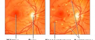
Sclerotic changes in the vessels of the retina indicate the irreversibility of pathological changes: hypertensive retinopathy, which occurs with the progression of arterial hypertension, leads to pronounced disorders in the fundus. With ophthalmoscopy, the following symptoms can be identified:
- increased unevenness of blood vessels (narrowing of capillaries and dilation of small veins);
- the appearance of a large number of convoluted vascular trunks;
- sclerosis and hyalinosis in the artery wall (symptoms of silver and copper wire);
- multiple manifestations of the symptom of arteriovenous chiasm;
- formation of microthrombi;
- pinpoint hemorrhages;
- changes in the optic disc area (waxy pallor).
The deformation of blood vessels, which the doctor will see, indicates a high risk of complications and inexorable progression of the disease.
Stage 3
Vasopathy caused by hypertension can cause complete loss of vision. Hypertensive macroangiopathy of the last stage, in addition to the usual symptoms of retinopathy, is manifested by the following symptoms:
- swelling and multiple hemorrhages in the retina;
- white foci of accumulation of exudate, similar to lumps of cotton wool;
- swelling and unevenness of the optic nerve head.
Damage to blood vessels and nerves in the last stage leads to significant deterioration of vision and a high risk of retinal detachment.
Diagnostics
Ophthalmologists diagnose and treat retinal angiopathy of the hypertensive type.
To identify the disease and determine its stage, it is necessary to undergo the following diagnostic measures:
- ophthalmoscopy – examination of the fundus using special instruments to evaluate the retina, optic nerve head, and blood vessels;
- fluorescein angiography to study the condition of blood vessels;
- tonoscopy, which allows you to determine blood pressure in the central artery and central vein of the retina;
- rheoophthalmography - study of blood circulation in the vessels of the eyes;
- Doppler ultrasound - to assess blood flow in the retina.
The use of magnetic resonance imaging makes it possible to detect changes in surrounding tissues.
When conducting diagnostics, it is necessary to exclude diabetic retinopathy, anemia, autoimmune diseases, and occlusion of the veins of the eye.
Traditional medicine
Source: tvoiglazki.ru
To treat angiopathy, the following methods are used by healers:
- First method
Take 100 grams of St. John's wort, chamomile, yarrow, birch buds, immortelle and mix everything. After this, 1 tablespoon of the mixture is added to half a liter of hot water and left for 20 minutes.
Then strain everything and add water to make half a liter of infusion. Drink 1 glass in the morning on an empty stomach and at night (after drinking a glass of infusion in the evening, you should not eat or drink). Take every day until the entire collection is used.
- Second recipe
Stir 1 teaspoon of white mistletoe, already ground into powder, in 250 milliliters of boiling water and leave to steep overnight. Take 2 tablespoons 2 times a day, 3-4 months.
- Third way
Take horsetail (20 grams), knotweed (30 grams), 50 grams of hawthorn flowers and mix everything. Then stir 2 teaspoons of the mixture into 250 milliliters of hot water and leave for half an hour. Take 1 tablespoon half an hour before meals 3 times a day for a month.
Regime and nutrition during illness
Patients are recommended to be active and prescribed a diet aimed at maintaining the normal functioning of the retina and reducing the risk of high blood pressure.
Foods with large amounts of carbohydrates, cholesterol and salt are excluded from the diet. The menu is designed in such a way that it is rich in vitamins, minerals and trace elements that are necessary for the retina, blood vessels and the entire body.
Exclude from the diet:
- fat;
- roast;
- salty;
- spicy;
- alcoholic drinks;
- fast food and deep-fried food;
- semi-finished products;
- flour products;
You need to use:
- dairy products;
- fruits and vegetables;
- greenery;
- eggs;
- various types of cereals;
- boiled, baked and steamed meat and fish of low-fat varieties.
- legumes;
- whole wheat bread;
- limit fluid intake.
Treatment of angiopathy is very simple. It comes down to the treatment of hypertension. Photo- and laser coagulation of newly formed vessels have a good effect in neutralizing the consequences of an advanced stage of angiopathy.
Neutron radiation is used experimentally on the hypothalamic-pituitary-adrenal system. Vitamins P, E, hormones, anti-sclerotic agents, oxygen therapy, and anticoagulants also help.
For prevention, you need to monitor the general condition of the body and lead a healthy lifestyle. No specific actions are required for the eyes, since retinal angiopathy is an echo of either a genetic abnormality, trauma, or congenital anomalies and vascular diseases.
One way or another, it is just an echo, a signal to listen to the general state of the body.
Mechanism of development of angiopathy
In the early stages, hypertensive angiopathy does not manifest itself in any way. This is one of the barriers to early diagnosis of the disease. Over time (when the disease enters phase 2), the patient begins to complain of:
- blurred vision;
- sensation of stars and spots before the eyes;
- eye strain;
- blurred image;
- disappearance of certain visual fields (this symptom can have short-term or long-term manifestations).
During this same period, high blood pressure is observed. Upon examination, the ophthalmologist notices that the arterial vessels of the retina are narrowed. At different stages of the disease, this symptom has different manifestations.
In addition, the lumen of the vessels itself changes. The blood supply to the organ becomes more difficult. In more advanced situations, blood circulation stops. The doctor diagnoses hemorrhage; blood accumulations in the form of extravasation are observed.
In the medical literature you can find information about the links (or, in other words, stages) of hypertensive angiopathy:
- Retinal artery spasm.
- Worsening of retinal vascular damage, their narrowing.
- The appearance of small blood clots in the retinal artery.
- The eye vessels begin to narrow like a glass tube. This process is already irreversible, and in medicine it is called hyalinosis.
- The vessels become fragile and often rupture, so the number of hemorrhages increases. Even a person without medical education can notice them.
- The blood supply to the entire retina is disrupted. The ophthalmologist diagnoses her with ischemia. The retina is destroyed to varying degrees. In this case, it is very difficult to save vision without surgery.
Methods for identifying and treating macroangiopathy
Diabetic or hypertensive angiopathy can be suspected based on symptoms, medical history, and patient complaints, but a complete picture of the pathology can only be formed through a full examination. For these purposes, consultations with narrow specialists are prescribed - an endocrinologist, angiosurgeon, neurologist, ophthalmologist, cardiologist, nephrologist and others. Laboratory tests are required:
- Blood sugar;
- Lipid spectrum;
- Coagulation parameters, platelets, fibrinogen.
- According to indications - adrenal hormones, thyroid hormones, kidney function tests, etc.
Instrumental diagnostics of macroangiopathy necessarily includes electrocardiography, 24-hour blood pressure profile, stress tests (treadmill, bicycle ergometry), ultrasound examination of the heart, large vessels, blood flow studies with Doppler, X-ray contrast angiography of the vessels of the brain, heart, and kidneys.
Treatment of macroangiopathy, regardless of its underlying cause, presents significant difficulties due to the irreversibility of changes, rapid progression of pathology, frequent combination with other chronic lesions of parenchymal organs and age-related factors. It should be comprehensive and aimed at the pathogenetic mechanisms of vascular disorders.
The goal of therapy for macroangiopathies is to stop the progression of complications that can lead to disability and death of the patient, because it is no longer possible to restore the normal structure of the arterial wall.
The main approaches in the treatment of macroangiopathy:
- Normalization of biochemical blood parameters - sugar, cholesterol and lipid fractions;
- Correction of hemocoagulation disorders;
- Reduction to normal values.
Diagnostic features
Hypertensive retinal angiopathy cannot be treated without proper diagnosis.
It can also be performed by an ophthalmologist. When coming for an examination, first of all, the patient must report symptoms and associated health problems, especially if they relate to the functioning of the cardiovascular system. One of the best diagnostic methods in this case is ophthalmochromoscopy. The essence of the method is a thorough examination of the fundus under white and red light. Arterial vessels in the red spectrum are not visible as well as in the white spectrum.
Therefore, narrowed vessels will be less noticeable than healthy ones. If you look at them through a red light, then they are completely undetectable. Ophthalmochromoscopy makes it possible to assess the condition of the blood vessels at the bottom of the eye.
A more complete picture of the blood supply to the eye can be obtained through ultrasound examination of the eyeballs. Using this method, it is possible to successfully determine the pathology of the eye structure. To assess the structure of the vessels themselves, the Doppler scanning method is most often used.
Doctors are able to see how blood circulates in the organs of vision thanks to x-rays, during which a contrast agent is used. It is the latter that ensures image quality and the ability to assess the condition of blood vessels, the presence of pathological formations in the blood supply, excessive vascular narrowing or dilation, and hemorrhages.
More accurate information about the structure of blood vessels, the characteristics of blood flow in the eye, and the state of the lumen of blood vessels can be obtained using MRI. Today this method is the safest and most accurate. That is why it is often used to diagnose angiopathy in children.
Stages of hypertensive retinopathy
Depending on the degree of damage to the retinal vessels, hypertensive angiopathy of the retinal vessels develops in several stages. And exactly what stage of the disease in a particular case can be determined by an ophthalmologist after examining the fundus. Depending on the degree of development of the pathology, certain disorders of the blood vessels of the eye occur. There are 4 stages of development of hypertensive retinopathy:
- first stage. Physiological changes occur, but there are no significant symptoms. The person does not experience discomfort and does not even realize there is a problem. Vascular spasm and expansion of the lumen of the ocular arteries occurs very slowly, so the disease can only be determined during an examination by an ophthalmologist;
- second stage. Organic changes occur in the walls of blood vessels. The symptoms become more pronounced and begin to cause inconvenience to the person, which is why the first complaints arise. During the examination, the doctor notices expansion, swelling of the venous network, pinpoint hemorrhages, shine of the vascular walls and waxy pallor of the fundus;
- third stage. In the structure of the retina and directly in the vessels, degenerative processes begin to occur with great intensity, which can provoke retinal detachment;
- fourth stage. This is the final stage of development of angiopathy of the ocular retina, which is accompanied by the release of fluid that accumulates in the fundus of the eye as a result of the inflammatory process and as a consequence of impaired blood circulation to an advanced degree.
At the first stage, it is possible to simply prevent the development of consequences, but if you start the development of the pathological process, then loss of vision is guaranteed. Therefore, you need to undergo a routine examination with doctors, including an ophthalmologist. And if a problem is detected, it is recommended to begin treatment as quickly as possible and follow the instructions of the attending physician.
Symptoms of angiopathy of one and both eyes
As a rule, hypertensive angiopathy of the retina of both eyes is diagnosed. In this case, the severity of changes in the eyeballs may vary. A person in the initial stage sometimes does not notice any symptoms. The first signs may appear precisely when blood pressure rises. When the numbers drop to normal, they go away.
At later stages of hypertensive angiopathy, organic changes in the retina develop. Due to poor blood supply, tissues experience oxygen deficiency, and degenerative changes in the sensitive epithelium occur. At the site of hemorrhages, foci of fibrous tissue are formed, in which there are no rods and cones that perceive light. As a result, the sensitivity of the retina gradually decreases.
Patients with hypertensive retinal angiopathy present the following complaints:
- decreased visual acuity. First, these changes are felt during a period of fatigue, after stress, at twilight, then the deterioration of visual acuity progresses, observed even in complete well-being, at rest, after rest;
- photopsia is possible - a person sees bright flashes, colored lines resembling a zigzag, lightning;
- many note the flickering of spots before their eyes, dark spots and dots, circles, all in fog, unclear outlines of objects;
- loss of visual fields;
- concentric narrowing of vision indicates serious disorders in the body;
- pain in the eyes.
If such symptoms appear, you should consult an ophthalmologist. Often people are referred for a fundus examination by family doctors and therapists when elevated blood pressure numbers are registered and typical complaints of hypertensive patients are identified:
- headaches - mainly in the back of the head;
- dizziness, nausea, tinnitus;
- vomiting is possible;
- facial redness. Feeling hot, restless, excited;
- blood pressure numbers are more than 140/90 mm Hg. Art.
Signs of visual impairment often appear as a harbinger of a hypertensive crisis. However, sometimes even a long course of arterial hypertension is not accompanied by visual symptoms. They appear already at the stage of organic changes, when the reverse development of the pathology is impossible.
Causes of the disease and mechanism of occurrence
Source: boleznikrovi.com
One of the main causes of the disease is a systemic disease such as hypertension. The connection between them is shown by Valsalva's experience. Measuring pressure in the central ophthalmic artery shows that in healthy people the pressure returns to normal after 10 minutes.
In patients with hypertension, normalization of blood pressure in the retinal vessels occurs only after at least half an hour. The duration of metabolic vasodilation was also observed. In healthy people it was no more than 2 minutes, in patients with hypertension - after 5-10 minutes.
Hypertensive retinal angiopathy occurs due to the following reasons:
- persistent and frequent increase in blood pressure;
- increased blood sugar, dyslipidemia;
- endocrinological pathologies;
- work associated with constant eye strain;
- excess body weight;
- magnesium and vitamin deficiency;
- frequent stressful situations;
- eating food with a lot of carbohydrates and fats;
- bad habits;
- pathologies of the structure of the blood vessels of the eye;
- increased intracranial pressure;
- spine disease;
- traumatic brain injuries;
- age.
Hypertensive retinal angiopathy has many clinical names, although even the term itself is now used only in the territory of the former USSR.
These names include albuminuric retinitis, arteriosclerotic retinitis, angiospastic retinitis, hypertensive retinopathy, arteriospastic retinitis, hypertensive angioretinoneuropathy, angio- or retin degeneration.
This diversity is associated both with the ambiguity of the symptoms of the disease and with the huge variety of its causes. There is no consensus on the classification of this disease. The fact is that according to some data it is classified according to the stage of development of hypertension, according to others - as renal hypertension and atherosclerotic changes in the blood vessels of the eye.
The pathohistological substrate of retinopathy is still not well understood. Active penetration, leakage, or in other words, extravasation of plasma into both the retina and the disc tissue undoubtedly takes place. Transudative fluid stratifies elements of different layers of the retina.
The accumulation of fluid is so great in some places that cyst-like spaces appear. The accumulation of fluid and fibrin in the inner layers of the retina ophthalmoscopically has the appearance of cotton wool-like foci. The shiny white spots that form a star shape are histologically lipid deposits.
Treatment
The main task in identifying hypertensive retinal angiopathy is treating the disease that caused it. If it is a hormone-producing tumor, surgery is performed. If there is an imbalance of nervous influences, medication and physiotherapeutic treatment are prescribed.
To stabilize blood pressure, the following is prescribed:
- calcium channel blockers;
- ACE inhibitors (angiotensin converting enzyme);
- antispasmodics;
- adrenergic blockers, sympatholytics;
- diuretics.
Drugs to stabilize blood pressure
Local treatment is prescribed by an ophthalmologist. Vitamins and antioxidants are usually prescribed. Vitamin C and rutin help strengthen the walls of blood vessels. To prevent dystrophic changes in the retina and reduce swelling, drops are prescribed:
- Emoxipin;
- Quinax;
- Taufon and others.
Sometimes, for greater effectiveness, drugs, for example, hormones, are injected into the periocular tissue or directly into the vitreous body.
Physiotherapeutic procedures are carried out:
- electrosleep;
- electrophoresis of vasoactive drugs;
- massage of the cervical-collar area.
If you suspect a retinal detachment, you need to act quickly. Detachment occurs if high blood pressure leads to a rupture of the vessel, blood gets between the retina and the choroid, disrupting the connection between them.
The retina can be fixed with a laser if you seek help right away. Other types of microsurgical intervention are performed according to indications:
- extrascleral filling of the retina (all layers of the eyeball are fixed from the outside with fillings);
- partial vitrectomy (removal of part of the vitreous body) followed by fixation of the retina.
A few hours after the detachment, the nerve cells and receptors die off, so it will be impossible to restore function, even if the retina is returned to its place. Therefore, anyone who has a tendency to hypertension should know the first signs of detachment:
- sudden loss of lateral vision;
- cloudiness, distortion of contours, vibration of objects;
- problems with coordination, orientation in space;
- photopsia, cobwebs, spots, rings, flashes that occur in bright light or when pressure is placed on the eye.
Peculiarities of symptoms of different forms of macroangiopathy
Both diabetic and hypertensive macroangiopathy cause similar manifestations of vascular pathology due to the stereotypical morphological basis, which is atherosclerosis. Depending on the location of the lesion, atherosclerosis of the coronary, renal, cerebral arteries, aorta, etc. is distinguished, that is, the forms of macroangiopathy in diabetes mellitus or hypertension will coincide with those in the case of ordinary atherosclerosis.
Symptoms of macroangiopathy include signs of ischemic damage to various organs, in severe cases - necrotic changes, and has some features:
- Coronary macroangiopathy forms the basis of coronary heart disease and causes angina pectoris, myocardial necrosis, arrhythmias, acute or chronic organ failure, sudden cardiac death, while in diabetes there are often atypical and painless forms of heart damage;
- In diabetics, repeated and recurrent heart attacks are more often diagnosed due to deep damage to the coronary vessels, as well as complications of cardiac infarction against the background of macroangiopathy (rhythm disorders, cardiac aneurysms, thromboembolic syndrome, cardiogenic shock, etc.);
- The risk of death from myocardial necrosis in diabetic macroangiopathy of cardiac vessels is twice as high as in patients without diabetes;
- Macroangiopathy of cerebral vessels occurs with symptoms of constant ischemia of brain tissue or infarction (stroke), and the combination of hypertension, diabetes and arterial damage increases the risk of cerebral vascular accidents several times;
- Every tenth diabetic develops an obliterating form of macroangiopathy of the lower extremities, with patients complaining of cold feet, impaired sensitivity, pain, swelling, trophic changes (hair loss, peeling of skin, cracks, ulcers);
- A specific sign of leg macroangiopathy in diabetes mellitus is gangrene, the likelihood of which is increased by concomitant changes in microcirculation and nerve trophism.
Neurological symptoms in cases of damage to the vessels of the neck and brain are manifested by sensitivity disorders, muscle weakness, and convulsions. Macroangiopathy of the neck vessels, characteristic of hypertension, is manifested by cranialgia, dizziness, memory impairment, hearing, and vision.
In case of damage to the aorta, diabetic and hypertensive patients complain of abdominal pain, constipation or diarrhea, symptoms of insufficient blood supply to the legs in the form of numbness, paresthesia, and pain.
Macroangiopathy of the renal arteries leads to their sclerosis, which further aggravates the signs of hypertension, which becomes insensitive to prescribed drugs. Against the background of renal macroangiopathy, edema increases, pallor and puffiness of the face appear, and chronic organ failure develops.
What is hypertensive angiopathy of the eye?
Hypertensive angiopathy is a disease of small ophthalmic vessels of the venous and arterial type. It occurs in people with long-term hypertensive problems, most often as a result of hypertension type I-II B. The lumen of the vessel in this case is deformed, and the vessels themselves expand and become curved. Since hypertension affects the vessels of the whole body, both eyes suffer from angiopathy.
Quite often the disease is diagnosed in young people. The disease is prone to progression, and therefore, if treatment is delayed, a number of negative changes may develop in the retina.
Some areas of the retina itself become cloudy. Often in this case the following manifestations are observed:
- blockage of the main vein in the retina;
- disturbance of blood supply to the optic nerve;
- obstruction of the patency of the main artery.
All this can be avoided with the help of modern treatment methods.
Angiopathy of the hypertensive type can be accompanied by serious damage to the blood vessels of the heart, genitourinary system, and central nervous system. The disease cannot disappear on its own. By delaying treatment, the patient only makes the situation worse. Although hypertensive angiopathy is identified in the medical literature as a separate disease, scientific discussions about its etiology are still actively underway.
Thus, some doctors define angiopathy as a certain pathological change or symptom of hypertension. It is customary to distinguish 3 stages of angiopathy:
- Initial. It does not make itself felt at all; only a specialist can notice vascular constrictions.
- This stage is characterized by swelling of the retina and minor hemorrhages.
- At this stage, exudate appears, which causes an inflammatory process in the organs of vision.
Treatment of the disease
Treatment should be carried out under the strict supervision of the attending physician.
Retinal angiopathy of the hypertensive type is a very serious disease and often leads to blindness, so you should not try to cure it at home yourself, but you must consult a specialist. The doctor will conduct a thorough examination and make a diagnosis. As therapy, medications are prescribed that lower blood pressure, anticoagulants and medications that improve metabolic processes in the retina. A complex of vitamins is also credited. It is imperative that each patient is given dietary nutrition, physiotherapeutic methods and folk remedies therapy.
Return to contents
Physiotherapeutic methods
Hypertensive angiopathy responds well to physiotherapeutic treatment methods. For this, laser therapy and exposure to a magnetic field are widely used. Laser therapy is based on the use of optical radiation of red and infrared radiation in pulsed or continuous mode. This method improves metabolic processes in the affected eye structures, and in combination with drug treatment helps preserve visual functions for a long time.
The procedure will help relieve spasms and improve blood microcirculation.
Treatment methods for hypertensive angiopathy
Treatment is primarily aimed at suppressing the main provoking factor - high blood pressure, as well as the consequences that it caused.
medications are indicated for patients diagnosed with hypertensive angiopathy :
- protecting the walls of blood vessels from damage and stimulating metabolic processes in the retina (Actovegin, Trental);
- dilating vessels (Xavin, Stugeron);
- blood thinners and prevent the formation of blood clots (Cardiomagnyl, Magnicor);
- promoting the resorption of exudate formations located on the retina (Wobenzym);
- having an antioxidant effect (ascorbic acid, Veteron).
Also, for hypertensive angiopathy, the patient is prescribed medications to correct blood pressure levels. Such drugs include diuretics (Clopamide), calcium channel blockers (Felodipine), alpha blockers (Atenolol), ACE inhibitors (Prestarium, Spirapril).
In addition, the patient should use eye drops Taufon, Aisotin, Quinax. They stimulate metabolic processes in the retina of the eye, provide nutrients, which improves blood circulation.
An integral part of the correction of the condition are physiotherapeutic procedures . In this case, laser therapy, magnetic therapy, and acupuncture are useful.
If there is a threat of retinal rupture, conservative treatment methods are not effective. Under such conditions, radical intervention is necessary. In most cases, the laser coagulation method .
Patients with hypertensive angiopathy are also recommended to follow a special diet. The point of the diet is to normalize the cardiovascular and digestive systems. In addition, correcting your diet will help you reduce body weight, which will also be beneficial: obesity often causes increased blood pressure.
It is necessary to exclude such foods and drinks from the patient’s diet as:
- red meat;
- sweets;
- baking;
- fatty and spicy foods;
- fatty dairy products;
- garlic;
- radish;
- spinach;
- legumes;
- industrial and animal fats;
- marinades and pickles;
- smoked meats;
- coffee, cocoa, tea.
The patient's diet should be based on fresh, stewed and boiled vegetables, fresh berries and fruits, grain bread, lean meat and fish, seafood, lean soups, dried fruits, and vegetable oils.
What you need to remember about the development of the disease
Hypertension is not a death sentence!
It has long been a well-established opinion that it is impossible to get rid of HYPERTENSION forever. To feel relief, you need to continuously drink expensive pharmaceutical drugs. Is it really? Let's figure out how hypertension is treated here and in Europe...
Retinal angiopathy is always a consequence of persistently elevated pressure that has not subsided for a long time. This condition leads to neuroregulatory dysfunction, dilation of the fundus vessels and small hemorrhages that are visible in the eyeball.
If the disease is left untreated, irreversible changes occur in the retina. Its areas become more cloudy, but timely and competent therapy can eliminate this symptom.
At the initial stage of development of angiopathy, when there are no characteristic symptoms, it is still possible to diagnose the presence of changes in the retina. This is done using fluorescein angiography, which shows the presence of changes in even the smallest vessels.
Hypertensive angiopathy is sometimes accompanied by damage to the vessels of the central nervous system, heart and urinary system. Often the vessels cannot get used to the excessive pressure, so they become brittle, causing bleeding in the heart and brain.
Neurological disorders occur due to cerebrovascular accidents:
- decreased mental activity;
- irritability;
- lack of concentration;
- emotional instability;
- bad memory.
Symptoms
Many people ignore obvious symptoms, not realizing that they signal a disease. Accordingly, if you do not want to lose your vision, you should pay attention to specific symptoms. If you begin to develop angiopathy, your vision will deteriorate slightly, and it will periodically become cloudy due to pressure changes in the body. Fatty spots may also appear on the eyes, and people suffering from myopia will notice that it begins to progress very quickly. Also symptoms are some things not related to the eyes, such as nosebleeds and gastrointestinal bleeding. You may also experience severe pain in your legs.
Hypertensive macroangiopathy
Hypertensive macroangiopathy develops against a background of chronically high blood pressure, but its manifestations are not immediately noticeable due to the large caliber of the affected arteries. The mechanism of atherosclerotic damage in hypertension comes down to a change in the inner layer of the arteries under the influence of turbulent blood flows and excess pressure load.
Lipids penetrate into the microtrauma zones of the endothelial lining, penetrate deeper and begin to form an atherosclerotic plaque. In the middle layer of the artery, hypertrophy of muscle fibers progresses during hypertension, and repeated spasm against the background of crises leads to infiltration of the vascular walls with plasma components - proteins and fats.
The targets for hypertensive macroangiopathy are the aorta, renal arteries, vessels of the brain, and heart. Arterial hypertension is characterized by macroangiopathy of the neck vessels with obstruction of the carotid arteries and the risk of thrombosis, the consequences of which are extremely destructive.
In addition to atherosclerotic changes in large arteries, as a result of hypertension, smaller vessels are also damaged (sclerosis, hyalinosis), so the symptoms of trophic disorders can be extremely diverse.
Kinds
The disease is divided into many types, taking into account the cause of the pathology, the location and size of the lesion.
Despite the broad classification of pathology, 2 or more forms can be simultaneously diagnosed in a given patient. Such vascular lesions occur in more than 50% of patients with angiopathy.
Some patients experience temporary damage to the vascular system. With timely diagnosis and monitoring of the course of the disease, specific treatment may not be required.
Hypertensive angiopathy
High blood pressure and progression of hypertension negatively affect the condition and functioning of the vascular system. As a result of pathological changes, the muscle layer hypertrophies, which entails the development of fibrosis.
Against the background of this condition, blood circulation becomes more complicated, the lumen of the vessels becomes thinner, and their blockage is possible. This is accompanied by the same high pressure, due to which blood effusions open.
In the hypertensive form of angiopathy, most patients suffer from the retina, blood vessels in the brain, renal arteries and coronary heart vessels.
Diabetic
This is a common angiopathy that progresses due to diabetes. With such a lesion, the capillary bed is transformed, the reason for this is the excess of sugar levels in the blood sample. Due to the way the capillaries are affected, the disease also reaches large vessels, which can make a person disabled in the future.
More often, this type of angiopathy affects the feet and the retina of the eye.
In the diabetic form of angiopathy, the greatest danger lies in the fact that the patient may have the affected limb amputated. When the disease affects large vessels, heart attacks, strokes and other pathologies are possible.
Hypotonic
Against the background of the hypotonic form of angiopathy, the peripheral bed suffers, which, due to loss of vascular tone, is filled with blood. As a result of this process, the permeability of the vascular walls increases, and blood cells are deposited in the lumen of the capillaries.
Such changes are more common on the legs, but the disease may develop on other parts of the body.
Traumatic
With compressive injuries of the chest and skull, patients' blood pressure often rises. At the same time, light spots appear on the retina, and some blood vessels become clogged.
Such pathological changes lead to loss of vision.
Even despite the timely provision of highly professional medical assistance, it is not possible to restore visual acuity.
Hypertensive macroangiopathy: signs, causes, diagnosis and treatment features
- June 22, 2018
- Cardiology
- Lenara Aminova
Hypertensive macroangiopathy: what is it? To fully understand this term, you need to know the differences between hypertension and angiopathy. These two concepts are related.
Hypertension and angiopathy
Angiopathy is a group of diseases associated with vascular damage due to increased nervous excitability of cells and neurons. Angiopathy is characterized by spasm and paresis of blood vessels, which contributes to increased permeability of the walls and frequent hemorrhage.
There are several types of angiopathy, it all depends on the size of the affected vessels and their condition.
- Hypertensive macroangiopathy, developing as a result of atherosclerotic lesions. With this disease, the patient’s condition is not the best; its severe course complicates it.
- Microangiopathy occurs as a result of necrosis, thrombosis, hyalinosis and fibrinoid swelling. With the development of this disease, damage to the eye vessels and capillaries of the kidneys is observed.
Hypertension is a condition in which a person's blood pressure increases. There is an increase in pressure on the walls of the inner choroid and a decrease in its diameter.
Hypertension is called the main disease of the 21st century. It develops rapidly and leads to death within 5 years.
The causes of hypertension include poor nutrition, bad habits, ecology, and stress.
Cause-and-effect relationships
With hypertensive macroangiopathy, the vessels change their structure and physical properties. With this disease, their appearance, function and structure are modified. The vascular walls are damaged and, as a result, their diameter changes.
Macroangiopathy often develops together with microangiopathy. This occurs due to rapid damage to small vessels at an early stage of the disease.
In a particularly severe case, hypertensive macroangiopathy of the vessels of the neck and brain is observed, which leads to early heart attacks and strokes.
In hypertensive diseases, a persistent increase in blood pressure is observed. The diameter of the vessel decreases, and greater blood pressure is exerted on the walls.
With a sharp increase, the vessels are damaged from the inside. In the place where the membrane is damaged, blood cells, lipids, and calcium accumulate, which leads to the formation of atherosclerotic plaques or blood clots. As a result, the lumen of the vessels disappears, they do not change their diameter, and the blood flow becomes vortex rather than linear.
Hypertensive macroangiopathy with the formation of arterial deformations is associated precisely with the formation of plaques, which create the risk of vascular rupture and blood clots. These blood clots can break off and clog blood vessels. As a result, myocardial infarction or stroke occurs.
In hypertension, macroangiopathy is inevitable, which determines the clinical manifestation of this disease.
Other reasons
Hypertensive macroangiopathy (what it is, we described above) occurs for many additional reasons:
- due to diabetes;
- due to age;
- with high intracranial pressure;
- as a result of blood diseases;
- due to injuries and osteochondrosis;
- as a result of harmful working conditions;
- from poisoning or intoxication;
- with weakened immunity;
- under the influence of ecology.
Macroangiopathy of the hypertensive type is the main symptom or sign of a disease such as hypertension.
Symptoms
Signs and treatment of hypertensive macroangiopathy depend on the main factors in the occurrence of the disease and the condition of the blood vessels in general. The types of damaged vessels are also important.
If the disease affects the limbs, the person feels pain, discomfort, swelling and bluish skin. It is even possible to develop gangrene if the vessels become clogged.
With macroangiopathy, the patient sometimes limps, and the gait changes.
If the disease affects the brain, then strokes and ischemic attacks occur, from which the brain suffers. Typically, such patients suffer from constant headaches, memory loss, dizziness to the point of fainting, pressure changes and darkening in the eyes. It is important to know that these are signs of hypertensive macroangiopathy.
If the disease affects the heart vessels, then ischemia and then a heart attack may develop.
A person suffers from shortness of breath from any physical activity, he suffers from constant pain in the back and chest, feels tired and overwhelmed. The symptoms of the disease worsen if the patient does not take any measures to cure it.
Further development of cardiac vascular macroangiopathy threatens myocardial infarction, severe pain and exercise intolerance.
If angiopathy affects the kidneys, then cure is almost impossible. The kidneys begin to suffer from edema, which impairs the electronic and water processes in them. As a result, kidney failure develops. So, we learned what hypertensive macroangiopathy is and the signs (symptoms) of this disease. How to identify it and with what?
Identification of the disease
The diagnostic process is a comprehensive examination of the patient’s body. This includes blood tests and reactions to treatment, and recording blood pressure numbers. After a detailed examination, it is established that these are signs of hypertensive macroangiopathy, and not another disease.
In case of damage to the vessels of the brain or neck, MRI and ultrasound examination are additionally prescribed. The work of the heart and the state of the vascular system can be studied by ECG, echocardiography, and coronary angiography. The condition of the kidneys is determined by ultrasound and urography.
An important stage of diagnosis is monitoring changes in blood vessels. Correct diagnosis and adequate assessment of existing disorders are the key to prescribing competent treatment. The result largely depends on the diagnosis.
Treatment of macroangiopathy
Hypertensive macroangiopathy is treated by normalizing blood pressure. Only through this can the risk of complications, such as paralysis, stroke or heart attack, be reduced. In addition, with proper treatment, attention is paid to the composition of the blood (thickness, fat level and viscosity).
With complex treatment, it is most often possible to eliminate all manifestations of macro- and microangiopathy, as well as find out the root cause of their occurrence and cure them. Experts prescribe patients to take anticoagulants that thin the blood.
The patient should take antispasmodics, which reduce pain and dilate blood vessels, strengthening them. In case of diabetes mellitus, the patient is prescribed the required dosage of insulin.
Unconventional treatment
There is also an alternative treatment for hypertensive macroangiopathy. This can include mud baths, needle injections and magnetic therapy.
The patient is put on a strict diet, fried and fatty foods are excluded from the diet, since first of all it is necessary to reduce the amount of cholesterol in the blood.
Preventive measures
You can protect yourself from such a disease - just listen to the following advice and follow them unquestioningly.
- The diet should include vegetables, fruits, fish and dairy products. We exclude fatty and fried foods. Salt is also an enemy, so it is necessary to reduce its amount.
- Bad habits are bad for your health. Alcohol and smoking are prohibited.
- The patient must monitor his mental state, avoid stress, depression, and be sure to follow the correct daily routine. At the request of the patient, the attending physician may prescribe sleeping pills or sedatives.
- If you have a sedentary lifestyle, you need to walk in the fresh air more often, engage in light sports: swimming, walking, physical therapy.
- Even if you don't have diabetes, it's a good idea to check your blood sugar levels daily.
Important
All treatment methods are important in the fight against a detected disease. Only a variety of medications that change blood pressure and reduce the likelihood of blood clots will help you cope with hypertensive macroangiopathy, the signs of which we listed above.
Only if the whole body is treated can the disease be overcome. An important condition is an integrated approach. If high blood pressure is observed along with an altered blood chemical composition, then the entire treatment plan is adjusted and complex treatment regimens are prescribed.
To identify the disease at an early stage of its development, you need to undergo a complete examination of the body at least twice a year by all specialists. Early treatment gives a good chance of a complete recovery for the patient.
Source: https://SamMedic.ru/330943a-gipertonicheskaya-makroangiopatiya-priznaki-prichinyi-diagnostika-i-osobennosti-lecheniya
Retinal angiopathy of the hypertensive type
Retinal angiopathy of the hypertensive type Source: youtube.com
Retinal vascular angiopathy is a pathology of the blood vessels of the eyes due to disturbances in nervous regulation, outflow or inflow of blood. This includes retinal angiopathy of the hypertensive type.
It occurs after a long course of hypertension. Hypertensive angiopathy occurs in children and adults, but often appears in people over 30 years of age. Doctors pay a lot of attention to angiopathy, because it can lead to vision loss.
When a pathology is detected, treatment must be started, as it progresses and can become chronic.
Since ancient times, people have attached special importance to vision. In his life, such sensations as smell, taste and others began to occupy an ever smaller part, and he began to pay more attention to vision, because the whole huge world of colors and colors that appeared before him became extremely important.
It has become extremely important to expand the scope of adaptive abilities. To do this, you need to quickly switch from near to distant objects and be able to distinguish between different colors, which was not possible for many animals. Therefore, a person takes special care of his vision.
If earlier this problem was practically unsolvable, now doctors look at these problems more optimistically. Timely diagnosis of the disease and the latest research have made it possible to effectively treat many eye defects that previously seemed incurable.
The main causes of eye defects are:
- circulatory disorders,
- innervation,
- properties of the eye muscles and biochemical properties of the lens or vitreous body, which can be caused by metabolic disorders.
After all, the eyes are a precisely balanced system that can be severely disrupted even with minor lesions.
Angiopathy is a disorder of the properties of blood vessels, in which they spasm or their lumen becomes smaller due to other reasons. In the eye, such causes may be congenital defects or metabolic disorders. The danger of this condition lies in two aspects.
Firstly, a spasmodic vessel delivers much less oxygen and nutrients to nearby tissues, which leads to malnutrition. Secondly, spasmodic veins can stratify, and aneurysms, expansion and pinpoint hemorrhages, and the formation of intervascular shunts will form.
This occurs under the influence of pressure, which creates mechanical stress on the vessel wall. This is especially dangerous for the retina, due to the constant metabolic processes that occur and require the maintenance of homeostasis, nutrition and oxygen supply.
Hypertensive retinal angiopathy is damage to the vessels of the retina due to hypertension, endangering blood vessels throughout the body. Currently, hypertension is called one of the main diseases of mankind.
Arterial hypertension is a chronic disease, the main symptom of which is a persistent and prolonged increase in blood pressure. Hypertension can be considered when systolic pressure is over 140 mm or when diastolic pressure is over 90 mm.
Against the backdrop of prolonged development of arterial hypertension without proper treatment, very serious complications subsequently arise, one of which is hypertensive angiopathy. More serious complications are myocardial infarction, stroke, and early mortality.
Hypertensive angiopathy of the retina develops as a result of a long course of hypertension. Vascular damage occurs as a result of constantly elevated blood pressure. The lesion progresses gradually over a fairly long period of time.

