Description
Mechanical injuries to the eyes include wounds and blunt trauma to the eyeball, its adnexa and bone bed.
Mechanical damage can be accompanied by hemorrhages into the soft tissues and structures of the eye, subcutaneous emphysema, loss of intraocular membranes, inflammation, decreased vision, and crushed eyes. Diagnosis of mechanical eye injuries is based on examination of the victim by an ophthalmic surgeon, neurosurgeon, otolaryngologist, or maxillofacial surgeon; radiography of the orbit, biomicroscopy, ophthalmoscopy, ultrasound echography and biometry, fluorescein tests, etc. The method of treating mechanical eye injuries depends on the nature and extent of the injury, as well as the complications that have developed.
Additional facts
Due to their superficial location on the face, the eyes are extremely vulnerable to various types of damage - mechanical injuries, burns, the introduction of foreign bodies, etc. Mechanical damage to the eyes quite often entails disabling complications: weakened vision or blindness, functional death of the eyeball. Severe eye injuries occur more often in men (90%) than in women (10%). About 60% of visual injuries occur in adults under the age of 40; 22% of injured people are children under 16 years of age. According to statistics, foreign bodies of the eye occupy the first place among injuries to the organ of vision; second – bruises, eye contusions and blunt injuries; third - eye burns.
Mechanical eye damage
Severity
There are four degrees of severity of eye contusion:
- 1st degree – transient traumatic failure of the eyeball. The organ is fully capable after therapeutic assistance. Varieties include corneal edema and retinal clouding.
- 2nd degree – persistent and more serious impairment of eye function, slightly reducing visual acuity. Varieties: corneal erosion, retinal tears, hemorrhages.
- 3rd degree - a strong blow to the eye, capable of causing more than half damage to vision. Varieties include: eyelid tears, retinal tears, lens pathologies.
- Grade 4 is the most severe form, which can cause 100% loss of visual function in the damaged eye.
Classification
In ophthalmology, mechanical eye injuries are classified according to the mechanism of occurrence, localization, and severity. According to the mechanism of traumatic action, wounds and blunt eye injuries (concussions, concussions) are distinguished. In turn, eye injuries can be non-penetrating, penetrating or through; uninfected and infected; with or without the introduction of a foreign body; with and without prolapse of the eye membranes. Based on the location of the damage, injuries to the protective apparatus of the eye (eyelids, orbit, lacrimal apparatus), eyeball, combined injuries to the appendages and internal structures of the eye, combined mechanical damage to the eyes and other areas of the face are distinguished. The degree of mechanical damage to the eyes depends on the type of traumatic object, as well as the strength and speed of the impact. According to the severity of eye injury, the following are distinguished: • mild injuries caused by foreign bodies entering the conjunctiva or the surface of the cornea, burns I-II, permanent wounds and hematomas of the eyelids, temporary ophthalmia, etc.
• moderate injuries, characterized by severe conjunctivitis and corneal opacities;
rupture or partial separation of the eyelid; burns of the eyelids and eyeball II-IIIA; non-perforating injury to the eyeball. • severe injuries accompanied by a perforated wound of the eyeball; wounds of the eyelids with significant tissue defects; contusion of the eyeball with damage to more than 50% of its surface; decreased vision due to rupture of the internal membranes, lens trauma, retinal detachment, hemorrhages into the eye cavity; damage to the bones of the orbit and exophthalmos; burns IIIB-IV. According to the circumstances and conditions of occurrence of mechanical eye injuries, industrial, agricultural, household, children's and military injuries are distinguished.
Causes of eyeball contusion
Violations of the integrity of the visual apparatus can be provoked by various factors:
- Ignoring safety during work.
- Irresponsible attitude towards your health.
- An accident, such as a blow, a fall or an attack.
- Antisocial behavior, alcoholism and drugs.
- Sports injuries due to insufficient protection.
- Presence in the combat area.
- Powerful changes in the atmosphere, gusty winds.
- A stream of water or gas.
Depending on the cause of its appearance, pathology is usually divided into two types:
- Direct . The occurrence is caused by the influence of an external force directly on the eye (for example, due to a bruise).
- Indirect . Appears when force is applied to the area adjacent to the eye, to the tissues surrounding it (bones of the skull, face, orbit) or to more distant areas of the body (compressed by the chest). As a rule, the consequences are observed in changes in the ocular structure.
Causes
Superficial eye injuries often occur when the eyelids, conjunctiva or cornea are damaged by a nail, contact lens, pieces of clothing, or tree branches. Blunt mechanical injuries to the eyes can occur when the eyeball or facial skeleton is hit by a large object (fist, ball, stick, stone, etc.) when falling on a hard object. Blunt injuries are often accompanied by hemorrhages in the tissue of the eyelids and eyes, fractures of the orbital walls, and contusions of the eye. Blunt mechanical injuries to the eyes are often combined with closed craniocerebral injury. Penetrating eye injuries are caused by mechanical damage to the eyelids or eyeball with sharp objects (stationery and cutlery, wooden, metal or glass fragments, wire). With shrapnel wounds, the penetration of a foreign body into the eye is often noted.
Mechanism of injury
At the moment of contusion, the eyeball is displaced and the structures that make it up are compressed.
At the same time, intraocular pressure increases and the pupil sharply dilates. Fluid rushing into the anterior chamber can cause tears in the iris along the pupillary edge or in the corners of the chamber.
The lens shifts, the ligament supporting it may not withstand the load and may break. Subluxation or dislocation of the lens occurs, and it falls out into the vitreous or anterior chamber. Ruptures of the lens capsule cannot be ruled out.
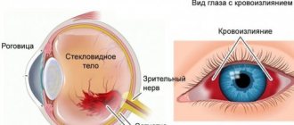
From vessels damaged by trauma, blood flows under the mucous membrane, chorionic membrane, retina, or into the chambers of the eye. The degree of hemorrhage that occurs varies.
Due to the tension of the structures at the time of injury, separation, ruptures or detachment of the retina is possible.
With severe injuries, rupture of the fibrous capsule of the eyeball may occur. More often this happens at the border of the limbus or at the attachment points of the extraocular muscles.
Ruptures or compression of the optic nerve are possible when the bone structures of the orbit are damaged.
Diagnostics
Recognition of the nature and severity of mechanical eye injuries is made taking into account the medical history, clinical picture of the injury and additional studies. For any eye injuries, it is necessary to conduct a survey X-ray of the orbit in 2 projections to exclude the presence of bone damage and the introduction of a foreign body. A mandatory diagnostic step for mechanical damage is to examine the structures of the eye using various methods (ophthalmoscopy, biomicroscopy, gonioscopy, diaphanoscopy), and measure intraocular pressure. When the eyeball protrudes, exophthalmometry is performed. For various disorders (oculomotor, refractive) the state of convergence and refraction is examined, the reserve and volume of accommodation is determined. A fluorescein instillation test is used to detect corneal damage. To clarify the nature of post-traumatic changes in the fundus, fluorescein angiography of the retina is performed. Electrophysiological studies (electrooculography, electroretinography, visual evoked potentials) in comparison with the clinic and angiography data allow us to judge the condition of the retina and optic nerve. In order to identify retinal detachment in case of mechanical damage to the eyes, assess its location, size and prevalence, an ultrasound of the eye is performed in A and B modes. Using ultrasound biometry of the eye, changes in the size of the eyeball are assessed and, accordingly, post-concussion hypertension or hypotonic syndrome. Patients with mechanical eye injuries should be consulted by an ophthalmologist, neurologist, neurosurgeon, otolaryngologist, or maxillofacial surgeon. Additionally, an X-ray or CT scan of the skull and paranasal sinuses may be required.
All about eye contusion: what it is, ICD-10 code, consequences, degrees, etc.
The culprit of concussion of the organs of vision can be a huge number of reasons.
According to ophthalmologists, it is this type of injury that contributes to the progression of numerous eye pathologies. The issue can be resolved quite easily if you contact a qualified specialist in a timely manner. In order for treatment of an eye injury to bring maximum effectiveness, you need to be able to correctly classify the type of injury and select the appropriate therapy. Now let's look at what eye contusion is and what are the causes of its occurrence.
What it is?
An eye contusion is an injury to the visual organ as a result of exposure to heavy blunt objects or intense mechanical force.
ICD-10 code
Eye contusion according to ICD-10 (international classification of diseases) is designated by code S05 and is one of the most dangerous injuries to the eyeball.
S05 Trauma to the eye and orbit Classification:
- S05.0 Conjunctival trauma and corneal abrasion without mention of a foreign body.
- S05.1 Contusion of the eyeball and orbital tissues.
- S05.2 Laceration of the eye with prolapse or loss of intraocular tissue.
- S05.3 Laceration of the eye without prolapse or loss of intraocular tissue.
- S05.4 Penetrating wound of the orbit with or without the presence of a foreign body.
- S05.5 Penetrating wound of the eyeball with a foreign body.
- S05.6 Penetrating wound of the eyeball without a foreign body.
- S05.7 Eyeball avulsion.
- S05.8 Other injuries of the eye and orbit.
- S05.9 Injury to unspecified part of the eye and orbit.
Causes of eyeball contusion
Violations of the integrity of the visual apparatus can be provoked by various factors:
- Ignoring safety during work.
- Irresponsible attitude towards your health.
- An accident, such as a blow, a fall or an attack.
- Antisocial behavior, alcoholism and drugs.
- Sports injuries due to insufficient protection.
- Presence in the combat area.
- Powerful changes in the atmosphere, gusty winds.
- A stream of water or gas.
Depending on the cause of its appearance, pathology is usually divided into two types:
- Direct . The occurrence is caused by the influence of an external force directly on the eye (for example, due to a bruise).
- Indirect . Appears when force is applied to the area adjacent to the eye, to the tissues surrounding it (bones of the skull, face, orbit) or to more distant areas of the body (compressed by the chest). As a rule, the consequences are observed in changes in the ocular structure.
Development order
As a result of contusion of the visual apparatus, disturbances occur leading to deformation of the ocular structures. Therefore, it is necessary to understand what processes occur in a short period of time of its formation.
The algorithm characteristic for the development of eye contusion is as follows:
- After mechanical impact, contusion of the eyeballs occurs.
- There is a short-term sharp jump in intraocular pressure.
- Then the visual apparatus returns to its original position.
- Due to processes that temporarily take place inside the eyes, blood circulation changes.
- Vessels and eye tissues are torn.
- Biochemical changes occur in the intraocular fluid.
- The development of a stress reaction is observed.
Severity
There are four degrees of severity of eye contusion:
- 1st degree – transient traumatic failure of the eyeball. The organ is fully capable after therapeutic assistance. Varieties include corneal edema and retinal clouding.
- 2nd degree – persistent and more serious impairment of eye function, slightly reducing visual acuity. Varieties: corneal erosion, retinal tears, hemorrhages.
- 3rd degree - a strong blow to the eye, capable of causing more than half damage to vision. Varieties include: eyelid tears, retinal tears, lens pathologies.
- Grade 4 is the most severe form, which can cause 100% loss of visual function in the damaged eye.
Consequences for the eyeball
The consequences of an eye contusion can be very serious, including complete loss of vision, without the possibility of recovery.
Corneas
Damage to the cornea is the most common consequence of eyeball contusion. Signs of such defects are erosion of varying depths and areas. They have the ability to change their size upward within a week.
The first indicators of corneal injury include:
- decreased visual acuity;
- unpleasant sensitivity to light;
- uncontrolled production of tears;
- feeling that something is in the eye;
- blepharospasms.
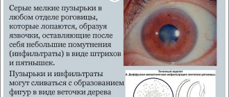
Blurred vision occurs if the central zones of the cornea are eroded. When the stroma is damaged, visual acuity noticeably decreases. Destruction of the endothelium leads to edema in the depths of the stroma, and opacification of the cornea in the form of stripes and latticework leads to the penetration of purulent composition into other areas of the stroma.
Important! In particularly severe cases of corneal defect, 3rd degree eye contusion may develop. At this stage, the eye becomes cloudy and takes on a gray tint.
Lens
Eye contusion is often a consequence of traumatic cataract. The reason is caused by clouding of the lens due to a change in its location (luxation). Due to the fact that moisture enters through microcracks in the capsule of the anterior chamber of the eye, the volume of fluid may increase with visible bruising.
Fibers in the form of a swollen mass fill the entire volume of the capsule if its cracks are significant. Sometimes the fibers block part of the anterior chamber, increasing pressure inside the eye and causing glaucoma. Lens luxation is characterized by deformation of the anterior chamber and displacement of the iris. The lens itself takes the form of a drop filled with fat.
Retina
Retinal injury is one of the most severe types of damage and the most difficult to treat. The cause is caused by retinal shock.
Damage may be accompanied by clouding of the eye, which in turn provokes inflammation of the internal structure of the retina; concentric narrowing of the field of view.
The color of such clouding ranges from light gray to milky, this is due to inflammation of the internal cells of the retinal structure.
With a minor injury, the structure of the eye and vision quickly and independently resume their functions. If it comes to severe cases (for example, 2nd degree contusion of the eyeball), then visual acuity is significantly reduced, and bleeding may develop:
- preretinal;
- retinal;
- subretinal.
Read more about the symptoms of various types of contusions in this article.
First aid and treatment
Before the ambulance arrives, you need to be able to properly assist the victim:
- Be sure to apply a bandage or gauze to the damaged area; if you don’t have anything at hand, any clean piece of fabric will do.
- The injured area must be cooled by placing a damp cloth on top.
- If the victim has nausea, it is necessary to sit him down with a cushion under his head and give him a sedative.
Attention! It is necessary to provide first aid to the victim and take him to the hospital. Doctors often allow bruise treatment at home.
Subsequent inpatient treatment includes:
- Anti-inflammatory drugs are usually prescribed by injection or infusion. The following medications are most often prescribed: Dexamethasone, Flosteron, Diprospan, Diclofenac, Indomethacin, Nimesulide.
- Antidepressants and tranquilizers, for example Tazepam, Elenium, Phenazepam.
- Antihistamines - HI receptor blockers: Loratadine, Tavegil, Suprastin.
- Enzyme preparations: Lidaza, Gemaza, Fibronolysin.
- Hemostatic agents: Dicynon, Vikasol.
- Diuretics such as Diacarb.
- Be sure to administer instillations into the conjunctival sac of the eye using: antibacterial drugs: Floxal or Vigamox, Antiseptics, Glucocorticoids.
- Non-steroidal drugs: Indocollir or Uniclofen.
Combination medications are often used, combining several active substances at once. In some cases, surgery is used. This type of treatment is used in the presence of various tears, ruptures, and anatomical defects. A prerequisite for the treatment of eye contusion is that the treatment must be prescribed by a medical specialist.
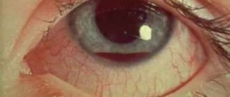
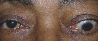
Photo based on the article
Several photographs illustrating eyeball contusion and first aid:
Conclusion
The onset of an illness such as a bruise or contusion of the eye is unpredictable and can come at any time. No one is immune from accidents.
Widespread damage to the organ of vision is not always dangerous and has disastrous consequences.
Do not forget that
measures not taken in due time can result in serious consequences, sometimes loss of vision.
Read more about the consequences of concussion in this article. It is important to carry out treatment in a timely and high-quality manner. There are many methods: this includes intensive therapy (in some cases the patient undergoes it at home) and various types of operations. Post-treatment prevention and rehabilitation include outpatient monitoring of eye health.
It is important not to self-medicate at home, and rather than delaying the problem, consult a doctor for qualified help. But the best thing is to prevent such injuries and take care of your health.
We also bring to your attention useful materials about contusions of the brain and ENT organs.
If you want to consult with the site’s specialists or ask your question, you can do it completely free of charge
Source: https://inBrain.top/bolezni/kontuziya/glaza.html
Treatment
The variety of factors causing mechanical damage to the eye, as well as varying degrees of injury severity, determine differentiated tactics in each specific case. In case of eyelid injuries with a violation of the integrity of the skin, primary surgical treatment of the wound is performed, and, if necessary, excision of crushed tissue along the edges of the wound and suturing. Superficial mechanical damage to the eyes, as a rule, is treated conservatively with the help of instillations of antiseptic and antibacterial drops and ointments. When fragments are introduced, the conjunctival cavity is jet washed and foreign bodies are mechanically removed from the conjunctiva or cornea. For blunt mechanical injuries to the eyes, rest, the application of a protective binocular bandage, and instillation of atropine or pilocarpine under the control of intraocular pressure are recommended. In order to quickly resolve hemorrhages, autohemotherapy, electrophoresis with potassium iodide, and subconjunctival injections of dionine can be prescribed. To prevent infectious complications, sulfonamides and antibiotics are prescribed. According to indications, surgical treatment is carried out (extraction of a dislocated lens followed by implantation of an IOL in an aphakic eye, suturing the sclera, vitrectomy for hemophthalmos, enucleation of an atrophied eyeball, etc.). If necessary, reconstructive operations are performed in the delayed period: dissection of synechiae, iris plastic surgery, reconstruction of the anterior segment of the eye, restoration of the iridolenticular diaphragm. For post-concussion retinal detachments, various surgical interventions are performed: scleroplasty, ballooning, laser coagulation, silicone injection, etc. If post-traumatic cataract develops, it is necessary to remove it using one of the accepted methods. In the case of optic nerve atrophy, drug and hardware therapy is performed aimed at stimulating the remaining nerve fibers (ultrasound therapy, electro- and phonophoresis of drugs, microwave therapy, hyperbaric oxygenation, laser, electrical and magnetic stimulation). For phacogenic glaucoma, antiglaucomatous surgery is required. Surgical treatment of orbital injuries is carried out jointly with otolaryngologists, neurosurgeons, and dental surgeons.
ICD code for eye contusion
Eye injuries may not be penetrating, but may be of the nature of a concussion - an intense mechanical shock or blow. However, more often a bruise of the eyeball is combined with violations of the integrity of the cornea - from a minor wound with damage only to the outer epithelial layer to serious injuries affecting the deep structures of the eye.
Since the sides of the eye are naturally protected by the thickened edges of the orbit, the vector of contusion is usually directed from below and anteriorly into the eye.
A bruise is a sharp and very rapid compression with a simultaneous peak-like increase in intraocular pressure. This development of events, even with a mild contusion, leads to damage to fragile intraocular structures.
If the impact force is strong enough, the dense outer capsule of the eye may even rupture.
Eye contusion - what is it?
An eye contusion is an injury to the visual organ as a result of exposure to heavy blunt objects or intense mechanical force.
Possible complications
Trauma to the visual system can provoke a number of dangerous consequences:
- Purulent inflammation followed by removal of the eye. Usually occurs when the treatment of wounds is delayed.
- Penetration of staphylococcus threatens blindness and even death.
- Inflammation of the cornea. Negatively affects visual acuity.
- Development of ophthalmia. Diagnosed some time after the contusion.
- Displacement of the optic nerve.
- Infection with further blood poisoning.
- Drooping of the upper eyelid.
- Brain abscess.
To avoid serious complications, it is extremely important to start treatment in a timely manner. Moreover, only a doctor should select the course of therapy!
Mechanism of injury
At the moment of contusion, the eyeball is displaced and the structures that make it up are compressed.
At the same time, intraocular pressure increases and the pupil sharply dilates. Fluid rushing into the anterior chamber can cause tears in the iris along the pupillary edge or in the corners of the chamber.
The lens shifts, the ligament supporting it may not withstand the load and may break. Subluxation or dislocation of the lens occurs, and it falls out into the vitreous or anterior chamber. Ruptures of the lens capsule cannot be ruled out.
From vessels damaged by trauma, blood flows under the mucous membrane, chorionic membrane, retina, or into the chambers of the eye. The degree of hemorrhage that occurs varies.
Due to the tension of the structures at the time of injury, separation, ruptures or detachment of the retina is possible.
With severe injuries, rupture of the fibrous capsule of the eyeball may occur. More often this happens at the border of the limbus or at the attachment points of the extraocular muscles.
Ruptures or compression of the optic nerve are possible when the bone structures of the orbit are damaged.
Diagnostics
The examination of the patient begins with collecting an anamnesis. It is extremely important for the doctor to obtain information about how the injury was caused. The ophthalmologist examines the cornea and retina, the lens, and the eyelid. Primary measures are taken to treat the damaged area, and if necessary, the foreign object is removed.
To make an accurate diagnosis, a number of procedures are required:
- Ultrasound of the visual apparatus.
- Visometry.
- Perimetry (analysis of optical fields).
- Radiography.
- Ophthalmochromoscopy.
- Measuring intraocular pressure.
- Rheography.