Types of fungal eye diseases
Oculomycosis is a fungal infection of the eyes. There are several types:
- Candidal blepharitis. The disease spreads only on the eyelids.
- Scleritis. A fungal infection affects the sclera and multiplies successfully in it.
- Conjunctivitis. The mucous membrane is affected.
- Keratitis. The disease affects the cornea.
- Endophthalmitis. The avascular gelatinous substance that fills the eye cavity suffers.
On a note! The most unsafe diseases of a vital organ are those that can enter the sclera and vitreous body. Microorganisms can infect fragile capillaries and nerve endings.
Types of blepharitis
In ophthalmology, there are several types of eye blepharitis; let’s look at the most common diseases:
Squamous blepharitis
Blepharitis squamosal is also called simple blepharitis. It causes the eyelids to itch, redden and thicken, photophobia (photophobia) develops, and the palpebral fissure decreases. At the base of the eyelashes, white or yellowish scales of dry epithelium and epidermis appear, tightly adhering to the skin. They are similar to dandruff. Blood vessels are often visible on the eyelids. The patient is sensitive to dust and wind. By the end of the day, the patient feels severe visual fatigue.
Ophthalmic rosacea
(Blepharitis rosacea) is similar to scaly blepharitis and develops against the background of dermatological diseases of the face. Most patients suffering from rosacea have its manifestations in the eyes. But serious damage to the eyelids, sclera and cornea of the eyes is observed in only 18 percent of them.
With mild ophthalmic rosacea, dryness and irritation of the eyes and visual disturbances may occur. In severe cases of the disease, lesions on the surface of the eyes appear. For example, keratitis.
Ulcerative blepharitis
(Blepharitis ulcerosa) is the most severe form of the pathology, usually observed in young people. It is characterized by severe pain. Dry crusts and ulcers oozing blood appear along the edges of the eyelids. The eyelashes peel off with crusts, their sockets fester.
New hair grows incorrectly, often causing bald eyelids. Rarely do the eyelids droop or become scarred. Without timely treatment, inflammation spreads to the cornea and conjunctiva.
Allergic blepharitis
often accompanied by conjunctivitis. Appears due to an allergic reaction to dust, pollen and fluff, cosmetics, wool, medications, perfume, and household chemicals. The patient experiences swelling and itching of the eyelids, lacrimation, pain in the eyes and photophobia.
Inflammation mostly occurs in both eyes. With chronic blepharitis of the eye, the itching becomes unbearable. In the spring, when the plants bloom, the patient experiences an exacerbation.
Meibomian blepharitis
(Blepharitis meibomiana). With this type of disease, the sebaceous glands (meibomia), located deep in the cartilage of the eyelids, swell. If you press them down, a yellow discharge appears. Pus accumulates in the corners of the eyes. The pathology often occurs against the background of conjunctivitis.
Demodectic inflammation
Caused by Demodex mites that parasitize the eyelash bulbs. All people have them, but they provoke eyelid pathology under certain conditions. For example, with immunodeficiency.
The disease causes itching of the eyelids, severe after a night's sleep. The eyes itch and discharge flows from them. Over time, the eyelids become thicker and redder. White flakes form at the base of the eyelashes.
Seborrheic blepharitis
caused by pathologies of the follicles and glands of the eyelashes.
It often occurs against the background of seborrhea of the sternum and head. Small gray sebaceous scales appear on the bases of the eyelashes. If they are removed, the blood-oozing ulcers characteristic of ulcerative blepharitis will not form. Rarely eyelashes fall out or turn gray. Based on location, eye blepharitis is divided into the following types:
- posterior marginal, inflammation affects the thickness of the eyelids and affects meimobia, a complication with this type is destruction of the conjunctiva and cornea;
- anterior marginal, pathology covers only the ciliary bases and is divided according to etiology into staphylococcal and seborrheic;
- angular, in this case the inflammatory process affects the corners of the eyes. {banner_horizontalnyy}
Causes
This type of disease can occur as a result of microorganisms from the environment entering a vital organ.
Also, infection from mycotic foci located on the surface of the body can penetrate into nearby tissues.
Routes of infection
Depending on how the fungi got into the eyes, there are 2 ways of infection:
- endogenous route of infection;
- exogenous route of infection.
The first route of infection is based on the fact that the pathogen spreads from other affected parts of the body. Distribution occurs through blood circulation. The endogenous or hematogenous route of infection is considered the most dangerous and is quite rare.
Causes:
- otitis;
- sinusitis;
- onychomycosis;
- frontitis.
The disease may be accompanied by candidiasis of the nasal and oral cavity.
Exogenous or contact occurs as a result of the penetration of the fungus into the eye tissue from the environment.
Sources of infection:
- use of unsterile instruments;
- mycosis in the eyebrow area;
- dirty hands;
- fungal infections in the form of inflamed spots on the surface of the body.
The household route of infection can occur as a result of the ingestion of spores of certain fungi.
Who is at risk
The distribution area of fungi is soil, so people living in villages and urban-type settlements are most often susceptible to fungal eye diseases. The risk group includes such occupations as workers in mills, elevators and feed mills. While drying grain, foreign bodies and dust may get into your eyes.
Teenagers and schoolchildren are also at risk because they are in large groups.
Now you will know what kind of mucus accumulates in your eyes in the morning!
When you wake up in the morning, you have probably more than once encountered a feeling of some discomfort due to strange dry grains of mucus that have accumulated in the corners of your eyes overnight. In this case, your hands involuntarily reach out to rub it because of the pain that arises, which you want to get rid of as soon as possible. People call these grains of sand differently: sour grains, dung, sleepy grits. Oddly enough, this substance performs an important function that you had no idea about. As you know, the tear film envelops the eyes and allows them to function normally. In land mammals, including humans, it has three layers.
The first layer is the mucin or hycocalyx layer, consisting of mucus. It covers the cornea of the eye, binds and retains moisture in it. The second layer consists of a solution of tears. Its thickness is only a few micrometers. This layer ensures constant hydration of the eyes, nourishes them with oxygen, removes dead cells and protects the eye from infection.
The tritium layer, the outer one, consists of an oily substance and is called the meibum or lipid layer. It contains products of the meibomian glands of the eyelids. At normal body temperature, the meibum substance is liquid and transparent. But if the temperature drops just one degree, it turns into a waxy substance, or rather into the dry eye mucus already known to us.
This mucus appears and thickens at night, because body temperature always drops at night. In addition, during sleep, the muscles that act on the channels of the meibomian glands relax. For this reason, there is an excessive secretion of meibuman on the eyelids and eyelashes. Here's the explanation for sleepy, droopy eyes in the morning!
Why is meibomian gland mucus so important for eye health? By holding back the tears in her eyes, she keeps them moist. In addition, blinking plays an important role in this process. When a person blinks, an additional amount of meibum is released in addition to the normal secretion of this substance.
When you blink, the mucus and water in your tears mix to form the tear film. If you don't blink for a long time, the film may break down into its component parts. As a result, the cornea of the eye will lose its natural protection from direct contact with air. This can lead to a feeling of dryness in the eye and even a burning sensation. In the worst case, keratoconjunctivitis sicca may develop.
Google Ads
Symptoms
The incubation period for the development of the disease depends on how strong the person’s immunity is. The rate of development is influenced by the general state of health and the presence of pathologies. It can take 10 hours from infection to manifestation of the disease, in some cases up to 3 weeks.
Signs:
- painful sensations inside the eye;
- presence of burning, may itch;
- sharp decrease in vision;
- Various types of ulcers can be seen on the eyelids, the organ also swells, and redness is often observed;
- purulent discharge.
A person may feel a haze before the eyes, which prevents them from clearly viewing surrounding objects. Redness of the white, various inclusions in the eyeball and strong tearing also indicate the penetration of a fungal infection.
In eyes
Mycosis of the eyeball primarily affects the conjunctiva. This is the entire surface of the eye, with the exception of the cornea. The fungus can be spread with dirty hands.
Symptoms of mycosis:
- noticeable redness in the eyeball;
- swelling;
- increased lacrimation;
- discharge with pus.
The cornea is a circular, avascular, transparent tissue. If toxins get onto the front of the eye surface, its surface immediately becomes red and an increased pulsation is felt, causing pain. As a result, vision decreases, a fear of light develops, and the cornea becomes cloudy.
For centuries
Fungal blepharitis or mycosis of the eyelids occurs as a result of the activity of fungi of the genus Candida. The disease most often affects a weakened body. Under the influence of fungi, the edges of the eyelids thicken, which leads to eyelash loss. A crust becomes noticeable along the edge of the eyelids, which peels off and falls off. When scratching, scars and ulcers can form.
The fact that fungus has entered the eyes is indicated by such signs as: rapid blinking, a feeling of dryness in the eye, frequent white discharge that is located in the corners of the eyes.
Eye diseases with discharge
An ophthalmic disorder is determined by the nature of the discharge. White mucus sometimes flows from the eyes in the morning without inflammation in a normal state. Regular discharge is accompanied by other complaints as diseases develop.
Possible pathologies:
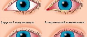
- White mucus is secreted during conjunctivitis and viral infections, after suffering from acute respiratory viral infections. The sclera and mucous membrane become inflamed, red, a lot of tears are produced, and sensitivity to light increases. The tissues swell, a cough and other signs of a cold appear. The virus initially appears in one eye and then spreads to the other.
- Dry eye syndrome is accompanied by overstrain and dryness. The disorder manifests itself after working for a long time at the monitor.
- Liquid transparent discharge appears in case of allergies from both eyes. Severe itching and redness occurs. Symptoms disappear after stopping contact with the allergen.
- A liquid transparent discharge appears upon contact with dust and foreign objects. This is a natural protective reaction of the eye, allowing small fragments to be washed out from the mucous membrane.
- Yellow mucus occurs in large quantities during their dacryocystitis. The disorder becomes more complicated when the injured eye is frequently pressed with fingers.
- Blepharitis is accompanied by greenish or yellow mucus with foam. The eyes itch, the eyelids swell, the discharge dries out and flakes. Sticky mucus accumulates in the corners of the eyes in the morning, sticking the eyelids together with a hard crust.
- Inflammation caused by the accumulation of a large number of leukocytes is accompanied by thick yellow and greenish discharge. Bacterial conjunctivitis, a virus, a fungus that affects the cornea and trachoma develops. The barley opens and dense crusts form that are difficult to remove. A film appears that impairs the clarity of vision. Redness occurs, the eyelids swell, a lot of tears are released, and sensitivity to light increases.
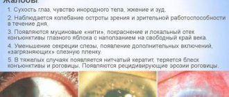
Thread-like discharge occurs at the initial stage of keratitis. Pathology appears when there is a problem with the functioning of the lacrimal glands. Dryness, burning, thread-like discharge occurs, and the sclera of the eyes turns red. The cornea undergoes dystrophic changes.
The anatomy of the body contributes to the accumulation of mucus in the corners of the eyes. These places go a little deeper in the orbit. Therefore, liquid flows there. Near the corners is the nasolacrimal duct.
What it is?
Fungal blepharitis Source: drvision.ru One of the varieties of various inflammatory eye diseases is blepharitis.
Accompanied by inflammatory processes along the edges of the eyelid. The disease affects various segments of the population, regardless of gender and race. It is difficult to cure and can last for many years. Manifests:
- peeling of the skin surface and the appearance of plaque on the skin of the eyelids;
- sticking together of eyelashes with yellowish or greenish discharge;
- increased lacrimation;
- the formation of peeling scales in the head and eyebrows.
The pathogenesis of the pathology consists of the development of numerous factors. Staphylococcal, fungal or tick-borne infections and microbial colonies on the surface of the eyelid. Their metabolic products - enzymes and toxins - cause tissue destruction.
Chronic meibomitis and seborrheic dermatitis aggravate this process. Pathology can be caused by many reasons, but the main causative agent is considered to be the eyelash mite, which lives on the skin and is part of the skin microflora - the acne gland (Demodex folliculorum).
The development of the disease and its progression leads to multiple pathologies:
- inflammation and loss of optical transparency of the vitreous body of the eye;
- formation of induced astigmatism; corneal perforation.
Inflammation of the eyelids
In order to understand what blepharitis is, you need to have a good understanding of how the human eyelid works and why, what functions it performs. The main task of the eyelid is to protect the eyeball from drying out and adverse environmental influences, such as wind, sun and even just dust.
This determines its structure:
- the upper layer of the eyelid is covered with skin, along the edge of which there are eyelashes;
- in the ciliary bulbs and between them there are exits of the sebaceous glands;
- the second layer consists of muscles, which allows you to close/open your eyes;
- The plate connecting the upper and lower eyelids is located even deeper;
- The plate contains the so-called lacrimal glands.
The skin around the eyes is so thin that its thickness is only 0.3 - 0.5 mm. Subcutaneous tissue is loose. This structure is designed to ensure the mobility of the eyelid, but because of this it is easily susceptible to swelling and injury. It is this feature that makes plastic surgery possible.
Xanthelasma
In some cases, the formation may not be a simple milia, but a xanthelasma. In this situation, a spot on the eyelid may indicate the presence of some pathological changes in the body.
Xanthelasma of the eyelids is a disease in which yellow, white, and sometimes orange spots appear on the skin of the eyelids. Along with pathology, liver dysfunction, hypertension, and diabetes mellitus are often diagnosed.
Why does a white spot form on the lower eyelid of the eye? Let's look further.
Diagnostics
You cannot independently determine the complexity of the lesion. Only a consultation with an ophthalmologist and dermatologist will help make an accurate diagnosis.
To identify the pathogen, doctors prescribe bacteriological culture.
An accurate diagnosis is made as a result of the following instrumental studies and diagnostics in laboratories:
- Refractometry. The optical system of the eye is examined. As a result of the study, the refractive power is determined. An automatic refractometer is used to determine it.
- Study of visual acuity. For diagnostics, an optotype table and a special projector are used. With fungal infection, visual acuity decreases below v=1.
- Ophthalmoscopy. A fundus lens or ophthalmoscope is used to examine the fundus of the eye.
- Biomicroscopy. As a result of a non-contact method using a binocular microscope and a slit lamp, individual ocular structures are examined.
- Angiography. Photography or video is being taken. As a result of the procedure, the quality of the vessels is assessed.
Treatment
Fungal infections are less treatable than bacterial infections. This is due to the structure of the fungal cell, which is in many ways similar to the structure of human cells. Fungi develop strong immunity to modern fungal drugs.
Do not think that antifungal drugs are completely harmless. Therefore, self-treatment is strictly prohibited.
Before drug treatment is prescribed, it is important to conduct a series of laboratory tests to accurately determine the diagnosis.
Whether the treatment was chosen correctly or not can be judged within a week from the start of treatment. Therefore, you must strictly follow the doctor's instructions.
Medication
Depending on the severity of the disease, doctors prescribe certain drug therapy.
Antibiotics are used to treat keratitis. In addition, the doctor prescribes antiviral and anti-inflammatory drugs.
Drug treatment is prescribed depending on what kind of infection has gotten inside.
Folk remedies
The remedies used by traditional healers are not inferior in quality to pharmacological drugs and are used to treat fungal diseases.
- Children often experience eye inflammation. For comfortable treatment, you can use herbs. A decoction of oak bark, St. John's wort, chamomile or calendula will help relieve inflammation. You need to rinse your eyes every hour.
- Baking soda will help quickly relieve the symptoms of inflammation, but it is powerless against mycosis. To wash the eyes, take 1/3 tsp. soda and dilute it in 100 ml of warm water.
- A bag of used tea leaves also helps in the fight against eye diseases. It should be applied warm to the closed eye. The procedure should be repeated at least 4 times a day.
To avoid diseases, it is important to adhere to preventive measures.
Inexpensive but effective drugs
To avoid relapse, it is important to strictly follow the recommendations prescribed by the ophthalmologist. Drug therapy is usually carried out at home. Patients with complications are admitted to the hospital.
Conservative treatment involves taking tablets orally and using drugs applied to the surface of the body. These include ointments and drops.
- Okomistin. A drug against mycosis. The composition includes miramistin. Drops against mycosis are prescribed mainly after planned operations. Also used in cases of infection. Treatment lasts a week. A similar drug is Dismistin.
- Floconazole ointment. Fungal infection of a vital organ can be treated with this ointment. It needs to be placed under the lower eyelid. Duration of treatment – 2 weeks.
- Clotrimazole ointment. Use late in the evening as short-term blurred vision may occur. A cotton swab is used to apply the ointment around the eyes.
Treatment of pathology
Having discovered discharge, you must consult a doctor who will identify the pathology and prescribe appropriate treatment.
Therapy is carried out after examination and an accurate diagnosis. The doctor prescribes medications, most often these are ointments, tablets, gels, and sprays. Among the popular products are “Vizin”, “Normax”, “Maxitrol”, “Levomycetin”, and many others. Almost all pharmaceutical medications have contraindications; the doctor will definitely take them into account and give recommendations on how to properly carry out the treatment.
Against the background of drug therapy, but not as the only and main type, it is recommended to use folk remedies from medicinal herbs. Self-medication is unacceptable, you should also remember about allergies. Decoctions, infusions, lotions, ointments, rinsing mixtures are easy to prepare at home. The raw materials are:
- chamomile;
- calendula;
- St. John's wort;
- sage;
- hop cones;
- plantain;
- series;
- birch buds;
- elderberry flowers and berries.
Women cry 47 times a year, and men - 7. From a physiological point of view, this is a very useful process that protects against eye infections.
Useful video
From this video you will learn more about how fungal skin diseases occur:
Whatever the cause of such a disease, it is necessary to begin treatment as quickly as possible, and it is not recommended to use any means at your own discretion: the entire treatment process must be supervised by specialists .
Fungus on the eyelids is a common disease that causes discomfort and discomfort in the eye area. It occurs for many reasons, one of which is poor-quality eyelash extensions and non-compliance with hygiene rules.
Forecast
The sooner you start treating the disease, the more effective the prescribed treatment will be and the result will not be long in coming.
You should prepare yourself for the fact that fungal therapy takes a long time; you should not count on immediate results.
Possible complications
If fungal eye infections have reached an advanced stage, the development of complications is possible.
In patients whose keratitis is not treated, corneal perforation may occur, followed by surgery to perform a corneal transplant.
Prevention and possible complications
The presence of fungus on the body is under the control of the immune system, but with the weakening of the body’s defenses, active reproduction begins, resulting in eye damage. The development of the disease can be prevented by preventive measures aimed at maintaining immunity:
- adhere to a healthy lifestyle;
- do not drink alcohol, cigarettes or drugs;
- proper nutrition;
- maintaining hygiene;
- walks in the open air.
In the absence of proper care and timely treatment, unpleasant consequences are possible, which will require more effort and time to combat. The following complications are possible:
- deformation and scarring of the eyelids;
- sepsis;
- loss of eyelashes;
- in rare cases, meningitis.
To avoid serious problems, treatment should be started on time. Self-medication is considered dangerous, as it aggravates an already serious condition.