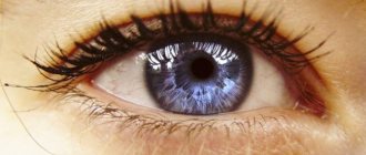
Surely many have noticed a white coating that appears in the corners of the eyes in the morning. Most people do not attach any importance to it and simply wash their face, but in fact it is a symptom indicating a disorder in the body. The reasons for its appearance are very diverse, so only a specialist can make a diagnosis.
Timely detection of the disease and proper treatment guarantee a quick recovery and the elimination of complications.
The most common causes of plaque, itching and similar phenomena in the corners of the eyes: purulent infection, conjunctivitis, inflammation, eye fatigue syndrome, blepharitis, chalazions, recurrent styes, otitis media, sinusitis, sinusitis, meibomitis, demodicosis, weakened immunity.
Treatment of eye diseases
The main directions of therapy are preventing the inflammatory process and suppressing infection. The doctor may prescribe antibiotics and combine them with anti-inflammatory drugs.
- If the plaque is caused by excessive stress and fatigue, special drops are used, for example, Visin, Normax, Maxitrol, Levomycetin.
- In some cases, massage of the eyelids and rinsing of the tear ducts are necessary.
- If white plaque appears due to meibomite, install a solution of dicaine (0.5%) or trimecaine (3-5%) into the conjunctival sac.
How to treat
If a white dot that appears in the eye does not provoke obvious deterioration in vision, then special therapy is usually not prescribed. In general, treatment tactics are selected taking into account the reasons that provoked the development of the pathology. Specialists at the New Look clinic offer several ways to solve the diagnosed problem:
- for cataracts and destructive changes in the cornea, surgical intervention is most often recommended;
- for inflammatory processes, special anti-inflammatory drugs are prescribed in the form of eye drops;
- when connective scar tissue forms, absorbable drugs are prescribed, for example, Hypromellose, Actovegin, Korneregel.
In this medical center, surgical correction is performed using professional modern equipment. Today, operations are quite affordable and have a short recovery period, which is very convenient.
So you should not try to treat your eyes with all sorts of alternative means, as well as various drops, without an established diagnosis. Before starting therapy, you should consult an ophthalmologist.
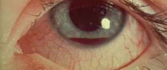
Causes of irritation, redness and bruising in the corners of the eyes
Discomfort and unpleasant phenomena do not always indicate the presence of pathology. Both plaque and bruising, redness, and itching can occur due to external irritants. Poor-quality allergenic cosmetics, certain eye drops, prolonged wearing of contact lenses, foreign bodies, overexertion and ordinary fatigue are the main external causes.
In these cases, as a rule, no treatment is required; it is enough to take the following measures:
- eliminate the source of irritation;
- good rest;
- change the brand of cosmetics, etc.
But if there is deterioration in vision or a person has allergies, you need to consult a specialist to conduct a diagnosis, identify the exact cause and prescribe adequate treatment.
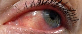
It is worth noting such a phenomenon as a crack in the corner of one eye or both. The wound may occur due to allergies due to frequent rubbing of the skin, or due to conjunctivitis.
At first glance, the crack does not pose a danger, but this is far from true, because a secondary infection can penetrate through the damage. So you need to quickly seek help from a doctor to prevent complications.
Itching, as already mentioned, can also be a symptom of conjunctivitis, but it is also characteristic of allergies. Usually it is accompanied by a runny nose, sneezing, and tearfulness. Demodectic mange should not be ruled out, although it usually occurs in people with weakened immune systems.
Those who work at a computer for a long time suffer from “dry eye syndrome,” so the appearance of irritation, itching and cracks in the corners indicate that you need to do special exercises and regularly be distracted by other activities. People who wear contact lenses and glasses should also remember that their eyes need to rest periodically.
White, yellow and black are symptoms of serious eye disorders
In rare cases, abnormal red reflexes can signal more dangerous eye conditions.
- Black reflex
An asymmetrical red reflex, where only one eye reflex appears red, or the red reflex of one eye is duller than the other, may be an indicator of strabismus or eye misalignment, a condition in which both eyes do not look in the same place at the same time.
Treatment for strabismus may include glasses, contacts, patches on the stronger eye, and eye muscle surgery. With early detection and treatment, strabismus can often be corrected with excellent results.
- White reflex
A white reflex that covers most of the pupil, also known as leukocoria, can signal the presence of several serious medical conditions, including cataracts, retinal detachments and intraocular infections.
It may also be a warning sign of retinoblastoma, an extremely rare and very serious childhood eye cancer. With early diagnosis and treatment, retinoblastoma is curable in 95% of cases.
- Yellow reflex
The yellow reflex may be a sign of Coats disease, in which the blood vessels inside the eye that supply the retina with blood and oxygen become distorted and leaky. This results in a blockage in the retina, which can lead to vision loss or retinal detachment.
Coats disease occurs primarily in boys under 10 years of age and usually affects only one eye. Treatment may include laser surgery, cryotherapy or, in later stages of the disease, more invasive surgery. Coats disease is difficult to distinguish from retinoblastoma in photographs because the white and yellow reflexes look similar.
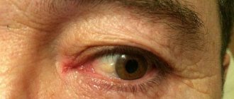
Drooping, swollen corners of the eyes, bags and bruises
Such defects may indicate overwork, stress, or serious health problems.
- First, it is recommended to rest well, get enough sleep, you can take sedatives, if necessary, and take a walk.
- When the work and rest schedule is normalized, but unpleasant phenomena do not go away, you need to contact a specialist. In this case, only he will be able to make a diagnosis, find out the cause and prescribe appropriate measures.
- It is worth noting that drooping corners, plaque and other phenomena may be associated with age-related changes. However, hormonal imbalances and vitamin deficiencies cannot be discounted. These two conditions provoke pigmentation of the skin and the appearance of bruises.
- Appearance can also deteriorate due to sudden weight loss. Problems with the heart and kidneys, exhaustion of the body after a protracted illness can also be provoking factors.
- When your eye hurts, there is a bruise in the corner or other unpleasant conditions, you need to consult a doctor to rule out chronic pathologies and active inflammatory processes.
Episcleritis of the eye
There are several more layers between the scleral “casing” of the eyeball and the orbit (as part of the skull). The membrane closest and directly adjacent to the sclera, like a packaging film, is the conjunctiva - a thin moisturizing mucous layer, the inflammation of which, apparently, every person has experienced at least once. If the inflammatory process develops “under” the conjunctiva, i.e. on any area of its surface contact with the sclera, such inflammation is called episcleritis - after the name of the outermost, relatively loose scleral layer, or episclera.
Everything said above about sclerites applies, in general, to episcleritis as a local variety; In many publications devoted to inflammation of the sclera, episcleritis is not even mentioned separately. However, there are differences, both histological and clinical.
The episclera is characterized, in addition to its looseness, by rich innervation and vasculature: nerve fibers and blood vessels of various sizes are concentrated in this layer. Pathogenic factors always penetrate into such areas faster, but both the body’s immune response and medicinal compounds are more easily transported through the bloodstream. In more than half of the cases (up to 60%), episcleritis does not even require ophthalmological intervention, does not last long, passes without consequences, and with a simple (see below) type of episcleritis, it is extremely rare that the process spreads to deeper cellular layers, turning into “true” scleritis
What does a wen in the corner or on the eyelid mean?
This cosmetic defect is especially common among newborns and the elderly. If in the former it is associated with a change in environment, then in the older it is usually caused by hormonal imbalances.
If a wen appears, and bags or bruises are added to it, you need to consult a therapist. The doctor, based on the patient’s medical history and complaints, will recommend treatment or refer you to specialized specialists, for example, an endocrinologist or gynecologist.
First you need to find out what reasons provoked this phenomenon. It is worth noting that this also applies to other cosmetic defects, including plaque in the corner of the eyes. If the wen is the result of a hormonal imbalance, efforts must be made to eliminate the latter, otherwise, even after removing the defect, it may appear again.
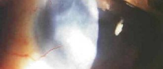
Causes of eyesore in humans
Cloudiness of the cornea in humans usually occurs as a result of inflammation of the cornea of the eye - deep or superficial keratitis. In this case, the reasons for the appearance of eyesores can be:
- mechanical (burn, superficial injuries caused by foreign bodies, chemicals or rays);
- infectious - caused by the influenza virus, Koch's bacillus, Pseudomonas aeruginosa, Staphylococcus aureus, fungi, amoebae;
- as a complication of conjunctivitis and any other diseases that can affect the layers of the cornea (for example, trachoma);
- In addition, there are known cases of cataract formation after suffering from syphilis and chlamydia , the fundamental factor being their inadequate or ineffective treatment.
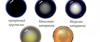
Stages of formation of an eyesore as a result of keratitis
People at risk of developing eyesores include:
- using lenses , especially if hygiene rules are not followed;
- having problems with intraocular pressure ;
- staying for a long time in the scorching bright sun;
- those who have undergone eye surgery, for example, removal of pterygium , cataracts, etc.;
- working with welding equipment;
- visiting saunas and bath complexes where high temperatures are used;
- genetic predisposition and congenital diseases that provoke clouding of the cornea, for example, blenorrhea .
Contacting a veterinarian
Often, dog owners seek qualified veterinary help too late, when the animal has already begun to lose its vision..
You must respond immediately if the following signs appear:
the animal's eyes become cloudy;- a white spot appeared on the eyeball;
- the dog is bothered by liquid or purulent discharge near the lower eyelid;
- There was an injury to the organs of vision - chemical or mechanical.
If the owner notices that the dog’s vision is deteriorating for some reason, especially at a young age, this is an alarming symptom that requires diagnosis.
Only in a veterinary clinic after a thorough examination and all research will an accurate diagnosis be made. Self-medication in this situation is not worthwhile.
Surgery
This treatment option is used if the spot on the pupil grows, cannot be treated by other means and can cause irreversible consequences. The methods of surgical intervention are determined by the attending physician, taking into account several factors.
Among them:
- patient's age;
- specificity of the clinical picture;
- presence of concomitant diseases;
- restrictions and contraindications.
Most often, surgical therapy is used for nevi and leukomas of the eye. They are removed through minimally invasive procedures using a laser or microscalpel. The radio wave method is widely used to remove tumors. For traumatic lesions, treatment is possible with a corneal transplant.
Diagnostics
Treatment begins with identifying the causes of eye disease. To do this, a scraping is taken from the cornea in the place where it is damaged. This is a rather outdated method that complicates the injury to the organ of vision.
In modern veterinary practice, the following diagnostic methods are used:
- Ultrasound of the eyeball;
- herpes test;
- electroretinography;
- serology;
- parcentesis.
As soon as the results of the study are ready, therapy begins, especially if the clouding has spread to the entire area of the eye.