Structure of the conjunctiva
The conjunctiva is a thin transparent mucous membrane. It covers the entire posterior surface of the eyelids, where it is tightly connected to the cartilage, and forms the upper and lower conjunctival fornix.
Fornixes are areas with relatively free conjunctiva, which looks like blind pockets that provide freedom of movement to the eyeball. Moreover, the upper arch is twice as large as the lower one. The conjunctiva of the fornix extends to the apple of the eye and is localized on top of the dense Tenon's membrane, close to the limbus. The epithelium of the conjunctiva, which is its surface layer, passes directly into the epithelium of the cornea.
The conjunctiva has two main functions: protective and secretory. The protective function is performed by a fairly significant coating of the eyeball. The secretory function is determined by a large number of glands localized in the thickness of the conjunctiva. The conjunctival cartilage contains goblet cells as well as Henle cells, which produce mucin. Goblet cells are also present in large numbers in the conjunctival fornix. Between the conjunctiva of the eyelids and the fornix there are additional Wolfring lacrimal glands: three at the top and one at the bottom. In the area of the vaults there are Krause's glands: approximately 40 at the top, 8 at the bottom. These glands are like the lacrimal gland in miniature; their daily active work can completely cover the need for hydration of the eyeball. The lacrimal gland begins to work only in the case of strong emotional reactions, irritation of the eye, etc. In the area of the limbus, the conjunctiva contains Becher and Manz cells, which also produce mucin, which, along with the tear fluid, is the main component of the tear film, which moisturizes the eye and serves as its protection.
The blood supply to the conjunctiva of the eyelids occurs through the same vessels as the blood supply to the eyelids. The conjunctiva of the eyeball includes the superficial and deep layers of blood vessels. The superficial one is formed by the perforating arteries of the eyelids and the anterior ciliary arteries. The anterior ciliary arteries enter the deep layer of vessels, forming a dense network that envelops the cornea.
The venous vascular system of the cornea corresponds to the arterial system. At the same time, the conjunctiva is rich in lymphoid tissue and lymphatic vessels. The lacrimal, subtrochlear and infraorbital nerves are responsible for the sensitivity of the conjunctiva.
Diseases
Some anomalies of ophthalmological origin can damage the mucous membrane of the eyes:
- pinguecula (wen);
- neoplasms;
- conjunctivitis (infectious or allergic);
- keratoconjunctivitis (inflammatory processes affecting the mucous membrane and cornea).
Pinguecula
The disease is accompanied by the formation of a benign neoplasm of a yellow hue. The growth slowly increases in size and is usually located in the area of the inner corner of the organ of vision. The disease is most often diagnosed in elderly patients. The formation does not have a negative impact on visual function and is classified as a cosmetic defect.
The growth cannot develop into a cancerous tumor, since it is formed as a result of an increased concentration of protein and cholesterol. At the same time, the pinguecula does not go away on its own, without treatment.
Despite the fact that pathology, at first glance, seems harmless, education should not be ignored. After all, it can occur with complications. For example, provoke the development of pterygium. This is a disease accompanied by the growth of a wing-shaped film over the mucous membrane. Such a growth impairs visual function.
The disease is characterized by the following symptoms:
- redness of the eyeball;
- increased lacrimation;
- itching and burning;
- sensation of the presence of a foreign body.
| The pinguecula has an elastic structure and extremely rarely appears in both eyes at the same time. Some patients claim that the formation of a growth occurs due to wearing contact optics for a long time. But this theory has no scientific evidence. |
According to studies, the disease develops as a result of wear and tear of the mucous membrane. For this reason, the disease is typical for older people. There are a number of other factors that can lead to the formation of a growth:
- prolonged exposure to dust, smoke;
- dry and hot climate;
- prolonged exposure to direct sunlight without protective optics.
If an increase in the size of the pinguecula is accompanied by the development of dry eye syndrome, then moisturizing medications, for example, Oxial drops, are introduced into the course of treatment. If irritation occurs, you should take antibacterial medications. During the period of therapy, you must stop wearing contact lenses.
Nevus
In appearance it resembles a mole. Most often, the pathology is diagnosed in fair-skinned and blue-eyed people. The growth is not equipped with nerve fibers, so it does not cause pain.
Usually a nevus appears from birth, but for a certain time it is not visible. Coloring of the spot occurs during puberty. Such a formation can develop into a cancerous tumor. If the disease progresses and grows, then there is a risk of loss of visual acuity and distortion of perception.
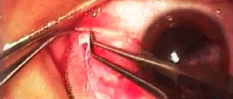
Patients with dynamically developing nevus are prescribed excision. If the tumor has reached an impressive size, standard surgical intervention is performed. Laser therapy is becoming increasingly popular. It is characterized by minimal trauma and the ability to get rid of formations in the most inaccessible places.
When performing electroexcision, the growth is removed using an electric scalpel. During the operation, it is possible to perform plastic surgery of the mucous membrane or eliminate corneal defects.
Conjunctivitis
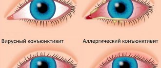
The cause of the development of the inflammatory process can be bacteria, fungi or allergens. The viral type of the disease is accompanied by increased lacrimation, itching and the appearance of non-purulent exudate. Therapy is carried out using interferon-based eye drops.
| With allergic conjunctivitis, patients suffer from severe itching, swelling of the eyelids and pain. Often the anomaly is accompanied by a runny nose and bronchial asthma. Antihistamines will help get rid of unpleasant symptoms. |
Pathology of bacterial origin is caused by streptococci, gonococci, etc. It is characterized by the appearance of purulent discharge of yellow color and viscous consistency. The basis of therapy is antibiotics. Return to contents
Keratoconjunctivitis
In most cases, the disease affects both eyes at once. Patients complain of intolerance to bright light, pain and discomfort. Swelling of the conjunctiva and redness of the cornea are also observed. With a pathology of viral or allergic origin, increased lacrimation and hemorrhage in the mucous membrane appear.
The main reasons for the development of keratoconjunctivitis:
- weakening of the body's protective barrier;
- deformation of the mucous membrane;
- non-compliance with the rules and terms of use of contact lenses;
- long course of treatment with corticosteroids;
- lack of vitamins;
- penetration of pathogenic microorganisms into the visual apparatus;
- getting a foreign object into the eye;
- chronic inflammation.
The prognosis for treatment of the disease is not always favorable. The course of treatment is selected based on factors. Provoking the development of the disease. If the anomaly has become severe, then even expensive therapy will not help restore full vision.
Symptoms of corneal damage in various pathologies
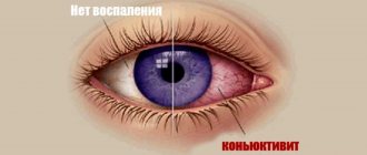
The conjunctiva, following the mucous membrane, reacts to all external irritation with inflammation. Irritants can be: temperature, chemicals, allergens, but in most cases, it is a bacterial or viral infection. The main manifestations of inflammation of the conjunctiva are lacrimation, burning sensation, redness, itching, dryness, pain when blinking or moving the eyes, which is caused by an increase in lymphoid tissue. When the cornea is involved in the process of inflammation, a foreign body sensation may occur. Inflammation of the conjunctiva is often accompanied by various discharges from the eyes. They can be either watery-mucous or purulent with crusts, which is due to the nature of the damaging irritant agent. Acute viral lesions are often accompanied by hemorrhages under the conjunctiva, which becomes swollen.
Insufficient function of the lacrimal glands can cause the conjunctiva to dry out, leading to degenerative conditions. The tissues of the conjunctiva of the eyeball, its fornix and eyelid can sometimes grow together, which limits the movement of the eyeball.
Under the physiological norm, the conjunctiva does not extend to the cornea, but in some people, under the influence of external factors (windy climate, dusty work), a slow growth of the conjunctiva into the cornea occurs. This growth is called a pterygium, and once it reaches a certain size, it can seriously reduce vision.
The conjunctiva may normally contain some pigment inclusions - brownish-dark spots, which must be shown to an ophthalmologist and observed for a while.
Symptoms of pathology of the conjunctiva of the eye
If the conjunctiva is exposed to negative environmental factors, it becomes inflamed. Such influences include:
- mechanical damage;
- allergen antigens;
- polluting objects (smoke, smog, chemicals);
- pathogens of a viral nature;
- bacterial infection.
After the formation of inflammation, cells of the immune system migrate to the lesion. They secrete inflammatory mediators, so the following clinical symptoms appear:
- edema;
- itching, burning, irritation;
- redness;
- pain at rest and when blinking;
- sensation of a foreign object under the eyelids;
- decreased production of tear fluid, which leads to dryness;
- discharge of clear or purulent exudate.
All of the above symptoms do not always appear. Their formation depends on the cause that caused the inflammation. Rarely, a small hemorrhage appears, which subsequently resolves on its own.
If there are no therapeutic measures, inflammation intensifies. Excessive drying contributes to microtrauma and irritation. With chronic damage, the conjunctiva can become fused to the eyeball, limiting its movement.
Diagnosis and treatment
A detailed examination of the conjunctiva requires an ophthalmologist to use a slit lamp (biomicroscopy). At the same time, he evaluates the conjunctiva of the eyelids, eyeball and fornix, the dilation of its vessels, identifies possible hemorrhages, swelling, the nature of the discharge, and the involvement of other eye structures in the inflammatory or degenerative process.
Treatment of conjunctival diseases is determined by the reasons that caused them.
In this case, therapeutic treatment (washing, antibiotics, hormonal drugs) and surgery, as with pterygium or symblepharon, can be prescribed. Yakovleva Yulia Valerievna
Diagnostic methods for damage to the conjunctiva of the eye
To determine the type of pathological process, doctors prescribe a detailed examination, which includes a number of procedures:
- Ultrasound analysis.
- Biomicroscopy using a slit lamp.
- Bacteriological examination of exudate to detect pathogenic microorganisms.
It is worth remembering that the conjunctiva of the eye is an important element of the optical system. Therefore, protect it from damage that could lead to serious vision problems.
Bacterial conjunctivitis
Acute epidemic Koch-Wicks conjunctivitis
Acute epidemic Koch-Wicks conjunctivitis is a fairly common disease and is observed in almost all countries of the world with a hot climate. Constant centers of annual outbreaks of epidemics of Koch-Wicks conjunctivitis are the republics of Central Asia and partly Transcaucasia and the North Caucasus. In the north the disease is rare. Bacterial Koch-Wicks conjunctivitis can occur in seasonal outbreaks in the summer and autumn. Infection occurs through dirty hands and airborne droplets. The source of infection can be food, water, or infected human conjunctiva. In typical cases, the onset of the disease is sudden. The incubation period ranges from several hours to 1-2 days.
| Koch-Wicks bacterial conjunctivitis is usually bilateral. The first symptoms of conjunctivitis manifest themselves in hyperemia of the mucous membrane of the eyelids, which quickly spreads to the transitional folds and to the mucous membrane of the eyeball. The greatest hyperemia and swelling occurs in the area of the lower transitional fold, which, when the lower eyelid is pulled back, appears in the form of a roller. In the first two days, mucopurulent or purulent discharge appears (see.
|
Blennorrheic conjunctivitis or gonoblennorrhea
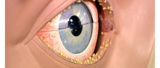
Gonoblenorrhea is caused by the gram-negative diplococcus Neisseria gonorrhoeae. Blenorrheal conjunctivitis can occur in newborns during the passage of the mother's birth canal or later, due to contact with a sick mother if she does not comply with the rules of personal hygiene, there are cases of intrauterine infection. In adults, when pus is introduced into the conjunctival cavity with gonorrheal urethritis.
| Medical personnel may become infected while treating such patients or during childbirth. Very rarely, gonoblennorrhea occurs metastatically. There are blenorrheal conjunctivitis in newborns, children and adults. Blenorrheal conjunctivitis in newborns occurs on the 2-3rd day after birth. As a rule, both eyes are affected at short intervals. Symptoms of conjunctivitis include severe swelling of the eyelids. The eyelids are swollen and dense to the touch. Only sometimes it is difficult to open them (see.
|
Adeno-pharyngoconjunctival fever (AFCL)
The onset of the disease is often acute with a rise in temperature, catarrhal symptoms, pain and swelling of the pre-auricular lymph nodes. Conjunctivitis begins in one eye, and after 2-3 days the second eye becomes ill. It occurs in three forms: follicular, catarrhal and membranous. Patients complain of redness of the eyes, foreign body sensation, and itching. The eyelids are swollen, there is slight blepharospasm, and slight mucopurulent discharge.
The membranous form of AFCL is characterized by the presence of gray, delicate, thin films that are easily removed from the conjunctiva of the eyelids and the transitional fold. Recurrences of membranous conjunctivitis are possible, but extremely rare. The outcome of the disease is favorable.
The follicular form has a less acute onset and is manifested by the formation of pinkish-grayish follicles and papillae against the background of hyperemic and edematous conjunctiva, mainly in the corners of the lower transitional fold. The duration of the disease is up to two weeks.
The catarrhal form of AFCL occurs, as a rule, unnoticeably and favorably. There is slight swelling of the eyelids and mild photophobia. The conjunctiva of the eyelids is hyperemic, slightly edematous, and there is a slight mucopurulent discharge. There are no follicles, papillae, films, or hemorrhages. The cornea is not involved in the pathological process. The average duration of catarrhal conjunctivitis is 10 days.
Each form of adeno-pharyngoconjunctival fever has three successively developing stages: 1) hyperemia and hypertrophy of the conjunctiva (follicles, papillae, films) mainly of the lower eyelid; 2) mild, painless swelling of the eyelids and conjunctiva; 3) reverse development of films, follicles and papillae, as well as resorption of hemorrhages in the conjunctiva and skin of the eyelids. A characteristic feature of all forms and stages of AFCL is a decrease in corneal sensitivity.
Parts of the conjunctiva
The conjunctiva is a continuous tissue that consists of two segments:
Bulbar conjunctiva
This is the part of tissue that covers the entire front region of the sclera, the white part of the eye. The bulbous conjunctiva extends to the area where the cornea is covered by the sclera, without covering the cornea.
Palpebral conjunctiva
This is the part that is responsible for covering and protecting the inner area of the lower and upper eyelids.
This part of the conjunctiva can be called the palmate conjunctiva.