A person cannot see in complete darkness. In order for a person to see an object, light must be reflected from the object and hit the retina. Light sources can be natural (fire, Sun) and artificial (various lamps). But what is light?
According to modern scientific concepts, light is electromagnetic waves of a certain (sufficiently high) frequency range. This theory originates from Huygens and is confirmed by many experiments (in particular, the experience of T. Jung). At the same time, the carpuscular-wave dualism is fully manifested in the nature of light, which largely determines its properties: when propagating, light behaves like a wave, when emitting or absorbing, it behaves like a particle (photon). Thus, light effects that occur during the propagation of light (interference, diffraction, etc.) are described by Maxwell’s equations, and the effects that occur during its absorption and emission (photoelectric effect, Compton effect) are described by the equations of quantum field theory.
To put it simply, the human eye is a radio receiver capable of receiving electromagnetic waves of a certain (optical) frequency range. The primary sources of these waves are the bodies that emit them (the sun, lamps, etc.), the secondary sources are the bodies that reflect the waves of the primary sources. Light from the sources enters the eye and makes them visible to humans. Thus, if a body is transparent to waves in the visible frequency range (air, water, glass, etc.), then it cannot be detected by the eye. In this case, the eye, like any other radio receiver, is “tuned” to a certain range of radio frequencies (in the case of the eye, this is the range from 400 to 790 terahertz), and does not perceive waves that have higher (ultraviolet) or lower (infrared) frequencies. This “tuning” is manifested in the entire structure of the eye - starting from the lens and vitreous body, which are transparent precisely in this frequency range, and ending with the size of the photoreceptors, which in this analogy are similar to the antennas of radio receivers and have dimensions that ensure the most effective reception of radio waves in this particular range.
All this together determines the frequency range in which a person sees. It is called the visible radiation range.
Visible radiation is electromagnetic waves perceived by the human eye, which occupy a region of the spectrum with a wavelength from approximately 380 (violet) to 740 nm (red). Such waves occupy the frequency range from 400 to 790 terahertz. Electromagnetic radiation with such frequencies is also called visible light, or simply light (in the narrow sense of the word). The human eye has the greatest sensitivity to light in the region of 555 nm (540 THz), in the green part of the spectrum.
White light divided by a prism into the colors of the spectrum [4]
When a white beam is decomposed in a prism, a spectrum is formed in which radiation of different wavelengths is refracted at different angles. Colors included in the spectrum, that is, those colors that can be produced by light waves of one wavelength (or a very narrow range), are called spectral colors. The main spectral colors (which have their own names), as well as the emission characteristics of these colors, are presented in the table:
The spectrum does not contain all the colors that the human brain can distinguish and they are formed from mixing other colors.[4]
What does a person see
Thanks to vision, we receive 90% of information about the world around us, so the eye is one of the most important sense organs. The eye can be called a complex optical device. Its main task is to “transmit” the correct image to the optic nerve.
Structure of the human eye
The cornea is the transparent membrane that covers the front of the eye. It lacks blood vessels and has great refractive power. Part of the optical system of the eye. The cornea borders the opaque outer layer of the eye, the sclera.
The anterior chamber of the eye is the space between the cornea and the iris. It is filled with intraocular fluid.
The iris is shaped like a circle with a hole inside (the pupil). The iris consists of muscles that, when contracted and relaxed, change the size of the pupil. It enters the choroid of the eye. The iris is responsible for the color of the eyes (if it is blue, it means there are few pigment cells, if it is brown, it means a lot). Performs the same function as the aperture in a camera, regulating the light flow.
The pupil is an opening in the iris. Its size usually depends on the light level. The more light, the smaller the pupil.
The lens is the “natural lens” of the eye. It is transparent, elastic - it can change its shape, almost instantly “focusing”, due to which a person sees well both near and far. Located in the capsule, held in place by the ciliary band. The lens, like the cornea, is part of the optical system of the eye. The transparency of the human eye lens is excellent, transmitting most light with wavelengths between 450 and 1400 nm. Light with a wavelength above 720 nm is not perceived. The lens of the human eye is almost colorless at birth, but becomes yellowish with age. This protects the retina from exposure to ultraviolet rays.
The vitreous is a gel-like transparent substance located in the back of the eye. The vitreous body maintains the shape of the eyeball and is involved in intraocular metabolism. Part of the optical system of the eye.
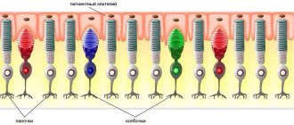
Retina - consists of photoreceptors (they are sensitive to light) and nerve cells. Receptor cells located in the retina are divided into two types: cones and rods. In these cells, which produce the enzyme rhodopsin, the energy of light (photons) is converted into electrical energy of the nervous tissue, i.e. photochemical reaction.
The sclera is the opaque outer layer of the eyeball that merges at the front of the eyeball into the transparent cornea. 6 extraocular muscles are attached to the sclera. It contains a small number of nerve endings and blood vessels.
The choroid - lines the posterior part of the sclera, adjacent to it is the retina, with which it is closely connected. The choroid is responsible for the blood supply to intraocular structures. In diseases of the retina, it is very often involved in the pathological process. There are no nerve endings in the choroid, so when it is diseased, there is no pain, which usually signals some kind of problem.
Optic Nerve - The optic nerve carries signals from nerve endings to the brain.[6]
A person is not born with an already developed organ of vision: in the first months of life, the formation of the brain and vision occurs, and by about 9 months they are able to almost instantly process incoming visual information. In order to see, light is needed. [3]
Accommodation of the eye
Accommodation of the eye provides a clear image even at different distances of objects. This function becomes possible thanks to the elastic properties of the lens, which freely changes curvature, and, consequently, refractive power. In this regard, even when the object moves, the rays reflected from it are focused onto the plane of the retina. When a person examines infinitely distant objects, the ciliary muscle is in a relaxed state, the ligament of Zinn, which is attached to the anterior and posterior lens capsule, is tense. When the fibers of the ligament of Zinn are stretched, the lens stretches, that is, its curvature decreases. When looking into the distance, due to the least curvature of the lens, its refractive power is also the least. As the object approaches the eye, the ciliary muscle contracts. As a result, the ligament of Zinn relaxes, that is, the lens stops stretching. In the case of complete relaxation of the fibers of the ligament of Zinn, the lens lowers by approximately 0.3 mm under the influence of gravity. Due to its elastic properties, the lens lens, in the absence of tension, becomes more convex, and its refractive power increases.
The contraction of the fibers of the ciliary muscle is responsible for the excitation of the parasympathetic fibers of the oculomotor nerve, which respond to the influx of afferent impulses into the midbrain area.
If accommodation does not work, that is, the person looks into the distance, then the anterior radius of curvature of the lens is 10 mm; with maximum contraction of the ciliary muscle, the anterior radius of curvature of the lens changes to 5.3 mm. The changes in the rear radius are less significant: from 6 mm it decreases to 5.5 mm.
Accommodation begins to work the moment the object approaches a distance of approximately 65 meters. In this case, the ciliary muscle moves from a relaxed state to a tense one. However, at such a distance of objects, the tension in the fibers is not high. A more significant contraction of the muscle occurs when the object approaches 5-10 meters. Subsequently, the degree of accommodation progressively increases until the object leaves the zone of clear visibility. The shortest distance at which an object is still clearly visible is called the point of closest clear vision. Normally, the far point of clear vision is infinitely far away. It is interesting that in birds and mammals the mechanism of accommodation is similar to that of humans.
With age, the elasticity of the lens decreases, and the amplitude of accommodation decreases. In this case, the farthest point of clear vision usually remains in the same place, and the nearest one gradually moves away.
It is important to note that when practicing at close range, approximately a third of the accommodation remains in reserve, so the eye does not get tired.
With senile farsightedness, the nearest point of clear vision is removed due to a decrease in the elasticity of the lens. With presbyopia, the refractive power of the crystalline lens decreases even with the greatest force of accommodation. At the age of ten years, the nearest point is located 7 cm from the eye, at 20 years old it shifts by 8.3 cm, at 30 years old - up to 11 cm, by the age of sixty it already shifts to 80-100 cm. Construction of an image on the retina
The eye is a very complex optical system. To study its properties, a simplified model is used, which is called a reduced eye. The visual axis of this model coincides with the axis of a regular eyeball and passes through the centers of the refractive media, ending up in the central fovea.
In the reduced model of the eye, only the substance of the vitreous body is classified as refractive media, in which there are no main points lying in the area of intersection of the refractive planes. In the true eyeball, two nodal points are located at a distance of 0.3 mm from each other, they are replaced by one point. A ray that passes through a nodal point must necessarily pass through its conjugate point, leaving it in a parallel direction. That is, in the reduced model, two points are replaced by one, which is placed at a distance of 7.5 mm from the surface of the cornea, that is, in the posterior third of the lens. The nodal point is 15 mm away from the retina. In the case of constructing an image, all points of the retina are considered as luminous. A straight line is drawn from each of them through the nodal point.
The image that is formed on the retina is reduced, inverse and real. To determine the size on the retina, you need to fixate a long word that is printed in small print. At the same time, it is determined how many letters the patient can distinguish with complete immobility of the eyeball. After this, use a ruler to measure the length of the letters in millimeters. Next, using geometric calculations, you can determine the length of the image on the retina. This size gives an idea of the diameter of the macula, which is responsible for central clear vision.
The image on the retina is reversed, but we see objects straight. This is due to daily training of the brain, in particular the visual analyzer. To determine position in space, in addition to stimuli from the retina, a person uses the excitation of proprioceptors of the muscular apparatus of the eye, as well as readings from other analyzers.
We can say that the formation of ideas about the position of the body in space is based on conditioned reflexes.
Light sensitivity of the human eye
The ability of the eye to perceive light and recognize varying degrees of its brightness is called light perception, and the ability to adapt to different brightness of lighting is called adaptation of the eye; light sensitivity is assessed by the threshold value of the light stimulus. A person with good eyesight can see the light from a candle at a distance of several kilometers at night. Maximum light sensitivity is achieved after a sufficiently long dark adaptation. It is determined under the influence of light flux in a solid angle of 50° at a wavelength of 500 nm (maximum sensitivity of the eye). Under these conditions, the threshold light energy is about 10−9 erg/s, which is equivalent to the flux of several optical quanta per second through the pupil. The contribution of the pupil to the regulation of eye sensitivity is extremely insignificant. The entire range of brightness that our visual mechanism is capable of perceiving is enormous: from 10−6 cd•m² for an eye completely adapted to darkness, to 106 cd•m² for an eye completely adapted to light. The mechanism for such a wide range of sensitivity lies in decomposition and restoration photosensitive pigments in the retinal photoreceptors - cones and rods. The human eye contains two types of light-sensitive cells (receptors): highly sensitive rods, responsible for twilight (night) vision, and less sensitive cones, responsible for color vision.
Normalized graphs of the light sensitivity of the cones of the human eye S, M, L. The dotted line shows the twilight, “black and white” susceptibility of the rods.
In the human retina there are three types of cones, the maximum sensitivity of which occurs in the red, green and blue parts of the spectrum. The distribution of cone types in the retina is uneven: "blue" cones are found closer to the periphery, while "red" and "green" cones are randomly distributed. The correspondence of cone types to three “primary” colors allows the recognition of thousands of colors and shades. The spectral sensitivity curves of the three types of cones partially overlap, which contributes to the phenomenon of metamerism. Very strong light excites all 3 types of receptors, and is therefore perceived as blinding white radiation.
Uniform stimulation of all three elements, corresponding to the weighted average of daylight, also produces the sensation of white.
Human color vision is controlled by genes encoding light-sensitive opsin proteins. According to proponents of the three-component theory, the presence of three different proteins that respond to different wavelengths is sufficient for color perception.
Most mammals have only two of these genes, which is why they have black and white vision.
The red light-sensitive opsin is encoded in humans by the OPN1LW gene. Other human opsins are encoded by the genes OPN1MW, OPN1MW2 and OPN1SW, the first two of which encode proteins that are sensitive to light at medium wavelengths, and the third is responsible for an opsin that is sensitive to the short-wavelength part of the spectrum.
line of sight
Field of view is the space simultaneously perceived by the eye with a fixed gaze and a fixed position of the head. It has certain boundaries corresponding to the transition of the optically active part of the retina into the optically blind. The field of vision is artificially limited by the protruding parts of the face - the back of the nose, the upper edge of the orbit. In addition, its boundaries depend on the position of the eyeball in the orbit. [8] In addition, in each eye of a healthy person there is an area of the retina that is not sensitive to light, which is called the blind spot. Nerve fibers from the receptors to the blind spot go over the retina and gather into the optic nerve, which passes through the retina to the other side. Thus, there are no light receptors in this location.[9]
In this confocal micrograph, the optic disc is shown in black, the cells lining the blood vessels in red, and the contents of the vessels in green. The retinal cells appeared as blue spots. [10]
The blind spots in the two eyes are in different places (symmetrically). This fact, and the fact that the brain corrects the perceived image, explains why they are invisible when both eyes are used normally.
To observe your blind spot, close your right eye and with your left eye look at the right cross, which is circled. Keep your face and monitor upright. Without taking your eyes off the right cross, move your face closer (or further away) from the monitor and at the same time watch the left cross (without looking at it). At a certain point he will disappear.
This method can also estimate the approximate angular size of the blind spot.
Technique for detecting a blind spot [9]
The paracentral parts of the visual field are also distinguished. Depending on the participation of one or both eyes in vision, a monocular and binocular field of vision is distinguished. In clinical practice, the monocular visual field is usually examined. [8]
Dead zone
The existence of the so-called dead zone is a big drawback of the human eye. Because of this, car accidents often occur (or maybe not because of this). What's the point? And the fact is that objects located next to each other, when viewing them, suddenly, in some incomprehensible way, begin to disappear. In fact, of course, they do not disappear: the eyes simply stop seeing them.
You can promote your pages on social networks or earn money here:
Here is a typical example: in the picture below we see a red cross and a blue dot. Close your left eye and look only at the cross with your right eye. With your peripheral vision you see the dot. And now we are slowly approaching the monitor. At some point in time the blue dot will disappear altogether!
Binocular and Stereoscopic vision
The human visual analyzer under normal conditions provides binocular vision, that is, vision with two eyes with a single visual perception. The main reflex mechanism of binocular vision is the image fusion reflex - the fusion reflex (fusion), which occurs with simultaneous stimulation of functionally unequal neural elements of the retina of both eyes. As a result, physiological double vision occurs of objects located closer or further than the fixed point (binocular focusing). Physiological double vision (focus) helps to assess the distance of an object from the eyes and creates a feeling of relief, or stereoscopic vision.
When seeing with one eye, the perception of depth (relief distance) is carried out by ch. arr. thanks to secondary auxiliary signs of distance (apparent size of an object, linear and aerial perspective, blocking of some objects by others, accommodation of the eye, etc..). [1]
Conducting pathways of the visual analyzer 1 - Left half of the visual field, 2 - Right half of the visual field, 3 - Eye, 4 - Retina, 5 - Optic nerves, 6 - Oculomotor nerve, 7 - Chiasma, 8 - Optic tract, 9 - Lateral geniculate body , 10 - Superior colliculus, 11 - Nonspecific visual pathway, 12 - Visual cortex.[2]
A person sees not with his eyes, but through his eyes, from where information is transmitted through the optic nerve, chiasm, visual tracts to certain areas of the occipital lobes of the cerebral cortex, where the picture of the external world that we see is formed. All these organs make up our visual analyzer or visual system.[5]
Color and twilight vision
The retina consists of two types of nerve receptors - rods and cones. Rods are responsible for night (black and white) vision, and cones allow you to see the world in all the splendor of colors. The number of rods on the retina can reach 115–120 million, the number of cones is more modest - about 7 million. Rods even react to individual photons. Therefore, even in low light we can distinguish the outlines of objects (twilight vision).
But cones can show their activity only with sufficient lighting. They require more energy to activate because they are less sensitive.
There are three types of light-perceiving receptors corresponding to red, blue and green.
Their combination allows a person to recognize the whole variety of colors and thousands of their shades. And their overlay gives white color. By the way, the same principle is used in color television.
We see the world around us because all objects reflect the light falling on them. Moreover, the wavelengths of reflected light depend on the substance or paint applied to the object. For example, the paint on the surface of a red ball can only reflect wavelengths of 0.78 microns, but green foliage reflects the range from 0.51 - 0.55 microns.
Photons corresponding to these wavelengths, hitting the retina, can affect only the cones of the corresponding group. A red rose illuminated by green light turns into a black flower because it is unable to reflect these waves. Thus, bodies themselves do not have color. And the entire huge palette of colors and shades available to our vision is the result of an amazing property of our brain.
When a light flux corresponding to a certain color falls on a cone, an electrical impulse is formed as a result of a photochemical reaction. The combination of such signals rushes to the visual zone of the cerebral cortex, building an image there. As a result, we see not only the outlines of objects, but also their color.
Changes in vision with age
Elements of the retina begin to form at 6–10 weeks of intrauterine development, final morphological maturation occurs by 10–12 years. As the body develops, a child’s color perceptions change significantly. In a newborn, only rods function in the retina, providing black and white vision. The number of cones is small and they are not yet mature. Color recognition at an early age depends on the brightness, and not on the spectral characteristics of the color. As the cones mature, children first distinguish yellow, then green, and then red colors (from the age of 3 months they were able to develop conditioned reflexes to these colors). Cones begin to function fully by the end of 3 years of life. At school age, the discriminating color sensitivity of the eye increases. The sense of color reaches its maximum development by the age of 30 and then gradually decreases.
In a newborn, the diameter of the eyeball is 16 mm, and its weight is 3.0 g. The growth of the eyeball continues after birth. It grows most intensively in the first 5 years of life, less intensively - up to 9-12 years. In newborns, the shape of the eyeball is more spherical than in adults; as a result, in 90% of cases they have farsighted refraction.
The pupil of newborns is narrow. Due to the predominance of the tone of the sympathetic nerves innervating the muscles of the iris, at 6–8 years the pupils become wide, which increases the risk of sunburn of the retina. At 8–10 years of age, the pupil narrows. At 12–13 years of age, the speed and intensity of the pupillary reaction to light becomes the same as in an adult.
In newborns and preschool children, the lens is more convex and more elastic than in an adult, its refractive power is higher. This allows a child to clearly see an object at a shorter distance from the eye than an adult. And if in a baby it is transparent and colorless, then in an adult the lens has a slight yellowish tint, the intensity of which may increase with age. This does not affect visual acuity, but may affect the perception of blue and violet colors.
Sensory and motor functions of vision develop simultaneously. In the first days after birth, eye movements are asynchronous; when one eye is motionless, the movement of the other can be observed. The ability to fixate an object with one’s gaze is formed between the ages of 5 days and 3–5 months.
A reaction to the shape of an object is already observed in a 5-month-old child. In preschoolers, the first reaction is caused by the shape of an object, then by its size, and lastly by color. Visual acuity increases with age, and stereoscopic vision also improves. Stereoscopic vision reaches its optimal level by the age of 17–22, and from the age of 6, girls have higher stereoscopic visual acuity than boys. The field of view increases rapidly. By age 7, its size is approximately 80% of an adult's visual field.[11,12]
After 40 years, there is a decline in the level of peripheral vision, that is, the field of vision narrows and the lateral view deteriorates. After about 50 years of age, the production of tear fluid decreases, so the eyes are less hydrated than at a younger age. Excessive dryness can be expressed in redness of the eyes, pain, watery eyes when exposed to wind or bright light. This may not depend on normal factors (frequent eye strain or air pollution).
With age, the human eye begins to perceive the surroundings more dimly, with a decrease in contrast and brightness. The ability to recognize colors, especially those that are close in color, may also be impaired. This is directly related to the reduction in the number of retinal cells that perceive shades of color, contrast, and brightness. [14,15]
Some age-related visual impairments are caused by presbyopia, which manifests itself as unclear, blurred images when trying to look at objects located close to the eyes. The ability to focus vision on small objects requires accommodation of about 20 diopters (focusing on an object 50 mm from the observer) in children, up to 10 diopters at age 25 (100 mm) and levels of 0.5 to 1 diopter at age 60 (ability focusing on an object 1-2 meters away). It is believed that this is due to a weakening of the muscles that regulate the pupil, while the reaction of the pupils to the light flux entering the eye also worsens. [13] Therefore, difficulties arise with reading in dim light and the adaptation time increases when there are changes in illumination.
Also, with age, visual fatigue and even headaches begin to occur more quickly.
Static and dynamic refraction of the eye
Refraction is the optical alignment of the eyeball when focusing on distant objects.
If the eye is normal, then a beam of parallel rays coming from an infinitely distant object is refracted in such a way that their focus coincides with the central fovea of the retina. Such an eyeball is called emmetropic. However, not always a person can boast of such eyes. For example, myopia is accompanied by an increase in the length of the eyeball (exceeds 22.5-23 mm) or an increase in the refractive power of the eye due to changes in the curvature of the lens. In this case, a parallel beam of light does not fall on the macula area, but is projected in front of it. As a result, already diverging rays fall on the retinal plane. In this case, the image appears blurry. The eye is called myopic. To make the image clear, you need to move the focus to the retinal plane. This can be achieved if the light beam has diverging rather than parallel rays. This may explain the fact that a myopic patient sees well at close range.
For contact correction of myopia, biconcave lenses are used that can move the focus to the macula area. This can compensate for the increased refractive power of the lens substance. Quite often, myopia is hereditary. At the same time, the peak incidence occurs during school age and is associated with violation of hygiene rules. In severe cases, myopia can cause secondary changes in the retina, which can be accompanied by a significant decrease in vision and even blindness. In this regard, it is very important to carry out preventive and therapeutic measures in a timely manner, including eating right, exercising, and following hygiene recommendations.
Farsightedness is accompanied by a decrease in eye length or a decrease in the refractive index of optical media. In this case, a beam of parallel rays from a distant object falls behind the plane of the retina. In the macula, a section of converging rays is projected, that is, the image turns out blurry. The eye is called farsighted, that is, hypermetropic. Unlike a normal eye, the nearest point of clear vision in this case is some distance away. To correct hypermetropia, you can use doubly convex lenses that can increase the refractive power of the eye. It is important to understand that true congenital or acquired farsightedness differs from presbyopia (senile farsightedness).
With astigmatism, the ability to concentrate light rays at one point is impaired, that is, the focus is represented by a spot. This is due to the fact that the curvature of the lens varies along different meridians. With a greater vertical refractive power, astigmatism is usually called direct; with an increase in the horizontal component, it is called reverse. Even in the case of a normal eyeball, it is somewhat astigmatic, since there is no perfectly flat cornea. If we consider a disk with concentric circles, then their slight flattening occurs. If astigmatism leads to visual impairment, it is corrected using cylindrical lenses, which are placed in the corresponding meridians.
Color perception
The psychology of color perception is the human ability to perceive, identify and name colors.
The perception of color depends on a complex of physiological, psychological, cultural and social factors. Initially, research on color perception was carried out within the framework of color science; Later, ethnographers, sociologists and psychologists joined the problem.
Visual receptors are rightfully considered “a part of the brain brought to the surface of the body.” Unconscious processing and correction of visual perception ensures the “correctness” of vision, and it is also the cause of “errors” when assessing color under certain conditions. Thus, eliminating the “background” illumination of the eye (for example, when looking at distant objects through a narrow tube) significantly changes the perception of the color of these objects.
Simultaneous examination of the same non-self-luminous objects or light sources by several observers with normal color vision, under the same viewing conditions, makes it possible to establish an unambiguous correspondence between the spectral composition of the compared radiations and the color sensations caused by them. This is what color measurements (colorimetry) are based on. This correspondence is unambiguous, but not one-to-one: the same color sensations can cause streams of radiation of different spectral composition (metamerism).
There are many definitions of color as a physical quantity. But even in the best of them, from a colorimetric point of view, the mention is often omitted that the indicated (not mutual) unambiguity is achieved only under standardized conditions of observation, lighting, etc., and the change in color perception when changing the intensity of radiation of the same spectral composition is not taken into account (Bezold-Brücke phenomenon), the so-called color adaptation of the eye, etc. Therefore, the variety of color sensations that arise under real lighting conditions, variations in the angular sizes of elements compared in color, their fixation on different parts of the retina, different psychophysiological states of the observer, etc., is always richer than the colorimetric color variety.
For example, in colorimetry some colors (such as orange or yellow) are defined equally, which in everyday life are perceived (depending on lightness) as brown, “chestnut”, brown, “chocolate”, “olive”, etc. In In one of the best attempts to define the concept of Color, which belongs to Erwin Schrödinger, difficulties are removed by the simple absence of indications of the dependence of color sensations on numerous specific conditions of observation. According to Schrödinger, Color is a property of the spectral composition of radiation, common to all radiation that is visually indistinguishable to humans. [6]
Due to the nature of the eye, light that causes the sensation of the same color (for example, white), that is, the same degree of excitation of three visual receptors, can have a different spectral composition. In most cases, a person does not notice this effect, as if “guessing” the color. This is because although the color temperature of different lighting may be the same, the spectra of natural and artificial light reflected by the same pigment can differ significantly and cause a different color sensation.
The human eye perceives many different shades, but there are “forbidden” colors that are inaccessible to it. An example is a color that plays with both yellow and blue tones at the same time. This happens because the perception of color in the human eye, like many other things in our body, is built on the principle of opposition. The retina of the eye has special opponent neurons: some of them are activated when we see the color red, and some of them are suppressed when we see the color green. The same thing happens with the yellow-blue pair. Thus, the colors in the pairs red-green and blue-yellow have opposite effects on the same neurons. When a source emits both colors in a pair, their effect on the neuron cancels out, and the person cannot see either color. Moreover, a person is not only unable to see these colors under normal circumstances, but also to imagine them.
You can see such colors only as part of a scientific experiment. For example, scientists Hewitt Crane and Thomas Piantanida from the Stanford Institute in California created special visual models in which stripes of “arguing” shades alternated, quickly replacing each other. These images, recorded by a special device at human eye level, were shown to dozens of volunteers. After the experiment, people claimed that at a certain moment the boundaries between the shades disappeared, merging into one color that they had never encountered before.
What is the sclera and why is it needed?
Sclera
is a dense, opaque part of the outer membrane of the eye. It is formed by collagen fibers, giving it density.
The sclera occupies most of the fibrous membrane of the eyeball. In different areas its thickness ranges from 0.3 mm to 1 mm.
The main functions of the sclera are support for the internal and external structures of the visual organ, protection from adverse factors, and protection of the retina from excess light. It also ensures the outflow of aqueous humor and regulates intraocular pressure.
How the structure of the eyes changes with visual impairment
Many pathologies lead to changes in the structure of the visual organ, which can be detected by an experienced ophthalmologist
:
- with myopia (myopia), the eyeball enlarges and takes on an elongated shape;
- with farsightedness (hypermetropia), the organ of vision becomes shortened;
- keratoconus causes thinning of the cornea, giving it a cone shape;
- cataracts, which often occur in old age, provoke clouding of the naturally transparent lens;
- retinopathy is accompanied by damage to the vessels of the retina, its “drying out”.
Disturbances in the structure of the retina are also provoked by common diseases - arterial hypertension, kidney pathologies. The retina is negatively affected by toxicosis that occurs in some women during pregnancy.
Differences in vision between humans and animals. Metamerism in photography
Human vision is a three-stimulus analyzer, that is, the spectral characteristics of color are expressed in only three values. If compared radiation fluxes with different spectral compositions produce the same effect on the cones, the colors are perceived as the same.
In the animal world, there are four- and even five-stimulus color analyzers, so colors perceived as the same by humans may seem different to animals. In particular, birds of prey see traces of rodents on the paths to their burrows solely due to the ultraviolet luminescence of the components of their urine. A similar situation arises with image recording systems, both digital and analog. Although most of them are three-stimulus (three layers of film emulsion, three types of cells in the matrix of a digital camera or scanner), their metamerism is different from the metamerism of human vision. Therefore, colors perceived by the eye as the same may appear different in a photograph, and vice versa. [7]
Structure and functioning of the iris
Iris
, or iris, is located behind the anterior chamber of the eye. It consists of two muscle groups.
The iris performs the following functions:
- regulates the amount of light entering the internal structures of the eyeball;
- separates the cornea and lens from each other;
- helps change the size of the pupillary opening;
- participates in the formation of a clear picture.
The degree of pigmentation of the iris determines the color of the eyes, which can be very diverse. Sometimes pigment cells are distributed unevenly in it, leading to the development of heterochromia.