Electroretinography technique
Electronic tonography allows you to obtain more accurate data on the hydrodynamics of the eye.
The method consists of conducting extended tonometry (4 minutes) using a special device (electronic tonograph). On the tonograph display, the researcher reads data on the true (not tonometric) intraocular pressure (P0), then, using special tables, calculates the main indicators of the hydrodynamics of the eye - the coefficient of ease of outflow (C), the minute volume of aqueous humor (F) and the Becker coefficient (P0/ratio WITH). Hydrodynamic parameters of the eye are normal
| Index | Range of values | Average value |
| True intraocular pressure (P0) | 10.48 –19 mm Hg. Art. | 14-16 mm Hg. Art. |
| Minute volume of aqueous humor (F) | 1.1-4.0 mm3 /min | 2.0 mm3/min |
| Ease of churn rate (C) | 0.14-0.56 mm3 /min/mmHg. Art. | 0.2-0.3 mm3 /min/mmHg. Art. |
| Becker coefficient (P0/C) | 30-100 | 60-70 |
The most convincing way to make a diagnosis of glaucoma is a combination of the results of tonometry, daily tonometry and perimetry. Thus, when the outflow ease coefficient decreases to less than 0.15 mm3 / min / mm Hg. Art., the pathological nature of the daily curve and the presence of characteristic defects in the visual field, the diagnosis is beyond doubt for the doctor.
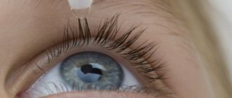
The electroretinography procedure lasts approximately 40-50 minutes. Women cannot undergo this procedure with cosmetics, so mascara and eye shadow will have to be washed off. For pain relief, a specialist instills lidocaine, or rather, its solution. After this, a special lens is put on the eye, which contains two electrodes that capture the light wave.
The patient places his chin on the stand, which is located in front of him, with his forehead resting on the bar. The patient needs to look at the light stimulus, and this must be done continuously. The irritant sends flashes of light to the retina of the eye. The electrodes record the retinal reaction, as well as the appearance of the potential. The specialist observes a retinogram that appears on a screen, which records the signals generated by the retina of the eye.
This examination is completely painless thanks to an anesthetic drug instilled into the eyes. After the procedure, the patient may experience a slight burning sensation or feel as if a small speck has gotten into the eye. Also, the eyes may water for a short time and feel discomfort. In a maximum of an hour and a half, all these consequences will pass, and everything will be the same as it was before the study.
Fluid constantly forms in the human eye. It is called aqueous humor and is located between the iris and cornea. The intraocular fluid leaves this part of the eyeball into the lymph at the same speed as it is formed. If the outflow process is disrupted, aqueous humor begins to put pressure on the walls of the eye.
- at rest, the pressure in the eye has a constant value, regardless of whether it is normal or increased;
- it increases when installing a tonometer with a weight that puts pressure on the eyeball;
- this leads to an accelerated outflow of aqueous humor and a simultaneous decrease in pressure;
- it continues to fall even after the device sensor (weight) is removed;
- Gradually, the flow of intraocular fluid is restored, and the pressure stabilizes.
This entire procedure allows you to measure IOP, determine the so-called “outflow ease coefficient” (OEF) and other parameters indicating the degree of development of glaucoma.
There are several methods for conducting this study: Maklakov method, electron tonography, pneumotonometry. Let's take a closer look at them.
This survey is state-of-the-art. In most cases, electronic tonography of the eye is prescribed. Before the procedure begins, drops with an anesthetic effect are instilled into the patient's eyes. The patient lies face up on the couch and focuses on a black dot that is painted on the ceiling.
After this, the research itself begins:
- the sensor of the electronic tonometer is held over the cornea for 20 seconds;
- then the device is lowered onto the eye for 5 seconds;
- then the sensor is removed for 30 seconds and again applied to the cornea for 4 minutes;
- the final stage is 3 pressure measurements with an interval of 5 s.
All data is transferred to the computer. They are deciphered by an ophthalmologist.
Using a tonograph, you can measure daily pressure fluctuations in the morning and evening, although even repeated pressure measurements at these hours do not always make it possible to suspect an increase.
Principles of daily changes in intraocular pressure:
- In healthy people, as well as in patients with glaucoma, pressure changes throughout the day.
- Most people have higher blood pressure in the morning.
- In patients, the magnitude of pressure fluctuations is greater.
It is best to track daily fluctuations from 7 to 8 o'clock in the morning and evening. Morning blood pressure measurements should be taken before a person gets out of bed. The normal fluctuation rate is 5 mmHg. Art. (for changes from evening to morning also). To diagnose glaucoma, you need to perform tonography for 10 days in a row.
Approximately 3% of the world's population suffers from glaucoma. This is a fairly severe pathology. Approximately 15% of all blind people lost their sight because of it. It manifests itself in various symptoms. They depend on the form of the disease. There are open-angle and closed-angle glaucoma. The first develops slowly, the second - rapidly.
- the appearance of rainbow circles before the eyes;
- decreased visual acuity;
- pain in the eyes and head.
If these symptoms occur, be sure to see a doctor. Sudden changes in pressure in the eyes can lead to damage to the optic nerve and the development of an atrophic process, during which the fibers die. Visual functions lost due to atrophy cannot be restored. Glaucoma occurs for a variety of reasons.
The patient's position during the tonographic examination is lying on his back, face up. To eliminate possible tension of accommodation, the gaze should be fixed at any distant point. Then local epibulbar (drop) anesthesia is performed 2-3 times with a solution of dicaine (0.5%). The eyelids are pulled apart and fixed in this position with a plastic ring. A tonograph sensor is installed on the immobilized eye.
Taking readings with a tonograph lasts about four minutes. All this time, the device graphically records changes in intraocular pressure. At the end of the procedure, the fixing ring is removed. For preventive purposes, it is recommended to instill 1-2 drops of an antiseptic (for example, sodium sulfacyl 30%).
The ophthalmological unit is equipped with the latest automatic eye tonograph, Glau Test 60. Before the study, it is calibrated to ensure maximum accuracy of the readings. Equipment of this class automatically records indicators and calculates the required values.
Alternative non-computer methods require manual calculations. For this, there are special tables and a tonographic curve. As a result, it is possible to determine the true value of IOP, average pressure, and volume of displaced fluid.
There are standard indicators for quantities that are assessed during tonography:
- for ECO (outflow ease coefficient), the range of 0.29–0.31 mm3/min/mmHg is considered normal;
- true intraocular pressure should be in the range of 15-17 mmHg. Art.;
- The minute volume of intraocular fluid in a healthy eye is 2.0 mm3/min.
Stages of the procedure
The patient is placed face up on the couch. He needs to calm down, relax and fix his gaze on a point that will be in the distance. Before tonography, 2-3% anesthesia (0.5% dicaine solution) is instilled into the eyes.
For convenience, the eyelids are separated with a special plastic ring. The tonograph sensor is carefully placed on the eye. The device graphically records the indicators for several minutes. The results should only be analyzed by a doctor.
After the procedure, the sensor is removed and the ring that fixed the eyelids is removed. To prevent infection and irritation, you need to drip a solution of sodium sulfacyl 30% (two drops) or any other suitable antiseptic into the conjunctival sac.
OCT indications
There are quite a few indications for electroretinography. If a person is suspected of having diseases of the nervous system, then there is a risk of vision deterioration, so this procedure is necessary. In case of congenital myopia in a person, electroretinography must be performed, and this does not depend on age.
The procedure will help determine the level of retinal damage, and it will also be possible to develop a treatment strategy. Electroretinography is also performed for amblyopia of any degree in order to determine whether treatment would be appropriate in this case or whether it is better to refuse it. If a person’s vision deteriorates at dusk or acquired myopia is diagnosed, electroretinography is performed to determine the degree of pigment sputtering on the retina.
There are several other indications for electroretinography. If a person has retinal dystrophy or is suspected of having this disease, then electroretinography is performed to monitor the dynamics of treatment. In cases where a person is suspected of having optic nerve atrophy, electroretinography is performed to monitor the development of the disease and determine the size of the affected area.
Electroretinography also has contraindications. If a person is diagnosed with pathological activity of the nervous system, then electroretinography is contraindicated.
- seizures;
- epilepsy;
- various other attacks.
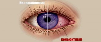
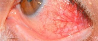
You should also not do electroretinography if you have allergies, conjunctivitis, or diseases of the cornea. Before carrying out this procedure, you should consult with an ophthalmologist or ophthalmologist in order to find out in advance about all contraindications and the possibilities of carrying out the procedure for a particular patient.
After the consultation, a decision will be made about what kind of electroretinography should be performed and whether it should be done at all, since the procedure can be harmful. If the procedure is contraindicated, but is performed, then a specific response can be obtained from the retinal cells, which will not provide any specific results regarding retinal disease.
Eye tonography is contraindicated in cases where:
- there are injuries to the cornea;
- inflammatory or infectious process in the membrane of the eye;
- the cornea has pathologies that can change the readings;
- individual allergic reaction to anesthesia, which is used at the beginning of the procedure.
Also, tonography of the eye is not performed if the patient has consumed alcohol or drugs. If the patient has mental disorders that do not allow his behavior to be predicted during the procedure, then tonography is not performed.
After signs of inflammation are eliminated, tonography can be performed. If antiseptics are used during the procedure, the patient will not have side effects or discomfort after the examination.
The indication for eye tonography is the need to measure the pressure inside the eye and is necessary in the following cases:
- if your eyes get tired very quickly;
- there is a feeling of heaviness in the eyes, and pain in the superciliary areas and eye sockets;
- after prolonged visual stress, the head begins to hurt;
- vision decreases;
- if, when looking at very strongly lit objects, rainbow circles appear.
These are the first signs of the disease. If you detect the disease in its early stages and start treatment on time, you can avoid complete blindness. In advanced cases, surgical intervention is used. If the disease is not treated, you can eventually lose your vision completely.
Also, signs of glaucoma can be small scotomas (dark spots) in front of the eyes, blanching of the optic nerve head, and asymmetry of the eyes.
Sometimes the cause of eye pain is trigeminal neuralgia. With glaucoma, the eyes hurt when the intraocular pressure is very high. This is possible during an acute attack. Tonography is carried out in the following cases:
- the patient has diseases of the cardiovascular and endocrine systems that impede the outflow of fluid;
- if the patient has relatives with glaucoma and is over 40 years old;
- The patient has neurological pathologies that can cause eye function.
The examination allows you to study the condition of the visual apparatus and identify possible pathologies. Any abnormalities of the cornea, retina, anterior chamber components, and optic nerve will be reflected in the examination results.
- sudden drop in vision;
- the effect of a veil before the eyes;
- increased pressure inside the eye;
- pain in the eye area;
- eye floaters syndrome;
- complete loss of vision;
- exophthalmos.
Optical tomography allows you to study the angle of the anterior chamber. In addition, the examination makes it possible to assess the condition of the drainage system and its functioning. All this is a necessary element in diagnosing glaucoma. OCT is mandatory in preparation for surgery.
- retinal detachment and other pathological changes;
- various types of tumors affecting the visual apparatus;
- rapidly developing myopia;
- thrombosis and other vascular pathologies;
- various types of macular damage;
- diabetic retinopathy;
- ulcer affecting the condition of the cornea;
- keratitis, deep type;
- proliferative vitreoretinopathy;
- various pathologies affecting the optic nerve head;
- epiretinal membrane;
- swelling of the macula, cystic in nature.
Optical tomography can detect even small changes in the condition of the organs of vision. As a result, it becomes possible: a quick and correct diagnosis, determination of the degree of damage to the elements of the visual apparatus, and the development of an effective therapeutic regimen.
- hypertension;
- diabetes, type mellitus;
- diseases of the vascular system.
- in the presence of diseases that do not allow you to focus your gaze on one point for 2-3 seconds;
- if the patient has mental pathologies;
- when the subject is unconscious;
- if the patient experiences confusion.
In addition, the examination procedure using a tomograph is not carried out in the presence of other diagnostic procedures. Ophthalmologists specifically set aside a separate day for OCT. After all, the contact medium is very sensitive to external influences. Therefore, doctors strive not to expose the visual organs to unnecessary stress, preferring to choose a separate day for optical coherence tomography.
Signs of glaucoma
Full and simplified tonography have no contraindications. They can be performed on absolutely all patients. To obtain a holistic picture of the patient’s condition, doctors prefer to analyze the daily results of tonometry, perimetry and tonography.
Confirmation of glaucoma will be an outflow ease coefficient of less than 0.15 in the presence of a scotoma closer to the center of the visual field, when the diurnal curve is clearly pathological. Patients with glaucoma often consult a doctor if they have small scotomas (dark spots); they are diagnosed with blanching of the optic nerve head and a decrease in the anterior chamber of the eye.
Pain is a sign of severely increased intraocular pressure during an acute attack of glaucoma. Pain in this disease often occurs due to trigeminal neuralgia, being not directly related to glaucoma.
Features of the procedure
Tonography is a method for analyzing the outflow of intraocular fluid, which allows for graphical recording of pressure indicators. Clinical tonography is carried out using impression and applanation devices. In the CIS countries, impression electrotonographs of Nesterov, Sakharov and others, a Schiotz tonometer temporarily attached to the cornea, and perilimbal suction cups are more common.
One tonography session takes approximately 4 minutes. The procedure makes it possible to determine the hydrodynamics of the visual system. Tonography shows the volume of fluid per minute and the coefficient of ease of outflow. The coefficient indicates the volume of fluid that flows out of the eye in 60 seconds under compression (force) pressure. The amount of filter pressure will be directly proportional to the outflow coefficient.
The procedure for measuring the outflow of intraocular fluid is carried out using a tonograph - a special electronic device. Simplified tonography is also possible, which involves the use of a small Maklakov tonometer.
Modern simplified tonography
The patient also needs to be placed on his back and anesthetized three times with a dicaine solution. The procedure uses a Maklakov tonometer, which is very light and compact (10 g of weight). To obtain the result, the eye is compressed with an ophthalmodynamometer or sclerocompressor for three minutes. After compression, intraocular pressure should be measured again.
When simplified tonography shows an intraocular pressure of 27 mmHg. Art. and above, you need to repeat the procedure after half an hour. Such manipulations will allow you to avoid mistakes and check the reliability of the first result.
Preparing for optical coherence tomography
The optical coherence tomography procedure does not require special preparation. However, to obtain the most complete clinical picture, it is necessary to artificially dilate the pupil. As a rule, they resort to the use of special drops, which give a short-term enlargement effect.
- Having a direct impact. Medicines of this type provoke contraction of the radial muscles. As a result, the pupil increases in diameter. Drugs of this type include: Tropicamide and Irifrin.
- Affecting indirectly. Such drugs affect a different type of muscle. They indirectly affect the diameter of the icon. Similar medications include: Cyclomed and Atropine.
Before using medications, you must carefully read the instructions. On the day of using mydriatic, you should not drive a car.
After instillation of special drops, the patient is examined using an OCT scanner.
- An ophthalmologist prepares the device for diagnostic procedures. At this time, the examinee is asked to sit on a chair near the tomograph.
- After bringing the equipment to a state of readiness, the diagnostician asks the patient to place his chin on a special stand. Then the subject must fix his gaze on a special object.
- The doctor moves the camera of the device towards the eye until the device produces a clear image of the retina. To obtain a high-quality picture, the distance between the eye and the camera should be 9 mm. When the greatest clarity of the image has been achieved, the camera is fixed in this position. Then the ophthalmologist performs calibration to achieve better picture quality.
- This stage includes the selection of the most informative images, allowing for the most complete clinical picture.
- After receiving the image, the ophthalmologist cleans the resulting images from various defects. Any artifacts and interference are removed.
- At the last stage of the survey, a comparative characteristic is carried out. It will include both the obtained images and images of healthy people in a similar age group. Scans of the patient himself, taken before the current examination, are also allowed for comparison.
Composition of aqueous liquid
The pathology disrupts the blood supply to the organs of vision. The chamber fluid is not similar in structure to blood plasma, although it is produced from it. The composition of moisture is adjusted as it circulates. If we compare the composition of plasma with the liquid of the anterior chamber, it can be noted that the latter has a number of distinctive features:
- increased acidity;
- predominance of sodium and potassium;
- presence of glucose and urea;
- low dry matter mass - almost 7 times less (per 100 ml);
- low percentage of proteins - does not exceed 0.02%;
- more chlorides;
- high concentration of acids - ascorbic and lactic;
- low specific gravity - 1.005;
- presence of hyaluronic acid.
The exact pattern of aqueous humor development is not yet fully understood. Anatomical facts, however, indicate that it is the processes of the ciliary body that produce this fluid. Passing its way from the posterior to the anterior chamber, it affects the following areas:
- ciliary body;
- posterior part of the cornea;
- iris;
- lens
Then the moisture seeps into the venous sinus of the sclera through the trabecular network of the angle of the anterior chamber of the eye. Following this, the fluid ends up in the vortex, intra- and episcleral venous plexus. It is also reabsorbed by the capillaries of the ciliary body and iris. Thus, for the most part, the chamber humor rotates in the anterior part of the visual organ.
The essence of the study
Tonography helps determine how difficult the circulation of intraocular fluid is.
Normally, the fluid contained between the cornea and the iris is constantly formed and flows into the lymphatic network at the same speed. Therefore, intraocular pressure remains unchanged. With glaucoma, the rate of fluid outflow decreases and its pressure increases. The degree of difficulty in the circulation of intraocular fluid can be assessed using tonography.
The method was proposed in 1950. Its essence is as follows:
- at rest, intraocular pressure may be normal or elevated, but it is a constant value;
- when a tonometer or weight is placed on the eye, the pressure in the anterior chamber increases;
- a violation of pressure leads to increased outflow of intraocular fluid, while the pressure gradually decreases;
- after the compression stops, the outflow still prevails over the secretion of fluid for some time, so the pressure curve drops below the isoline;
- then the inflow is gradually restored, which leads to stabilization of intraocular pressure.
The rate of pressure reduction is primarily determined by the state of the outflow tract and decreases in glaucoma. The rate of restoration of initial parameters is determined by the production of intraocular fluid.
When calculating using a tonometric curve, it is taken into account that for every millimeter of additional compression there is a certain amount of fluid flowing from the eye. This value is called the “ease of outflow coefficient” (ECR). It indicates how severe the impairments in glaucoma are.
Tonography is not an ideal research method. It gives errors that are associated with the following factors:
- the formation of intraocular fluid depends not only on its direct production in the tissues of the eye, but also on filtration through the walls of blood capillaries; during tonography, liquid does not enter the eye by ultrafiltration, its total amount decreases, and therefore an impression is created of a better outflow state than in reality (“false outflow”);
- during the test, blood flows out of the eye tissue, and its amount increases with glaucoma; this also creates the idea of good outflow of intraocular fluid.
To correct these errors, it is recommended to carry out tonography using an electronic device, as well as use functional tests.
Interpretation of OCT results
Decoding the results of electroretinography is quite simple; if the electrodes cannot track the reaction and waves, then in this case it is possible to diagnose a complete detachment of the retina or a severe inflammatory process. Then you need to urgently begin treatment; you may have to resort to surgical treatment.
If too strong wave fluctuations are detected, the specialist will diagnose either optic nerve atrophy or rupture of this nerve. It is worth noting that fluctuations in adults and children cannot be compared, since they will differ in any case.
Electroretinography is one of the most highly informative methods for determining and diagnosing diseases. Such research is carried out mainly to recognize diseases of the outer layers of the retina of the human eye, that is, the work of photoreceptors and bipolar cells of the retina is monitored.
- study of morphology - the shape and cut of the profile are considered, the clarity of the contours is assessed;
- quantitative analysis - through examination, all tissue changes are recorded, not only the fact of thinning or thickening is considered, but also the degree of changes;
- study of reflectivity - the degree of reflection of the sent signal from tissues is assessed.
The process of interpreting optical tomography results is closely related to color codes. They provide an opportunity to learn about the condition of tissues.
- Warm. Colors with a warm temperature indicate the presence of areas with thin tissue. For example, black and blue shades will indicate areas that are dangerously thinning.
- Cold. Colors with a cool temperature indicate areas that will be characterized by thickening. For example, areas colored in yellowish or reddish tones will show areas of greatest thickness.
Today, technologies make it possible to create three-dimensional images. The latest generation tomographs easily display a 3D model of the examined area for study. Such capabilities allow us to draw the most complete conclusion about the state of health of the patient’s visual system.
Price
Often, to undergo an examination, the patient has to go to a specialized medical center. Regular district clinics do not have the necessary equipment. As a rule, ophthalmology offices are poorly equipped and can only offer outdated diagnostic methods.
Thus, the person being examined needs to visit a private institution providing medical services. Perhaps residents of small settlements will have to visit large cities. As a rule, in large cities there is an abundance of ophthalmology offices with an OCT scanner.
- settlement;
- prestige of the medical center;
- doctor's degree of training;
- brands of equipment used to conduct the survey.
On average, the price of coherence tomography fluctuates from 1500 to 2000 rubles per procedure.
Pneumotonometry
This is another modern method of measuring pressure in the eye. It is used quite often in the diagnosis of glaucoma. Langham developed the method and proposed using a computerized device with a pneumatic pump. Preparation for the examination is no different from previous methods. First, an anesthetic is instilled into the patient's eyes, after which a silicone nozzle is placed on the cornea.
It is disposable, its diameter is 5 mm. There is no need to remove the attachment from the eyeball, as happens during examination using the Maklakov method and electronic tonography. In this case, the device measures the pressure in the eye at a rate of 40 times per second. The patient can lie down or sit. All information is sent to the computer monitor.
What affects intraocular pressure
The amount of intraocular pressure is almost the same in the eyes of one person. The permissible difference should not exceed 3-4 mm. When the difference exceeds 4-5 mmHg. Art. (even with normal ophthalmotonus values), glaucoma can be suspected.
In newborns, the pressure is the highest. It gradually decreases to 10 years. At the age of 20, most people experience a slow increase in blood pressure; after 70, a slight decrease is acceptable. The maximum age difference in ophthalmotonus indicators is 1.5 mm Hg. Art.
Women's blood pressure is usually higher than that of the opposite sex. The difference can be 0.5 mmHg. Art. National and racial characteristics, as well as living conditions do not affect the pressure in the eye. Sometimes with moderate activity, ophthalmotonus decreases.
Climate has a very slight effect on intraocular pressure: a decrease with an increase in altitude above sea level, at lower temperatures. Seasonal differences are more pronounced: in summer the pressure is lower than in the cold season. The difference can be 1 mmHg. Art. from the norm.
The pressure is greatly influenced by the position of the body: vertically, a decrease of 1-4 mm Hg is permissible. Art. from the horizontal norm.
Factors that can change tonography results:
- Water. When drinking a liter of water, the eye increases by 4.4 mmHg. Art. and retains size for 2.5 hours.
- Coffee. When drinking a cup of coffee, the pressure increases by 4 mm and remains for 1.5 hours.
- Alcohol. When drinking alcoholic beverages, blood pressure increases by 3.7 mm and persists for 1 hour.
- Physical activity. Exercises dramatically reduce intraocular pressure by 4.3 mm. It takes 65 minutes to recover.
It can be concluded that single tonography is an unreliable method for diagnosing glaucoma. If there is one test result (exceeding the norm by 2-4 mm Hg), it is impossible to accurately diagnose in the absence of other abnormalities. Even the most modern measuring instruments are imprecise, and tonography methods are subjective due to a large number of individual factors.
Which doctor should I contact?
To conduct tonography and other methods of diagnosing glaucoma, you must contact an ophthalmologist. It is advisable to perform these studies on modern electronic equipment, which increases their accuracy.
When detecting glaucoma, the tonography method is used. A feature of glaucoma is a decrease in the ease of outflow of intraocular fluid. Tonography makes it possible to determine the coefficient of such deviations. Tonography is also suitable as a way to monitor the effectiveness of the chosen treatment.
Efficiency of tonography
Modern tonography makes it possible to identify some pathological and physiological features of the course of glaucoma, but its significance in assessing the patient’s condition is insignificant. Tonography results are variable and may change.
Nesterov himself, who diligently developed tonography techniques, considered such diagnostics auxiliary. It helps to complement the picture painted by other studies, but is by no means an absolutely reliable method for measuring eye performance in glaucoma.
Modern medicine tends to believe that each patient has his own norm of intraocular pressure, ophthalmotonus, and individual tolerance of pressure by the optic nerve. This greatly complicates the diagnosis of glaucoma using tonography.
Sources used:
- Bezdetko, P.A. Diagnostic reference book for an ophthalmologist / P.A. Bezdetko, S.F. Zubarev, N.V. Panchenko. - M.: Phoenix, 2006.
- Ermakov, V.P. What and how children from birth to 10 years old with intact and impaired vision see. Diagnostics, development and vision training / V.P. Ermakov. - M.: Vlados, 2015.
- Morozov, V.I. Diseases of the visual pathway. Clinic. Diagnostics. Treatment / V.I. Morozov, A.A. Yakovlev. - Moscow
- Website of the Federal State Budgetary Institution "NIIGB"
Disadvantages of tonography
This method is not ideal, as there are errors. Firstly, the formation of aqueous humor depends on various factors: from its filtration through the walls of blood vessels, from direct production in the tissues of the eyeball. Its formation decreases during the procedure itself, which may give the impression that its outflow is good.
- Water and drinking. The patient needs to drink 1 liter of water. After about 45 minutes, the examination is repeated. Due to drinking water, the volume of intraocular fluid increases and its outflow decreases. Tonography data changes by 30%.
- Water-dark. The subject drinks 200 ml of water and goes into a dark room for 1 hour. After this, the pressure in the eye is measured again.
There are a number of other factors that affect IOP. In newborns it is elevated, but gradually decreases and returns to normal by about 10 years. In women it is usually slightly higher than in men. In summer the pressure is lower than in the cold season. The difference, as a rule, does not exceed 1 mm Hg. Art. This indicator is also influenced by body position. A decrease of a few mmHg is acceptable. Art. in a vertical position.
All these factors are natural and are taken into account by the doctor during the examination. There are other reasons that may negatively affect the results of the examination. As such, it does not require any preliminary preparation. However, the ophthalmologist warns that you should not drink coffee on the day of the examination. It is also contraindicated to drink alcohol two to three days before measuring blood pressure.
A single tomography may not provide an accurate picture of the nature of the disease. If glaucoma is suspected, several tests may be performed. The need for repeated measurements of intraocular pressure may already occur when the diagnosis is established. Tonography will help establish the degree and form of pathology.
Normal eye tonography
During tonography, the normal pressure inside the eye is 15-17 mm Hg. If these indicators are equal to 20 mm, then the likelihood of glaucoma increases, and an indicator at the level of 24 mm indicates the presence of glaucoma.
The CLO indicator should be from 0.11 to 0.6 mm3 per minute. The average value is 0.3 mm3 per minute. If glaucoma is at an early stage of development, then these indicators are closer to the lower limit of normal - from 0.12 mm3 to 0.2 mm3 per minute. If such results are obtained as a result of the study, then it is necessary to undergo a more thorough examination and be constantly monitored by a doctor. If there is glaucoma, then the CLO indicator will be 0.1 mm3/min or lower.
The rate of formation of intraocular fluid is another tonography indicator. It varies from person to person, but on average it ranges from 1.5-4.5 mm3/min. It is very important that the indicators in the right and left eyes differ from each other. If they exceed 0.8 mm3, then this can also be considered a sign of glaucoma.{banner_gorizontalnyy2}
The Berker coefficient (KB) is the ratio of the initial indicator to the maximum result obtained during tonography. Its norm should not exceed 100, but with glaucoma, especially in the early stages, this figure is much higher.
KB varies depending on age. At the age of up to 30 years, it does not exceed 90; from 30 to 39 years, the indicator is 100; for the next two decades, 5 units are added. At the age of 60 years and older, normal BC levels are 135 units.
If intraocular pressure readings exceed 27 mm, then to eliminate errors, a re-diagnosis is carried out after 30 minutes. This will help avoid measurement errors.
The highest intraocular pressure is observed in infants. Also, women, compared to men, have slightly higher rates. Also, pressure is slightly affected by weather changes, as well as body position.