Optics Basics
Let's remember the school physics curriculum. Many teachers showed students an entertaining trick: two rooms with low levels of light, but one of them has small holes in the walls. Behind them is a strong light source, such as the sun. In some cases, instead of the pinholes used to illuminate the room, a small flashlight was used.
If an object made of opaque material is placed between a point light source and the second hole in the wall, then its image will appear on the partition located behind the second hole, inverted one hundred and eighty degrees.
A converging lens performs a similar trick with light rays. The reason lies in the fact that every microscopic point of any object, when illuminated, itself becomes a source of light, reflecting particles that fall on it in all directions.
| White products absorb practically nothing from the visible range; they reflect all the light that hits them into the environment. Black objects, on the contrary, use any source of energy for heating. |
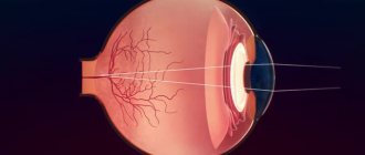
The structure of the optical system of the eye
The main indicator of its operation is the refractive power, which reflects the degree of adjustment of the angle of incidence of the light beam. Refraction takes place four times in the system: in the anterior and posterior chambers, the lens, the cornea and a little in the liquid medium of the eye. The greater the refractive characteristics of the organ of vision, the higher the degree of refraction of rays. On average, this figure is sixty diopters.
The optical system includes two main axes:
- Visual. The distance between the visible object and the core of the fovea. The maximum difference should be five degrees;
- Optical. It is the distance between the far points of the eyeball and the chambers of the eye, passing through the center of the lens.
The length between the anterior pole of the visual apparatus is sixty millimeters, this allows people to see the world in 3D.
Below we will take a closer look at the structure of the optical system and analyze each of its elements in detail.
Cornea
It is a transparent “detail” of the organ of vision, curved in cross section. More than 2/3 of the entire optical power of the eye falls on the cornea, which contains several layers covered with the thinnest tear film. The front part of the element is in constant contact with air, therefore it is more curved and has greater refractive power compared to the back.
Front camera
98% consists of intraocular fluid. Provides a degree of refraction equal to 1.33 D. If there is a deviation in the functioning of the organ of vision, the recesses of the camera are adjusted, as a result, for every millimeter there is an increase in refraction by 1 D.
Iris and pupil
The muscle fibers of the iris are responsible for changing the size of the pupils, i.e. regulate how much light passes through the optical system. In good lighting conditions, they are narrowed, as a result, direct rays fall directly on the central fovea. In this case, as a rule, visual acuity increases in people suffering from astigmatism. If, when the pupils constrict, problems with the eyes appear, then we can talk about pathological processes in the macula.
In low light conditions, the pupils increase in size, this leads to the following effects:
- The optical system receives a greater number of light fluxes, as a result, visual acuity increases, and a person can distinguish objects even in the dark;
- Direct rays fall on a significant part of the surface of the retina, i.e. Photoreceptors are involved in the process.
| With strong emotional shocks, the optical system loses the ability to self-regulate. A similar reaction is observed when taking a certain group of medications or drugs. In this case, vision suffers. |
When the pupils are greatly dilated in people diagnosed with astigmatism, the image turns out blurry, since the process involves areas of the cornea that have different degrees of refraction. Return to contents
Lens
One of the most complex elements of the optical system, consists of a large number of cells that have lost their nuclei. Performs two main functions: refraction of light and focusing of the image. Accommodation occurs as follows:
- When the ciliary muscles contract, the zonules that support the lens relax;
- It takes on a rounded shape, becomes thicker in the center, and its curvature changes;
- At the last stage of focusing, the depth of the front camera decreases.
The lens grows throughout a person's life. New fibers grow on top of the old ones, so the element gradually thickens. If at birth this figure is 3.5 millimeters, then in an adult it increases to 5 mm.
Vitreous body
It closes the optical system and performs a large number of important functions. It has good throughput, but at the same time it has weak refractive characteristics, so it does not take part in creating the image.
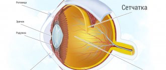
Retina
One of the most complex elements in the visual apparatus. It is she who is responsible for the perception of color and light. It has high sensitivity and is covered with the thinnest film. The retina is supported by epithelial ligaments, and the vitreous body presses it. The optical system uses an element to capture the image and transmit information along the optic nerves to the corresponding parts of the brain.
You will learn more about the structure of the system from the video
Properties of vision
The optical system of the eye provides a unified visual perception of both eyes, this property of vision is called binocularity. This property is due to a natural reflex to ensure the fusion of images received by two eyes into a single picture.
Due to the fact that the neural elements of the retina of the two eyes are different, when each eye receives an image, physiological double vision of objects occurs, depending on the degree of their distance from us.
This property of vision makes it possible to independently assess at what distance an object is located, as well as evaluate its relief. This feature of vision is called stereoscopicity. Moreover, stereoscopicity is only available when looking at an object with both eyes at the same time. If you look at the image with one eye, the relief effect becomes unavailable.
Here, it is worth noting that in the process of vision, the two eyes play slightly different roles. The element of the visual system that takes more part in the process of image formation is called the leading eye, and the second is called the slave eye. To check this property of an optical system, it is enough to look at any image through a hole in a dense screen with two eyes in turn; for the leading element the image will stand still, and for the slave it will move slightly.
Path of light rays and magnitude
The refraction of light in ophthalmology is called refraction. The rays falling on the optical axis change and meet at the main focus of the organ of vision. They are reflected from infinitely distant objects, so the role of the central focus is played by a point located on the optical axis.
Light rays reflected from objects at a final distance are combined at an additional focus. It is localized further than the main one, since the process of concentrating diverging rays takes place using additional refractive power.
Eye muscles: structure and functions
The muscles of the eye ensure coordinated movements of all components of the optical system and ensure the quality of vision.
If even one muscle is damaged, it will affect not only the motor ability of the eye and nearby muscles, but also the quality of vision.
There are six muscles in total that are responsible for eye movement, of which four are rectus muscles and two are oblique muscles. The work of all these muscles is controlled by the corresponding cranial nerve fibers - oculomotor, abducens and trochlear.
Thanks to the large number of nerve endings in each fiber, clear and efficient movements are ensured.
Functions of the eye muscles
As mentioned above, the eye muscles are divided into rectus and oblique. This classification is based on the principle of movement of these muscles and their location.
The four rectus muscles are located on the internal, external, inferior and superior surfaces.
The two oblique muscles of the eye line the upper and lower parts of the visual organ.
All muscle groups have an almost similar structure, except that they differ in the design of the lower part of the oblique muscles.
Such muscles begin in a dense ring that surrounds the optic nerve. Next comes the zone of the muscular “funnel”, from which each process continues its motor activity in a given direction. This direction leads to the tendon, into which the processes pass, attaching to the eyeball.
All movements of the eyeball are determined by the attachment point of each muscle.
Vertical and inward movements are performed by the superior and rectus inferior muscles. The oblique muscles are responsible for more complex and varied eye movements. So, the superior oblique muscle lowers the eye down and turns it outward. And the inferior oblique muscle, accordingly, raises the eye and also moves it outward.
Top
Accommodation
To obtain a clear image, the optical system must focus; for this, one of two methods is used:
- The lens moves relative to the retina;
- The degree of refraction increases.
The ability of the human eye to adapt to different distances and see objects located far away or nearby is called accommodation.
| When you concentrate your gaze on a near point, the refraction of the lens increases, it becomes round, and when viewing an object in the distance, the element takes on a flat shape. During accommodation, the curvature of the lens and its refractive properties change. |
Optical instruments for the eye
The human eye, despite its natural perfection, is far from ideal universal optical instruments in its properties. Therefore, it is necessary to use optics that equip the human eye with new abilities. When considering various devices, it should be remembered that in each case they and the organ of vision form a single optical system, the most important element of which is the lens.
If we talk about the eye as an optical device in physics, it generally helps to obtain an image of a particular object on the retina, and its apparent size is estimated by a person based on the size of this image.
A feature of the optical system, which includes the eyes, is that the parameters of such a system can change due to changes in the focal length of the lens during accommodation. Such considerations make it easy to study the action of a magnifying glass, which is an ordinary convex lens.
The same instruments, only more complex in structure and functioning, are the microscope, telescope, etc.
Physiological role of the optical system of the eye
It performs several important functions:
- Sets the required degree of refraction of light rays;
- Focuses the image and objects in the plane of the retina;
- Creates the required axle length.
As a result of the operation of the optical system, a person clearly distinguishes objects and their color. It also has the following characteristics:
- Binocularity. The ability to perceive a three-dimensional image simultaneously with both eyes, while the image does not double;
- Stereoscopic. A person can visually determine the approximate distance to an object and evaluate its outline;
- Visual acuity. This concept hides the ability to distinguish between a pair of points located at a certain distance from each other.
Return to contents
Features of visual perception
First of all, the optical system of the eye is designed to obtain information about the world around us through vision. This concept has many characteristics and features.
The sense of light allows the human eye to perceive daylight and artificial light, as well as to distinguish the degree of its intensity. And thanks to the natural adaptation of the eyeball, the optical system is able to independently adapt to illumination of varying brightness without outside help. Light sensitivity is determined by the natural threshold of light stimuli. Few people know that a person with good eyesight can see even a small light at a distance of several kilometers.
The sensitivity of the visual apparatus primarily depends on many factors, such as the intensity of the light source, its angular size and wavelength, as well as the time that the light stimulus acts on the eye. Due to the deterioration of the optical characteristics of the sclera with age, the sensitivity of the eyeball can greatly decrease.
Human optical system: stereoscopic or 3D vision
This concept comes from the Greek words “stereo” (solid) and “opsis” (look). It is used to denote depth of perception and three-dimensional structure derived from visual information from the eye.
Since the eyes are located on the lateral planes of the skull, the image is projected onto the retina in different ways, and there is a difference in the horizontal position of objects relative to each other.
| In medicine, this phenomenon is called binocular inequality. The differences are processed in the brain, and then the optimal depth of the picture is formed. |
Symptoms of damage to the optical system of the eye
Any deviation in its work will lead to vision problems. Signs signaling the development of pathological processes:
- Fast fatiguability;
- Constant headaches and stress;
- Split image;
- Blurred vision;
- Decrease in visual acuity;
- Blurred outlines of objects. A person cannot see objects located far away or nearby.
Any of the listed symptoms indicates the need to visit a doctor to find out the cause of the developing pathology.
Diagnostic methods for damage to the optical system of the eye
To evaluate the operation of the system, it is initially necessary to establish which eye is the slave and which is the master. For this purpose, basic testing is used; it can be done at home. Look through a piece of thick paper where a small hole has been made in the center, first with your left eye, then with your right eye. If the eye is leading, then the picture remains in a static state. At the slave's it begins to move.
To identify deviations in the operation of the optical system, the following examinations are used:
- Visometry. Used to determine visual acuity;
- Ophthalmometry. Determines the refractive ability of the cornea;
- Skiascopy. Helps to obtain objective information about the degree of refraction;
- Pachymetry. Measuring the thickness of the cornea;
- Ophthalmoscopy. Used for analysis of the fundus and retina;
- Biomicroscopic examination;
- Keratoscopy. Analyzes the condition of the cornea through a special lens;
- Ultrasound examination of the eyeball.
| Early diagnosis will allow you to identify deviations at the initial stage and avoid the development of complications. In some cases, delay in treatment can lead to blindness. |
Diagnostic and treatment methods
To identify existing diseases of the optical apparatus, patients are prescribed a detailed ophthalmological diagnosis. The examination consists of several informative procedures:
- visometry, which specifies the level of visual acuity;
- ophthalmometry necessary to determine the refractive abilities of the cornea;
- ophthalmoscopy, which examines the condition of the retina and fundus of the eye;
- keratoscopy, which examines the cornea of the eye;
- tonometry, which helps determine intraocular pressure indicators;
- pachymetry, which consists of measuring the thickness of the ocular cornea;
- skiascopy, which helps to obtain information regarding refraction.
- biomicroscopy, which studies in detail the superficial and deep structures of the eyeball.
If necessary, standard examination of the visual apparatus is supplemented with modern methods such as ultrasound, CT, and MRI.
The approach to the treatment of ophthalmological diseases is determined by the type of diagnosis the patient has. In the treatment of myopia, hypermetropia, and astigmatism, spectacle or lens correction methods are predominantly used. In case of infection of the eyeballs, anti-inflammatory drops, antiviral or antibacterial ointments are prescribed. Hardware procedures help enhance the effectiveness of the main treatment course. In the event of the development of serious pathologies, specialists decide on the need for surgical intervention.
The optical system of the eye is the most important part of the human body, allowing us to daily perceive the world around us and enjoy its beauty. It must be remembered that the organs of vision are highly sensitive and require constant care. Even minor disturbances in the functioning of the optical system should not be ignored and require timely medical attention.
Wonders of vision in nature
Snakes have unique eyes that can perceive infrared radiation. Thanks to this ability, they successfully hunt warm-blooded animals even in zero light conditions.
Butterflies have a different feature: wonderful creatures perceive part of the ultraviolet sector, so it is not difficult for them to detect pollen in flowers.
Geckos are famous for their excellent night vision. Moreover, they see in the same spectral range as people. It’s just that their retina is three hundred and fifty times more sensitive to light rays. A real night vision device!
The chameleon deserves special attention. He doesn’t need to turn his head to see all three hundred and sixty degrees of the environment. He is able to measure the distance to an object with one eye.
The giant squid boasts the largest eyes on the entire planet. He lives in the depths of the ocean, at its very bottom. There is almost never sunlight here, but the mollusk is able to see its enemy at a distance of a thousand meters.