If a person has good vision, then when looking at a distant object, the parallel light rays emanating from it and hitting the retina are precisely focused using refraction. The normal state of refraction with relaxed visual organs is called emmetropia. The emmetropic eye is able to function for a long time without getting tired, providing a person with good visibility both near and far. However, with age, refractive error often occurs and ametropia develops.
Definition
Emmetropia is a vision condition characterized by the fact that passing light rays are focused by refraction precisely on the retina of the eye, provided that it is completely relaxed. This is a completely normal manifestation, so a person sees very clearly even very distant objects.
Emmetropia of the eye occurs when the refractive power of the cornea and the axis of the eyeball are stabilized, allowing light to be accurately focused on the retina. With this phenomenon, a person has the clearest vision.
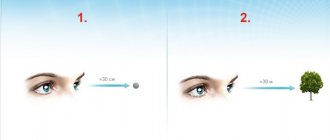
He sees equally well near and far from himself. When reading, if the object is located at a distance of approximately 30-33 cm, you need to make a minimum of effort, as a result of which the eyes get tired much later than in any other condition.
Emmetropia is a proportionate refraction in which the ratio of refractive power and the axis of the eye is optimal. When the indicators deviate in any direction, ametropia occurs.
Forms of the disease
Ametropia is divided into four categories:
- Refractive, caused by a violation of the refractive power index.
- Axial, caused by a violation of the length of the optical axis of the eye.
- Mixed, in which there is a simultaneous violation of the refractive power and the optical axis.
- Combined, in which each indicator corresponds to the norm, but their combination causes refractive error.
Refractive errors are ultimately determined by the location of the focus of light rays and cause various difficulties when viewing objects. There are four forms of refractive error:
Myopia or myopia is the most common type of ametropia, characteristic mainly of children and adolescents. With myopia, light rays are focused in front of the retina, resulting in poor visibility of distant objects. The occurrence and progression of myopia can be facilitated by eye strain as a result of prolonged work at the computer, in an incorrect position, or in poor lighting. Sometimes myopia is congenital. The danger of myopia lies in the high probability of its progression associated with stretching of the sclera.
Hyperopia is otherwise called farsightedness, in which case focusing occurs behind the retina. People with this condition have difficulty seeing near objects clearly, but can see distant objects well. Hypermetropia is always present in newborns in the first year of life. Subsequently, as a result of the final formation of the eyeball, vision is normalized, and this type of ametropia disappears.
Astigmatism is characterized by uneven refraction of rays coming from different directions, so the patient sees objects not only blurry, but also slightly elongated. Astigmatism is caused by curvature of the cornea and pathological changes in the lens. With astigmatism, both near and far vision are impaired in a person, and the person may not be aware of the existing refractive error. Symptoms of astigmatism resemble ordinary eye fatigue due to overwork, however, if dry mucous membranes, blurred vision, or migraines appear, then this should be a reason to consult a doctor.
Congenital astigmatism also occurs. Astigmatism cannot be treated, only its correction is possible, and the earlier this defect is detected, the more effectively it will help prevent a decrease in visual acuity.
In ophthalmology, astigmatism is divided into several types:
- Lenticular, associated with a change in the shape of the lens.
- Hypermetropic, with pronounced signs of hypermetropia.
- Myopic, accompanying myopia.
- Simple, in which the defect is present in only one meridian of the eye.
- Difficult when the refractive error of the same name exists in two perpendicular meridians.
- Mixed, in which hyperopic astigmatism develops in one meridian, and myopic astigmatism in the second meridian.
Presbyopia is an age-related disorder that occurs in people over 40 years of age and is caused by a decrease in the elasticity of the lens. This condition is often called age-related farsightedness. .
For all of the listed types of ametropia, weak, medium, and strong stages of the disease are distinguished - depending on how many diopters it is necessary to reduce or increase the refractive power of the eyes in order to correct vision.
There is uncomplicated and complicated ametropia of the eye. In the first case, visual impairment can be corrected, in the second, changes occur in the visual analyzer, including the retina and optic nerve.
Emmetropization
Many patients who have been diagnosed with this are interested in what it is - eye emmetropia. The description of this condition suggests that this condition is considered normal, but requires medical supervision.
Emmetropization is the process of emmetropia, which is controlled by incoming signals. The mechanisms that control this process are not yet precisely understood. Presumably, it is influenced by a genetic factor.
Emmetropization occurs as a result of ongoing active and passive processes. Passive processes involve enlarging the eyes to the required size as the child grows. The active process implies mechanisms for regulating the length of the axis of the eyeball.
Prevention
Ophthalmologists always draw the attention of patients and, especially parents of children with congenital forms of ametropia and hereditary predisposition, to the importance of preventive measures. First of all, this means maintaining visual hygiene:
- Moderate visual stress.
- Breaks from work to rest your eyes.
- Good workplace lighting.
- Perform daily eye exercises to strengthen the eye muscles and improve blood circulation in the eye tissues.
- Regular visits to an ophthalmologist to monitor your vision.
- Correct correction of visual impairments.
- Training of the eye muscles involved in the process of accommodation.
- Supportive use of dietary supplements containing lutein and zeaxanthin, vitamin and mineral complexes, the use of eye drops that improve metabolic processes in the tissues of the eye.
A healthy lifestyle, a healthy balanced diet with a high content of vitamins and minerals, exercise, and walks in the fresh air are very important for eye health. I recommend to my patients to quit smoking and reduce to a minimum and completely abstain from drinking alcohol.
For amblyopia, when one eye sees better than the other, it is recommended to cover the better-seeing eye at home with a bandage for several hours. At the same time, for the worse-seeing eye, there is an incentive to work and possible improvement of vision. Occlusion is very effective in children, but in adulthood its relevance decreases.
Emmetropic disorders
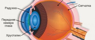
Disorders of emmetropia are called ametropia. In this case, light rays do not reach the retina. Ametropia is also called refractive error and emmetropia. Such disorders include farsightedness, nearsightedness and astigmatism. The ability of the eyes to focus light on the retina is based on such anatomical features as:
- eyeball length;
- curvature of the lens;
- curvature of the cornea.
If the eye has an excessively elongated axis, then the light flux is focused in front of the retina, which provokes the occurrence of myopia. If the light reaches the retina without focusing, then this is farsightedness.
The curvature of the cornea is also of great importance, since if it does not have an ideal spherical surface, then the light flux is refracted with disturbances and is focused rather unevenly, which provokes the occurrence of astigmatism. If the lens has an excessively curved shape, then this becomes the main cause of myopia, and if it is flat, then farsightedness.
What changes occur in the organs of vision
To correct anything, it is important to understand what negative processes occur in the eyes.
Presbyopia (age-related farsightedness) is the inability of the eyes to clearly distinguish nearby objects; only what is far away is clearly visible. This is explained by a violation of visual accommodation.
What structural changes do the visual organs undergo:
- Sclerotic degeneration of the lens: compaction, decreased elasticity of the nuclei and capsules. The lenses are not able to acquire normal shapes, which causes incorrect projection of images onto the retina of the eyes.
- Degradation of the ciliary eye muscles: loss of extensibility, mobility, as muscle fibers are replaced by connective tissues.
- Or, due to the constant tension of the accommodative muscles, their ability to hold and direct the lenses for the required curvature is significantly weakened.
As a result, images of objects are recorded not on the retina, but behind it. The letters in the book blur, the features of your beloved face are difficult to distinguish up close. (But you can clearly see the number of the regular bus, which is located at the end of the block).
Is it possible to correct vision, what should I do?
First, the cause of the progressive decrease in accommodation should be determined and, if possible, excluded.
What leads to age-related degradation of the eye organs, if presbyopia does not occur in every second person, there are people with excellent vision who have passed their 80th birthday.
Factors provoking pathological farsightedness:
- Bad habits (poisoning with toxins from tobacco, alcohol, blood cell tablets and eye tissue).
- Untimely treatment of infectious diseases (poisons of bacteria, viruses and other parasites poison the body).
- Metabolic disorders (diet, diseases of internal organs, vitamin deficiency, diabetes). The nutrition of eye cells deteriorates, and tissue degeneration begins.
- Physical inactivity - blood supply to brain and eye cells decreases, oxygen starvation of tissues begins.
- Overstrain of the visual organs from continuous work at the monitor is a factor that can be put at the head of the causes of presbyopia.
If you exclude all of the listed factors, as well as improve the nutrition of the cells of the visual organs, and also perform special therapeutic exercises daily to train the ciliary muscles, believe me, the lost functions will be restored.
Ametropia in children - causes
In pediatric ophthalmology around the world, ametropia is the most important problem today. As a result of refractive error, difficulties arise in the child’s adaptation to society. Regarding the spread of this disease, 6% of patients are noted in the structure of visual disability. In specialized boarding schools for the blind and visually impaired, more than 45% of children are diagnosed with ametropia.
The most common cause of low vision throughout the world is precisely the refractive error of the eye. If doctors talk about myopia, astigmatism (impaired shape of the lens, eye or cornea), farsightedness or strabismus, they mean ametropia. The cause of decreased visual acuity is uncorrected ametropia. In such children, the function of the muscular-accommodative apparatus of the eye is impaired, neuroses and a delay in general development appear.
Doctors and social workers are concerned about the problem of eye refractive errors. Having discovered ametropia in a child, it is necessary to carry out timely correction. In the Russian Federation, more than 40% of visits to eye offices are from patients with refractive error. Ten years ago, the problem of such diseases was included in the Program for the Elimination of Low Vision and Avoidable Blindness.
Today, there is an urgent need to create a safe and effective treatment method for children with this pathology. It should be accessible to all segments of the population; it allows radical correction of the optics of the eye at an early age. Regarding the surgical correction of ametropia, in children it is carried out in order to eliminate ametropia and carry out a possible step-by-step additional correction.
What pathologies are there?
Emmetropia is higher visual acuity, which is considered normal. A person with such indicators has very clear vision and sees objects well within 1.5 m without eye strain. However, it is worth noting that in some cases various types of violations may occur, which include:
- physiological astigmatism;
- ametropia;
- amblyopia;
- myopia;
- farsightedness.
Astigmatism is an anomaly, which consists in the fact that the rays of the light flux are collected at one point, as a result of which a circle is formed on the retina. The larger it is in diameter, the less visual acuity. The depth of the lesion directly depends on the width of the pupil.
Ametropia is a disproportionate refraction in which the focus of the rays does not coincide with the retina. Emmetropia is a proportional refraction, and it belongs to the most perfect and harmless variety. Visual acuity in this case is 1.0 or more.
Myopia - the light flux is concentrated in front of the retina, resulting in the image being somewhat blurred. Such a person sees poorly at a distance but well up close. With farsightedness, the picture is blurry, as if in a fog.
Amblyopia is a serious pathology in which light does not reach the retina, which can occur with cataracts, cataracts on the cornea and irreversible disorders in the vitreous body.
Basic information
Patients often ask what farsightedness is. The ophthalmological terminology for this disease is hypermetropia, which is considered a disease of old age, but this is only partly true. The disease occurs in young people and even children. Farsightedness is a refractive error in which the quality of vision decreases when viewing objects that are close to the eyes. This is caused by the fact that the image is focused not on the retina, as in normal vision, but behind it.
Despite the widespread prevalence of farsightedness, I am often asked questions, and this indicates a lack of awareness among people. Not everyone knows that a person is born farsighted. Natural farsightedness in children is associated with some delay in the development of the ocular apparatus, which, in the vast majority of cases, disappears as the child grows, and visual function stabilizes and begins to correspond to the norm.
Acquired hyperopia at the initial stage can only be detected by chance, since it occurs in a latent form, so farsightedness in adults is usually diagnosed when the defect reaches a moderate degree and it is no longer possible to restore vision to normal. That is why there is an opinion that no one can avoid this disease, although early diagnosis can prevent the progression of the disease and further deterioration of vision. There is a myth that a nearsighted person is protected from farsightedness. This is wrong. A number of patients have farsightedness in one eye and nearsightedness in the other, and astigmatism can be conditionally represented as a combination of farsightedness and myopia.
Without timely correction, farsightedness can lead to serious complications. Constant tension of the eye muscles disrupts microcirculation in the structures of the eyeball, which causes diseases such as conjunctivitis and blepharitis.
Moderate hypermetropia in a child without the use of corrective lenses is often complicated by convergent strabismus, spasm of accommodation, myopia, and amblyopia. Therefore, doctors strongly recommend that parents regularly bring their children for examination to a doctor and not ignore the child’s complaints and characteristic symptoms.
Degrees of the disease
When diagnosing hypermetropia and choosing a treatment method, the doctor is guided by several criteria, the most important of which is the degree of this refractive error.
Farsightedness is classified according to severity as follows:
- Mild hypermetropia – up to two diopters.
- Moderate hypermetropia – up to four diopters.
- High degree of hypermetropia - over four diopters.
A mild degree of the disease often does not manifest itself in any way, and the person may not be aware of the existing problem. The quality of visual functions is compensated by the intense work of the eye muscles, and there is no deterioration in the general condition. As the compensatory mechanism is exhausted, the symptoms increase.
Many older people know what grade 2 hypermetropia is. They notice blurred vision near and, to a lesser extent, distance, blurred vision in a darkened room, and other symptoms indicating farsightedness. In moderate cases, the patient realizes the need to see a doctor and begins to use glasses or contacts.
With a high degree of farsightedness, difficulties arise when viewing both close and far objects.
The weakening of visual function occurs gradually as the body ages. Elderly patients are characterized by the presence of hypermetropia in both eyes.
Ametropia of the eyes: general concepts
Ametropia is the general name for diseases of the visual organs in which refractive errors in the eyeball are pronounced. Refracted light rays are focused either behind the retina or in front of it, whereas in the normal state they should fall into the central part (macula). Due to such disorders, a person cannot see the world around him clearly enough - objects and objects look blurry. Often the situation is saved by modern means of correction (contact lenses, glasses), but sometimes these methods are not enough.
In medical practice, ametropia of the eyes is common. The disease can be acquired, but the hereditary factor plays an important role - the pathology is often congenital. The reasons may also be negative factors to which the fetus was exposed during intrauterine development.
- Ionizing radiation;
- Viral infections that the expectant mother has suffered from (chicken pox and influenza are especially dangerous for a pregnant woman);
- Alcohol consumption;
- Smoking;
- Use of narcotic drugs;
- Ecological conditions of the region in which the pregnant woman lives.
In addition to those listed, there are several other reasons for the formation of ametropia - traumatic injuries to the eyes, age-related changes in the tissues of the organs of vision, systematic high visual load, chronic vitamin deficiency and unbalanced nutrition. In the ICD 10 classification, ametropia has an indicator of 7.
For reference: ICD 10 refers to the serial number 10 of the revision of the International Classification of Diseases, which was carried out by representatives of the World Health Organization (Geneva, October 2, 1989). ICD 10 was approved by the 3rd World Health Assembly in May 1990. Since 1994, states that are members of the WHO have gradually begun to implement it. In the Russian Federation, the provisions of ICD 10 came into force according to the order of the Ministry of Health of 1997, issued under number 170.
Causes of macular degeneration
The cause of age-related macular degeneration is degeneration of the central part of the retina - the macula, which is expressed in damage to its cells. At present, the exact cause of the development of this pathology has not yet been established. Like all chronic age-related diseases, macular degeneration of the retina is a multifactorial disease. The progression of the disease is influenced by the following circumstances:
- Hereditary predisposition.
- Age over 55 years.
- Belonging to the female gender.
- Active smoking.
- Unbalanced diet.
- Deficiency of vitamins and antioxidants.
- Prolonged exposure to intense sunlight.
- Overweight.
- Presence of cardiovascular diseases - hypertension, atherosclerosis.
- The influence of bad ecology.
- Frequent eye strain.
- Eye diseases and injuries.
- Advanced retinal diseases.
- Peculiarities of work activity, exposure to laser and ionizing beams.
Carrying out diagnostics
Refraction can be determined after using special means in the form of drops, since only in this case is it possible to obtain the most accurate information without distorting the result. In addition, deviations can be detected using special lenses. If objective methods for determining refraction are additionally required, then skiascopy and refractometry are used.
It is very difficult to recognize the presence of abnormalities in a newborn child, so the examination is limited only to detecting the presence of visual functions.
Treatment of anisometropia
The main method of treatment is surgery. It is resorted to only in cases where the correction has not produced results or the disease is in a very advanced stage. Laser vision correction occurs. Such measures are taken only after the recommendation of a specialist.
It is also worth considering that the operation has contraindications and limitations. If corneal diseases are present, laser vision correction is not possible. During the operation, vision is corrected using special equipment. After surgery, you should not put strain on your eyes. Concussions and injuries should be avoided, otherwise the disease will progress.
Before starting treatment for anisometropia, you should consult a specialist and get tested. It is very important to follow all recommendations of specialists in the postoperative period. This will increase your chances of recovery.
Clinical picture
The listed types of ametropia differ in their characteristic features. However, there are symptoms that are present with all of these abnormalities. They may appear as:
- Headache.
- Double vision.
- Burning sensation and discomfort in the organs of vision.
- Fatigue too quickly during visual stress.
- Periodically occurring dizziness.
- Decreased visual function.
- Photophobia.
- Increased lacrimation.
The clinical picture of ametropia depends on the type of refractive error.
Thus, people with myopia experience a deterioration in visual perception when viewing distant objects; when trying to look at an object, they have to squint. Prolonged visual stress in such patients causes photophobia, pain and discomfort in the frontal area and eye sockets. Patients with myopia have problems driving a car and watching movies when the screen is far away.
It should be noted that with age (after 40 years), such people experience changes in accommodation, as a result of which visual indicators improve.
People with farsightedness have problems only when reading or using a smartphone, but they see distant objects well. Patients with hypermetropia often experience increased fatigue of the eye muscles and headaches.
How to maintain good vision
Emmetropia is a normal state of refraction, with the clearest image and the greatest range of vision, tending to infinity. However, it is worth remembering that refraction may deteriorate over time, which is why you need to follow the recommendations of an ophthalmologist and undergo periodic examinations.
To maintain healthy vision for a long time, you must take into account such aspects as:
- nutrition;
- visual stress;
- stress;
- mental stress;
- vitamins.
Nutrition is very important, since the daily diet must contain many foods rich in vitamins and nutrients. It is necessary to minimize the amount of stress, reduce visual and mental stress.
Ways to preserve emmetropic vision
The tendency to ametropia is often inherited. If both parents had refractive problems, it is unlikely that the child will have emmetropic vision. But even with favorable heredity, you can lose adequate refraction. To prevent this from happening, you should follow certain rules:
- eat a balanced diet - useful vitamins for the eyes are retinol, tocopherol, calciferol. B vitamins can stop age-related degenerative processes. They can be obtained from natural foods: blueberries, spinach, carrots, broccoli, kiwi;
- take complex vitamins – emmetropia is a gift of nature, and to preserve it you will need a course of vitamins 2-3 times a year. “Blueberry Forte”, “Vitrum Vision Forte”, “Complivit Oftalmo”, “Vitalux Plus” have proven themselves well;
- do eye exercises - it is enough to spend 5-10 minutes on training daily to maintain visual acuity for many years. Eye gymnastics includes turning the eye sockets in a circle in both directions, being in the dark with your eyes covered with your palms, and alternately looking at near and far objects.
Norm for eye tonometry
The IOP norm depends on the research method; for children and adults under 60 years of age it varies between 10-21 mm Hg; if weights were used, the indicator increases to 25 mm; the most favorable indicators are considered to be 15-16. After 60 years, the limit of normal IOP is 26 mm.
In the morning, IOP is higher than in the evening, but the difference should not exceed 3 mm. If the level exceeds 21 mm for several days, but there is no pathology of the optic nerve, the specialist suspects ocular hypertension, which can develop into glaucoma in the absence of adequate treatment.
When the indicators exceed the norm by several units, the patient is referred for additional examinations; in men the indicators are always higher than in women.
A measurement result of 26 mm indicates glaucoma; a reading of 27 units indicates an advanced stage of the disease.
Increased IOP
- Rupture of blood vessels in the eye, resulting in red spots appearing on the whites of the eyes;
- Blurred vision after sleep;
- Pain in the eyebrows and temples;
- Eye strain (reading, watching TV).
In this case, the nature of high blood pressure can be transient (one-time increase in IOP for a short time), labile (periodic increase), stable (regular increase).
In such cases, increased IOP may indicate both the development of glaucoma and indicate the presence of optical neuropathy (damage and atrophy of the optic nerve) or retinal detachment.
Reduced IOP
- The presence of infection in the body;
- Diabetes;
- Anomalies in the development of the eyeball;
- Dehydration of the body;
- Surgical interventions;
- Microtraumas of the eye shell.
The patient notes discomfort, dry mucous membranes, frequent blinking occurs, the organ of vision begins to decrease in size, and visual acuity decreases.
A decrease in indicators also indicates the beginning of a pathology of the optic nerve and the need for dynamic observation.
Symptoms
In addition to the main symptom of the disease (poor distance vision), there are also accompanying symptoms of the disorder. A person sees objects unclearly, the world around him at a distance appears blurry.
The main feature of myopia is a clear vision of objects near, but if a person sees poorly in front of him, we are talking about another visual impairment.
There is mild and severe myopia, in which the degree of discrimination of objects differs. In advanced cases
, a person clearly sees objects only “in front of his nose,” and in order to read anything, he has to bring a sheet of paper close to his eyes.
For mild cases
impairment, a person clearly sees nearby objects and can assume that he is far away, but sees the image blurry.
There is also malignant myopia (ICD-10 H44.2), but it is considered in a different way.
Myopia can be combined with astigmatism
, then there are the following symptoms:
- splitting of objects;
- image distortion;
- straight contours appear curved.
There are different degrees of myopia
:
- weak – up to 3 D (diopter);
- average – from 3.2 to 6 D;
- heavy – more than 6.2 D.
The first degree of myopia is characterized by an elongation of the eyeball by 1.5 mm more than normal. At the same time, a person sees everything up close, but in the distance the outlines of objects are lost and the picture becomes blurry. With an average degree, the eyes are 2-3 mm longer. In this case, the vessels and membrane are significantly stretched, and retinal dystrophy is observed. A person can distinguish objects no further than half a meter.
A high degree can reach even 30 diopters, it is characterized by various changes in the eyes. The bottom is thinned, the sclera is visible through the choroid and retina.