Many people who suffer from hypertension or hypotension do not know that blood pressure and vision are interrelated. Due to strong pressure surges, the blood vessels in the eyes are loaded. This causes blurred vision, as well as the development of glaucoma, retinal detachment and hemorrhages. Without proper treatment, pathological conditions lead to complete blindness. Therapy is mainly carried out through medications, and diseases such as glaucoma and retinal disorders require surgical intervention.
How does high blood pressure affect vision?
High blood pressure impairs blood circulation throughout the body.
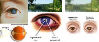
It has a negative effect on the blood vessels in the eyes. 80% of people with high blood pressure develop hypertensive retinopathy. Due to jumps in blood pressure, the capillaries in the retina of the eye swell, the distance between them becomes smaller, and visual abilities deteriorate. The organs of vision are subjected to heavy stress, as a result of which the walls of the vessels may not withstand and burst, causing hemorrhage in the eye. Due to the influence of high pressure and blood entering the retina, the optic nerve swells. Without treatment, it can atrophy, leading to glaucoma. Some people develop a mild form of this disease called ocular hypertension. Without proper treatment, it leads to clouding of the lens and blindness.
A consequence of hypertension can be a stroke.
Consequences may also be:
- stroke;
- thrombosis;
- loss of vision;
- retinal detachment.
Complications of the eyes due to hypertension
Arterial hypertension accompanies a group of diseases (Scheme 1), in which, as a result of at least three measurements at different times, systolic blood pressure (BP) is or exceeds 140 mmHg. Art., diastolic - 90 mm Hg. Art. It is more common between the ages of 40 and 69 years.
Scheme 1. Diseases of the cardiovascular system with damage to the organ of vision
Classification
. The European classification of hypertensive retinopathy distinguishes four stages of the disease:
Stage 1 - there are no changes in the fundus.
Stage 2 - narrowing of the arteries.
Stage 3 - the presence of symptoms characteristic of stage 2, in combination with retinal hemorrhages and/or exudate.
Stage 4 - the presence of symptoms characteristic of stage 3, combined with papilledema.
In the CIS countries they use the classification of M.L. Krasnova (1948), who distinguishes three stages of development of fundus changes in arterial hypertension, gradually transforming into one another (Scheme 2):
I. Hypertensive angiopathy - functional changes in retinal vessels.
II. Hypertensive angiosclerosis is organic changes in the vessels of the retina.
III. Hypertensive retino- and neuroretinopathy damages not only blood vessels, but also retinal tissue and the optic nerve.
Scheme 2. Stages of development of fundus changes in arterial hypertension
Clinical picture and diagnosis
. An examination by an ophthalmologist for arterial hypertension is mandatory and includes viziometry, measurement of intraocular pressure, perimetry and ophthalmoscopy (with fundus lenses), fluorescein angiography (FAT) and optical coherence tomography (OCT) of the retina, rheoophthalmography, Dopplerography of the vessels of the brain and spine.
At the stage of angiopathy (functional changes in retinal vessels), a decrease in central and peripheral vision is not observed; it is characterized by narrowing of the arteries, dilatation of the veins and tortuosity of the retinal vessels. In this regard, the normal ratio of the arteries and veins of the retina (2: 3) is disrupted, increasing to 1: 4. The Salus-Hun symptom of the 1st degree is characteristic (symptom of arteriovenous crossover, Fig. 1) - a slight narrowing of the vein under the pressure of the artery at the place of their intersection. In the central sections, around the macula, a corkscrew-shaped tortuosity of small venules appears (Gwist's symptom).
Rice. 1. The phenomenon of pathological decussation of retinal vessels: a - Salus-Hun I symptom; b — Salus-Hun II symptom; c — Salus-Hun III symptom
The stage of angiosclerosis (organic changes in the vessels of the retina) corresponds to stages IIA and IIB of hypertension; a decrease in central and peripheral vision is not typical (Scheme 3). When examining the fundus, narrowing, uneven caliber and the appearance of “side stripes” along the retinal arteries are observed. The vessels appear as if they are double-circuited due to thickening and decreased transparency of the vascular wall. The central reflex along the arterioles becomes wider and acquires a golden hue - a symptom of copper wire.
Scheme 3. Changes that occur during angiosclerosis
This picture is explained by lipoid infiltration of the vascular wall with protein deposits. With organic degeneration of the vessel wall (fibrosis, deposits of hyaline, amyloid, lime), the silver wire symptom occurs in the form of a bright white vascular reflex. The veins are dilated and tortuous. Characteristic symptoms are Salus-Hun II (a symptom of a venous arch; it consists of partial compression of the vein and an arc-shaped displacement to the side and into the thickness of the retina) and Salus-Hun III (visible “break” of the vein under the artery; Fig. 2).
Rice. 2. Hypertensive angiosclerosis - copper wire symptom, retinal hemorrhages, Gwist's symptom, Salus-Hun symptom II, III
The stage of retino- and neuroretinopathy (organic changes in the retina and optic nerve) is observed in stages IIIA and IIIB of hypertension. It is always a marker of severe complications of arterial hypertension, in particular kidney pathology. Visual acuity, as a rule, decreases with damage to the macular area (ischemia, hemorrhage, edema) and in the late stage of neuroretinopathy.
Perimetry in modern conditions (computer static perimetry) allows us to identify early functional changes in the visual analyzer: decreased light sensitivity, expansion of the blind spot, as well as the presence of scotomas at the stage of retinopathy and narrowing of visual fields. At this stage, obstruction of precapillary arterioles and capillaries with the appearance of ischemic zones and disruption of the blood-retinal barrier lead to the occurrence of areas of exudation, hemorrhages, edema of the retina and optic nerve head, and less commonly, newly formed vessels and microaneurysms.
Hemorrhages, depending on their location relative to the areas and layers of the retina, can be in the form of streaks, stripes, flames or spots. Preretinal hemorrhages can also be detected. Along the vascular arcades, as a result of ischemia and plasmorrhagia, gray-white foci resembling lumps of cotton wool - the so-called cotton wool exudates (see Fig. 2). “Hard” exudates appear as small foci with clear boundaries of white (ischemia + protein infiltration) or yellow (lipids + cholesterol) color.
They appear more often in the central sections and form a “star figure” in the area of the macula (Fig. 3). In hypertensive crises or malignant hypertension, the choroid may be involved in the pathological process: focal infarctions (Elshing's lesions) and fibrinoid vascular necrosis (Sigrist's lines).
Rice. 3. Stage of retinopathy - formation of edema and deposition of exudate in the vice figure of the star in the macular area: a - hemorrhage; b - “hard” exudates (indicated by an arrow)
An increase in the size of the optic disc, blurring of its boundaries and protrusion into the vitreous body, as well as the appearance of a waxy tint are characteristic of papilledema (neuroretinopathy).
During fluorescein angiography, local areas of choriocapillary occlusion can be seen, especially in malignant hypertension. It should be noted that the manifestations described above may be preceded by changes in the retina.
Differential diagnosis
The identified changes should be carried out with congestive optic disc, with retinopathy due to diabetes, collagenosis, blood diseases, and radiation damage.
Complications
from the eyes in hypertension are: spontaneous recurrent subconjunctival hemorrhages, thrombosis of the central retinal vein or its branches, acute obstruction of the central retinal artery or its branches, microaneurysms of the retinal arteries, anterior ischemic optic neuropathy, hemophthalmos, secondary vascular glaucoma.
Symptoms of the disease
Under the influence of high blood pressure, spots begin to float before the eyes, the image becomes unclear, and the field of vision decreases. Patients feel pain in the organs when in contact with light. Additional signs are rapid heartbeat, dizziness, temple spasms, increased sweating, and tinnitus. Some people complain of panic and nausea when their blood pressure rises. Symptoms of low blood pressure include:
Apathy is one of the possible manifestations of reduced blood pressure,
- headache;
- dizziness;
- apathy;
- weakness;
- insomnia;
- dyspnea.
Non-infectious epidemic
Elena Amanova, AiF Health: Is it true that of all the concomitant diseases, arterial hypertension with COVID‑19 is the most dangerous?
Alla Pogozheva : That's true. No wonder 72.6% of hospitalized patients are hypertensive. Despite the fact that COVID‑19 is a respiratory virus, the main target of the disease is blood vessels. Therefore, the presence of chronic obstructive pulmonary disease in a patient, for example, increases the risk of a fatal outcome by only 4%, cancer by 2%, and arterial hypertension by as much as 24%. In second and third places are diabetes mellitus and coronary heart disease (16 and 8%, respectively).
– At the same time, hypertension is a very common disease. Is not it?
– Over the past 40 years, the number of people suffering from arterial hypertension has doubled and reached 1.1 billion people. There are many reasons for this, as well as risk factors. The main ones are: genetics, physical inactivity, stress, smoking, alcohol abuse and, of course, poor nutrition. After all, hypertension, like a number of other nutrition-dependent diseases, including obesity, atherosclerosis, hyperlipidemia, diabetes mellitus, osteoporosis, gout and even cancer, can be associated with a deficiency or excess of certain nutrients.
Blood pressure pills are suspected of causing new side effects. What you need to know Read more
Treatment of the disease
People with hypertension and hypotension need to constantly maintain normal blood pressure, monitor blood pressure monitor readings daily and lead a healthy lifestyle. In addition to drug therapy aimed at restoring blood pressure, ophthalmological problems are treated. To restore the functioning of the optic nerve, medications are taken that improve blood circulation in the organs of vision. Thrombosis can be treated within a few months. To do this, doctors prescribe drops that help dilute the blood clot. They also give injections to strengthen blood vessels and relieve swelling.
The hemorrhage goes away in 2-3 days if you give your eyes a rest. Glaucoma and retinal detachment are more difficult to treat. The latter disease requires surgical intervention to restore the ability to see. Glaucoma is also treated with lens replacement surgery. It is impossible to completely stop clouding of the lens using medications. But you can stop the development of the disease with eye drops.
Stages of treatment and prevention
Therapy involves several types of treatment:
- stationary;
- outpatient clinic;
- sanatorium-resort.
After treatment at a day hospital, the patient is transferred to a local doctor, who monitors the further dynamics of the patient’s recovery. It is useful to undergo a course of therapy in a cardiac rehabilitation department, created on the basis of health resorts. Since hypertension cannot be treated, the patient must follow a certain daily routine and diet. At the end of the working day, a 2-hour rest is required. The duration of night sleep should be at least 9 hours, and daytime sleep - 2 hours. It is important to avoid stressful situations, conflicts at work, etc. All this leads to increased blood pressure. Following these rules will reduce the risk of vision impairment.
Source
Preventing ocular high pressure
What to do if you are at risk? First of all, adjust your diet, thus promoting IOP stability. It is necessary to minimize the consumption of salt, sugar, fast carbohydrates and include the following foods in the daily diet: dark chocolate, nuts, eggs, red vegetables and fruits. It is also necessary to maintain a sufficient amount of vitamins E, ascorbic acid and beta-carotene in the body.
In addition to nutrition, you also need to follow simple recommendations from doctors, adhering to a healthy lifestyle: spend more time in the fresh air, give up smoking and alcohol, do not eat foods containing high amounts of cholesterol, do not spend long periods of time looking at gadgets and computer screens, and do special exercises for eyes. The eyes are our window to the world; with their help, a person perceives up to 90% of the surrounding information, which is why it is important to maintain their health in order until old age. If you experience discomfort in your eyes, you should not hesitate and make an appointment with a doctor. We wish you good health and good vision!
Source
How can high blood pressure affect eye health?
- Retinal detachment
The entry of intraocular fluid between the layers of the retina can lead to its partial detachment. The first symptoms of this pathological condition are: the appearance of black dots, flashes of light and “floaters” before the eyes. The advanced stage of the disease is characterized by the appearance of a veil, partially narrowing the viewing angle. An advanced degree of detachment leads to retinal death and complete blindness.
- Retinal vascular thrombosis
Blood stagnation (thrombosis) in hypertension increases vascular permeability, which, in the absence of drug treatment, contributes to the occurrence of retinal edema. In this case, the use of drugs aimed at resolving blood clots, as well as drops that reduce intraocular pressure, is indicated.
- Retinal hemorrhages
An increase in blood pressure leads to rupture of blood vessels, as a result of which blood penetrates into the retina, thereby blocking some nerve impulses. In this case, symptoms such as blurred or double objects, as well as lack of clarity of vision, are observed. If the affected area is large, visibility may become completely blurred.
- Angiopathy
Failure of blood microcirculation caused by a persistent increase in blood pressure can lead to disruption of the blood supply to the optic nerve. This pathology is asymptomatic and can only be detected by examining the fundus of the eye. Angiopathy is dangerous because it can lead to partial or complete loss of vision.
- Ocular hypertension and glaucoma
Hypertension can cause increased intraocular pressure, which, if not treated in a timely manner, provokes the development of glaucoma. This serious disease manifests itself as pain in the eye area, a sharp deterioration in vision, the appearance of a light spot and other unfavorable symptoms.