With ametropia, refractive error occurs in the eye. As a result of the pathology, the focus of the image is formed not in front of the retina, but behind it, which is why a person sees all objects in the form of a blurry spot. The most common types of ametropia are myopia and farsightedness. A person with this disease sees images blurred and loses the ability to read small print or look into the distance. The cause of ametropia may be a genetic predisposition, poor lifestyle, constant eye strain, or poor quality correction.
Depending on the extent and type of disease, treatment may be medication or surgery. In this article we will look at what ametropia is, its main types and degrees, causes of occurrence, diagnosis and treatment methods.
Ametropia
Ametropia Source: present5.com The human eye is designed in such a way that light rays, passing through the lens, cornea and vitreous body, are refracted and connected on the surface of the retina.
And with the help of the visual pathways, we see a clear image of the world around us. However, there are many different pathologies of the visual organs, including malignant neoplasms.
Among all diseases, ametropia is the most common. This concept means a violation of the refraction (refractive power) of the eye. In simple terms, in an ametropic eye, the image is focused either in front of or behind the retina, which is why instead of a clear object, a blurred spot is visible.
Thus, the main types of ametropia are myopia (myopia) and farsightedness (hypermetropia). In a myopic eye, reflected rays from a distant object converge in front of the retina and then diverge.
Thus, an object located far away is not visible, but an object located nearby at a certain distance is visible. Quite the opposite happens in the farsighted eye. This limiting distance at which good visibility is maintained is called the furthest point of the eye.
The inverse distance from this point to the surface of the organ (in meters) is the value of ametropia - dioptre.
Refraction of the eye is the process of refraction of light rays in the optical system of the eye. The optical system of the eye is quite complex; it consists of several parts: the cornea, the aqueous humor of the anterior chamber, the lens and the vitreous body.
Light, passing through all components of the optical system of the eye, hits the retina - the inner layer of the eye, the cells of which convert light particles into nerve impulses, thanks to which an image is formed in the human brain. The refraction of the eye is measured in diopters - these are units of measurement of the refractive power of the lens.
Ametropia is a refractive error of the eyeball, which is characterized by failures in focusing light rays on the retina. Normally, light rays are focused on the retina, but when the focus is lost, this process occurs behind the retina or in front of it. As a result, a person sees the world around him with distortions, blurry and unclear.
This ophthalmological pathology occurs very often in humans, regardless of gender and age. There are several varieties of this disease, which is diagnosed during examination by an ophthalmologist.
Important
Depending on the degree and type of the disease, therapeutic measures will be carried out, lenses and drops will be prescribed. Methods are selected individually; the use of surgical correction is possible.
The main symptom is blurred vision. Specific complaints depend on what particular form of ametropia began to develop. A person loses the ability to see clearly into the distance, has difficulty reading small text, and sees surrounding objects slightly blurred.
This is what makes him see a doctor for an accurate diagnosis. The treatment prognosis is favorable - it is possible to normalize existing visual disturbances.
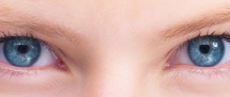
Emmetropic eye
Gullstrand, in his diagram of the optics of the eye, assigned to each of its parameters the average of the measured or otherwise found values of this parameter for real human eyes.
The eye parameters of each person may differ greatly from those indicated in the diagram. For example, the length of the eye can be more or less than 24 mm. However, such a difference does not necessarily lead to worsening vision.
A longer eye may have less optical power, while a shorter eye may have more power. As a result, a clear image of distant objects can be obtained on the retina in all cases and ensure their good visibility.
In these cases, changes in parameters compensate each other, the eye remains proportionate or, to use the accepted term, emmetropic.
PATIENT EXAMINATION PLAN FOR MYOPIA
- History taking
Taking an anamnesis precedes the examination of the patient. The specific direction of the examination and its detail depend on how completely and efficiently this stage was completed. The accumulated information allows the doctor to get an idea of the patient’s subjective feelings, the tolerability of the existing correction, as well as the need to prescribe correction agents.
- Exploratory survey
- Determining the position of the dominant eye
- Measuring monocular interpupillary distance for distance
Etiology
Source: augenlaserteam.ru Ametropia in children can be congenital or acquired; it occurs very often in children 3–5 years old.
Congenital pathologies are associated with unfavorable conditions for the correct formation of the visual apparatus in the embryo. Such conditions are considered to be: previous infectious diseases of the pregnant woman, lack of nutrients, stress. The disease is discovered in childhood after visiting an ophthalmologist. Acquired pathology can relate to different age groups.
The following causes of the disease are identified:
- all kinds of eye injuries;
- hereditary predisposition;
- inflammatory processes;
- eye overload;
- improper eye care;
- poor nutrition;
- lack of light.
It is very important to detect pathology in time and take measures to eliminate it, which will preserve vision.
When considering the pathogenesis of the disease, primary and secondary induced ametropia can be distinguished. The first case involves the formation of an optical defect associated with the long anteroposterior axis and refraction of the cornea. The second case of pathology formation is associated with pathological changes in the axis or cornea.
Secondary pathology develops due to changes in the refraction of rays or changes in the anteroposterior axis. The refraction of the cornea changes due to disturbances in its functioning due to inflammation, deformation after injury or dystrophy.
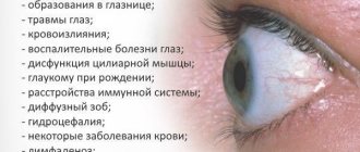
Causes and risk factors
The disease can be either congenital or acquired.
The causes of congenital ametropia are presumably unfavorable factors affecting the fetus during intrauterine development. These include:
- viral diseases of a pregnant woman (flu, chicken pox);
- ionizing radiation;
- smoking, drinking alcohol or using drugs during pregnancy;
- bad ecology.
The main manifestations of ametropia are decreased visual acuity, deterioration in its quality and clarity. The causes of acquired ametropia can be traumatic damage to the structures of the eye, inflammatory processes.
But most often acquired ametropia develops due to age-related changes in eye tissue or as a result of prolonged and frequent visual strain.
Refractive error, in which focusing on an object is impaired, is primarily due to the congenital weakness of the refractive function of the eye. This type is called axial ametropia.
Another group of reasons relates to anatomical factors. This group includes an increase or decrease in the size of the eyeball, although the refractive function remains normal. This type is called refractive ametropia.
Kinds
Source: glazalik.ru Experts distinguish 4 forms of ametropia:
- Myopia or nearsightedness. Light rays are fixed in front of the retina. A person has difficulty seeing objects located in the distance. Myopia is most often diagnosed in school-age children. This is due to intense visual stress.
- Hypermetropia or farsightedness. The rays are fixed behind the retina, which makes it difficult to perceive objects that are close, while a person sees well into the distance.
- Astigmatism is a visual defect in which the rays lose the ability to converge at one point. As a result, all surrounding objects appear blurry or deformed.
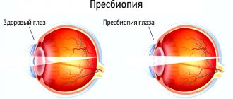
- Presbyopia or age-related farsightedness. This type is mainly diagnosed in older patients. Presbyopia is associated with a decrease in the elasticity of the lens, when it cannot fully perform its functions.
For all of the listed forms of ametropia, weak, medium, and strong stages of the disease are distinguished - depending on how many diopters it is necessary to reduce or increase the refractive power of the eyes in order to correct vision.
Farsightedness and myopia are divided into several degrees of ametropia, depending on the number of diopters: weak - no higher than 3; average – no higher than 6; strong – above 6.
Astigmatism is measured by slightly different indicators: weak – up to 2; medium – up to 4; strong – more than 4. There is complicated and uncomplicated ametropia of the eye.
In the second case, the disease manifests itself in the form of a decrease in uncorrected perceptual acuity, but the ability to correct it remains.
If a pathological condition develops, then the disease becomes complicated - the visual analyzer changes. Such pathological processes occur when strabismus and asthenopia are diagnosed - the retina and optic nerve may change.
There are stationary and progressive ametropia, the latter includes myopia, which can be aggravated due to stretching of the scleral membrane and an increase in the length of the anterior-posterior axis.
Astigmatism
This is another type of ametropia. It can be caused by the refraction of the eye, which makes it difficult for the image to focus on the retina. Similar to myopia and farsightedness, three degrees of the disease are distinguished, only the division occurs along +4D and +2D.
Astigmatism is not cured, but corrected. But for this, its timely detection is important. Otherwise, visual acuity may sharply decrease, and even strabismus may occur. In children, astigmatism can be observed even in infants up to one year old. There are known cases of congenital forms.
Reference
It should be noted that a similar defect causing astigmatism is observed in all people, but it is within the normal range - 0.5D.
With this disease, the eyes become red and watery. A headache may occur. In children, this problem can be identified by observing their behavior. With astigmatism, the child will squint to see objects.
Contact lenses and glasses are used for treatment. Laser correction can be used.
Forms and degrees
There are the following forms of the disease: Mixed - the values of the optical axis and refractive power are outside the normal range. Combined - the indicators are normal, but their combination has a negative effect on refraction. Refractive – normally only the length of the optical axis.
Axial - on the contrary, only the magnitude of the refractive force is normal.
There are three degrees:
- more than -6D – strong;
- up to -6D – average;
- up to -3D – weak.
As a rule, myopia results in an enlarged eyeball.
Myopia
Myopia is the most common form of pathology, which usually begins to develop in childhood. There are two main reasons for the development of this type of ametropia.
- Axial myopia. It develops when the eyeball is enlarged along the anteroposterior optical axis, as a result of which light rays do not reach the retina when focusing.
- Refractive myopia. Caused by irregular curvature of the cornea or lens. When refracted, the rays are scattered in the wrong way, also not reaching the central part of the retina. As a result, the image turns out fuzzy and blurry.
Due to the listed physiological reasons, a person has difficulty distinguishing between objects located at a distant distance, but clearly sees close objects. As a rule, acquired myopia develops due to non-compliance with the rules of visual hygiene. Schoolchildren spend the vast majority of their time with gadgets, and they also have high visual stress at school. In addition, a prerequisite for the development of this type of eye ametropia, especially with a hereditary factor, can be poor nutrition, ophthalmological and traumatic brain injuries and other factors.
In medicine, there are three degrees of myopia:
- weak from 0.5 to 3 diopters;
- average from 3 to 6 diopters;
- strong from 6 diopters or more.
A high degree of myopia can reach very significant values - up to 30 diopters. In this case, surgery is usually recommended, and if there are contraindications, soft or hard lenses (including orthokeratological ones) can be prescribed. It is difficult to wear glasses with a strong negative optical power of the eyes: thick glasses magnify them and distort the outlines of objects, severely limit peripheral vision, and besides, the eyes in such glasses quickly get tired, and the head begins to hurt. With significant myopic ametropia, degenerative changes in the retina, its detachment, and other disorders in the eye structures can also be observed.
Causes of ametropia
The reasons may be the following factors:
- Poor nutrition. The body may simply lack some elements that play an important role in the development of scleral tissue.
- Genetic predisposition. If parents have myopia, children suffer from this disease in 50 percent of cases.
- Statistics say that only 8 percent of children with healthy parents have this diagnosis.
- Eye strain. Constantly working in front of a monitor or watching TV for too long, poor lighting - all this puts a lot of strain on the visual organs.
- Poor quality correction. If there was no timely treatment or errors occurred during its implementation, then significant deviations may be observed.
- Weakness of the muscles of the organs of vision. This is mainly an innate feature.
- Increasingly, myopia is observed in children, even at a younger age. Moreover, this disease develops much faster in them than in adults.
Development of myopia
It is known that by the end of school, myopia develops in 20-30% of schoolchildren, and in 5% it progresses and can lead to low vision and blindness. The rate of progression can range from 0.5D to 1.5D per year. The greatest risk of developing myopia is between the ages of 8 and 20 years.
There are many hypotheses about the origin of myopia, which connect its development with the general condition of the body, climatic conditions, racial characteristics of the structure of the eyes, etc. In Russia, the most widespread concept of the pathogenesis of myopia, proposed by E.S. Avetisov.
The root cause of the development of myopia is recognized to be weakness of the ciliary muscle, most often congenital, which cannot perform its function (accommodate) for a long time at close range. In response to this, the eye lengthens along the anteroposterior axis during its growth. The reason for the weakening of accommodation is insufficient blood supply to the ciliary muscle. A decrease in muscle performance as a result of elongation of the eye leads to an even greater deterioration in hemodynamics. Thus, the process develops like a “vicious circle”.
The combination of weak accommodation with a weakened sclera (most often this is observed in patients with myopia, which is inherited, an autosomal recessive type of inheritance) leads to the development of progressive high myopia. Progressive myopia can be considered a multifactorial disease, and at different periods of life, one or another deviations in the state of both the body as a whole and the eye in particular are important. Great importance is attached to the factor of relatively increased intraocular pressure, which in myopes in 70% of cases is above 16.5 mm Hg. Art., as well as the tendency of the myopic sclera to develop residual microdeformations, which leads to an increase in the volume and length of the eye with high myopia.
Symptoms
Source: Glaza.guru Symptoms of visual impairment include the following:
- eyes get tired quickly;
- double vision occurs;
- the child looks at objects from a close distance, but does not pay attention to distant ones, or vice versa;
- blurring the contours of an object;
- headaches due to eyestrain;
- nausea, motion sickness in transport.
All of the above symptoms require immediate response and appropriate measures to improve and normalize vision.
Symptoms of myopic ametropia in children
A child, especially a preschooler, is not always able to understand what is happening to his vision. Parents should be attentive to his behavior. Here are some signs that may indicate the development of childhood myopia.
- Frequent complaints of headache. This symptom is attributed to fatigue, and at this time myopia progresses.
- Irritation and eye fatigue. With constant tension in an attempt to see distant objects, a spasm of accommodation occurs (false myopia). Pain when blinking, dryness and irritation of the mucous membrane may occur.
- Increased tear production. With myopia, the eyes are more sensitive due to the dilated pupil. More light enters it, and lacrimation is the body’s protective reaction to bright light.
The listed signs should be an alarming signal for parents. If ametropia is not corrected in a timely manner, negative consequences may occur, because other eye structures inevitably suffer.
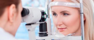
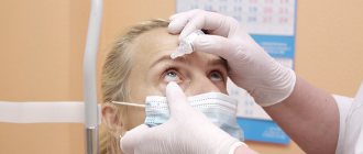
Diagnostics
Source: mednow.ru Ametropia is diagnosed using special ophthalmological examinations. These include:
- Visometry - determination of visual acuity using a letter table.
- Automatic refractometry – determination of the form of ametropia using a refractometer.
- Ophthalmometry is the study of the curvature of the cornea and its refractive power.
- Ophthalmoscopy is a study of the condition of the fundus of the eye.
- pachymetry - ultrasound examination of the thickness of the cornea of the eye;
- eye biomicroscopy - a diagnostic method using a slit lamp - a special ophthalmological microscope combined with a lighting device;
- skiascopy is a method for determining the refraction of the eye, based on observation of the movement of shadows in the area of the pupil when the eye is illuminated by light reflected from the mirror;
- vision test on a phoropter - determination of refraction using a special device - a phoropter;
- computer keratotopography – a method for studying the condition of the cornea using laser beams;
- ophthalmoscopy - examination of the fundus of the eye using a special mirror - an ophthalmoscope. Simple to implement, but very informative research. Allows you to assess the condition of the retina, optic nerve head, and fundus vessels;
- selection of suitable glasses (lenses).
All procedures take little time and are performed on an outpatient basis.
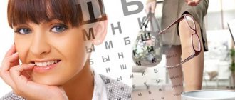
Visometry is a method of determining visual acuity using special tables. In Russia, Sivtsev-Golovin tables are most often used; letters of different sizes are written on these tables, larger ones at the top, small ones at the bottom.
With 100% vision, a person sees the 10th line from a distance of 5 meters. There are similar tables, where instead of letters there are rings, with breaks on a certain side. The person must indicate which side the gap is on (top, bottom, right, left).
Automatic refractometry is a study of the refraction of the eye (determining the point of the ideal image relative to the retina) using a special medical device - an automatic refractometer.
Cycloplegia is a drug-induced shutdown of the accommodative muscle of the eye in order to detect false myopia. A person with normal vision will experience “physiological” myopia caused by spasm of the ciliary muscle.
Ophthalmometry - measurement of the radii of curvature and refractive power of the cornea (the transparent membrane of the eye).
UZB - Ultrasound biometry (USB), or A-scan - ultrasound examination of the anterior segment of the eye. The technique presents the obtained data in the form of a one-dimensional image, which allows one to estimate the distance to the boundary of media (organism structures) with different acoustic (sound) resistance.
Allows you to assess the condition of the anterior chamber of the eye, cornea, lens, and determine the length of the anterior-posterior axis of the eyeballs.
Objective methods
Objective methods by which ametropia is detected include:
- skiascopy or the so-called shadow test. Using skiascopy, the degree of myopia is determined;
- refractometry. An effective method for determining clinical refraction by examining the reflected mark from the surface of the fundus. There is a mechanical method of determination and an automated one;
- ophthalmometry. It is used to determine the refractive power of the cornea.
Treatment methods for ametropia
Source: present5.com The most common way to improve vision is to wear glasses with specially selected lenses or use contact lenses.
However, this means wearing them frequently or constantly, which is not always convenient. Therefore, other methods have been developed. These are ophthalmic surgeries that often use a laser. Subsequently, wearing glasses is no longer required.
Treatment involves correction with contact lenses and glasses. At the same time, there is no talk of eliminating the problem. These devices only improve the standard of living, but do not eliminate myopia.
One of the methods can also be used:
- keratoplasty;
- lens replacement;
- introduction of lenses;
- radial keratotomy;
- correction using laser.
Indications for correction of ametropia:
- the presence of ametropia (impaired refraction of the eye or accommodation (the ability of the eye to change the refractive power of its optical system for a clear and distinct perception of objects located at different distances from it)) - decreased visual acuity near and/or far;
- deterioration in quality of life due to decreased visual acuity;
- inability to perform certain professional or educational activities due to decreased visual acuity; the lack of timely and correct correction of ametropia leads to its progression, and, therefore, to a further decrease in visual acuity.
Measures to prevent vision deterioration can be carried out only when an accurate diagnosis has been made and the type of disease has been established.
It often happens that astigmatism and myopia are diagnosed in one person, which can cause certain difficulties in the correct selection of treatment methods.
Correction of ametropia can be carried out using the following methods:
- with the help of lenses - they do not limit the visual space, do not put pressure on the bridge of the nose, are prescribed only to adults, so as not to injure the eye or cause infection;
- with the help of glasses, when ametropia of a high degree of change is observed, glasses with collective lenses are used, and in case of myopia with divergent lenses, glasses are worn constantly if the deviations in vision are higher than 3D (for astigmatism, spherical and cylindrical lenses are used);
- using a laser, restoration is prescribed only when the cause that provoked the deviation has been eliminated, however, this type of operation is not performed until the age of 18, and if the pathology worsens, laser therapy is not prescribed;
- by surgery - surgical correction is performed using photoreactive keratectomy or laser keratomileusis. Restoring or improving vision using surgical methods is a lengthy procedure.
Other methods of surgical vision correction
Further methods of refractive surgery are special methods of cutting the cornea, corneal implantation and lens surgery with implantation of artificial intraocular lenses.
- Special methods for cutting the cornea
If the patient's corneal curvature is weakly expressed, in certain cases it can be corrected using special loosening incisions. They can also be performed during cataract surgery to optimize the resulting refraction.
- Corneal implants
If you have a corneal disease that causes the cornea to bulge, under certain conditions it may be helpful to stabilize the cornea with tiny spacers. Thanks to this, the deterioration in visual acuity due to corneal deformation will be at least partially compensated.
- Lens surgery with implantation of artificial intraocular lenses
Not all degrees of nearsightedness or farsightedness can be treated with laser. If, for example, too much corneal tissue has been removed by a laser, under certain circumstances there may be too little tissue left in the cornea, rendering the eye unstable.
Therefore, for people with severe myopia or farsightedness, it may be advisable to eliminate ametropia by implanting an artificial lens. In this case, either an additional artificial lens will be placed in the eye, or the patient’s own lens will be replaced with an artificial one.
- Lens surgery: implantation with a high degree of ametropia
If an additional artificial lens is placed in the eye, in addition to its own, it is called a “phakic intraocular lens.” It is used primarily for very severe myopia, possibly in combination with severe astigmatism.
In case of farsightedness, on the contrary, it is used less often. The additional lens can be placed in the anterior chamber of the eye (between the cornea and the iris) or attached to the iris itself.
Correction with glasses or contact lenses
Source: bigpicture.ru
- Glasses.
This is the most popular optical device for vision correction. Special bifocal lenses help the light rays to focus at the desired point.
- Contact lenses.
Their operating principle remains the same as that of glasses. The lens is placed directly on the eyeball.
The selection of glasses and contact lenses should be carried out by a qualified specialist after diagnosis. Lenses must be properly cared for - kept in a special solution when not in use.
Before the operation, you need to visit a therapist, take urine and blood tests, and during the rehabilitation period you must strictly follow medical recommendations.
Spectacle correction - glasses are a special medical optical device consisting of lenses and frames. Depending on the type of refractive error, different types of lenses are chosen; for ametropia of varying degrees, the optical power of the lens also changes.
With hyperopia or farsightedness (a form of refractive error in which rays of light, passing through the optical system of the eye, focus (converge) not on the retina (the inner layer of the eye, the cells of which convert light rays into nerve impulses.
Thanks to which an image of objects is formed in the brain), and behind it - a person sees fuzzy, blurry near, and with a high degree of hypermetropia both near and far) they use collective (positive) lenses, passing through which the light is focused on the retina.
For myopia or myopia (a form of refractive error in which light rays are focused in front of the retina; a nearsighted person typically sees well near and blurry at a distance), diverging (negative) lenses are used to help focus light correctly on the retina.
For mild myopia (up to -3 diopters (a unit of measurement for the otic power of a lens)), glasses are prescribed only for distance, i.e. a person puts them on when he needs to look far enough away from himself.
At higher degrees of myopia, glasses are prescribed for constant wear, while complete correction of myopia is not performed due to the possibility of overcorrection and deterioration of visual acuity.
In some cases, for myopia, two types of glasses are prescribed - for near and for distance, with different optical powers of the lenses.
The selection of spectacle correction for astigmatism (a form of refraction in which there is a combination in the eye of varying degrees of the same refraction (myopic or hyperopic) or different types of it (mixed astigmatism)) is quite complicated - cylindrical and spherical lenses can be used.
Without proper correction, visual function with astigmatism is significantly reduced. Anisometropia (the presence of different types of refraction or different degrees of one type of refraction in the same person) is corrected with glasses if the difference between the optical powers of the eyes does not exceed 2 diopters.
If the difference is greater, it is recommended to wear contact lenses or special glasses with two lenses. Today, spectacle correction is the most common and accessible type of correction due to its relative cheapness and ease of use.
But there are situations when only spectacle correction is recommended or, conversely, they are advised to choose another type of correction.
Indications for spectacle vision correction:
- high degree of myopia;
- high degree of hypermetropia;
- astigmatism from -6 to +6 diopters;
- presbyopia - a decrease in near visual acuity that occurs after 40-45 years, is considered a natural sign of aging;
- childhood;
- intolerance to contact lenses;
- impossibility of surgical (laser) correction of ametropia.
Contraindications to spectacle correction:
- risk of possible eye injury (sports, outdoor games);
- professions that require a fairly large field of view, such as pilots or firefighters;
- anisometropia (with a difference of more than 2 diopters between the eyes);
- individual intolerance to glasses.
Contact (lens) vision correction is a change in refraction using contact lenses. They are called contact because they are in direct contact with the cornea - the transparent membrane of the eye.
A contact lens is a small, cup-shaped lens that is inserted behind the lower eyelid and rests against the cornea.
Currently, contact correction has become very widespread, because it freely allows you to engage in active sports, does not limit the field of view, and does not put pressure on the bridge of the nose and ears, unlike glasses.
There is an opinion that lens correction is only suitable for myopia, but this is not true. Any refractive error can be corrected with contact lenses. It’s just that for some disorders, for example, presbyopia, their use is inappropriate, because patients suffering from presbyopia wear glasses only for near work or for reading.
Currently, lenses are made from high-quality material (silicone hydrogel), which freely passes oxygen to the cornea of the eye.
The patient can choose lenses that are suitable specifically for him: hard or soft (mostly soft ones are used), and lenses are also divided depending on the period of wear. One-day lenses are widely used, which a person takes off in the evening, throws away, and puts on other lenses the next day.
A person chooses a convenient mode for wearing lenses: daytime wear (the lenses are removed at the end of the day), flexible wear (if necessary, you can leave the lenses on for 1-2 nights), extended wear (the lenses are not removed for several days in a row), continuous wear (the lenses are used within 30 days).
Contraindications to wearing contact lenses:
- high-grade refractive errors;
- chronic conjunctivitis (recurrent inflammation of the conjunctiva - the mucous membrane of the eye);
- chronic blepharitis (recurrent inflammation of the eyelid margins);
- demodicosis of the eyelids (presence of mite parasites).
Correction with glasses consists of the use of the following types of lenses:
- spherical lenses mainly have a positive effect in the treatment of presbyopia;
- cylindrical lenses are prescribed for regular use in cases where the correct form of astigmatism is detected;
- prismatic elements correct heterophoria, the effect of double vision in pathologies of the muscular system and strabismus;
Degrees of astigmatism
There is a classification of this eye pathology depending on its severity:
- weak to 3D. Subject to contact or spectacle correction, and also corrected using microsurgery;
- average from 3 to 6D. With such values, it is best to correct vision with toric lenses or undergo surgery;
- strong - over 6 diopters. This high degree of astigmatism occurs as a result of significant changes in the surface of the cornea. It is recommended to correct this form of pathology with rigid gas-permeable lenses or laser correction. In this case, it is very difficult to use glasses, the eyes get tired, and peripheral vision is narrowed.
Many people do not even suspect that they are developing astigmatism, since its symptoms resemble eye fatigue from visual strain. Meanwhile, here are some manifestations that may indicate it:
- impairment of near vision clarity;
- asthenopia - rapid eye fatigue;
- difficulties with orientation in space;
- frequent headache;
- dry mucous membrane.
If the listed symptoms recur with a certain frequency, then this is a reason to visit an ophthalmologist to find out the cause of the discomfort. Such phenomena may well indicate developing astigmatism, in which case it will be necessary to take measures to correct it. In most cases, types of ametropia can be corrected. There are different ways to treat this form of pathology.
Surgical interventions
If the desired effect from the selection of auxiliary optical systems is not achieved, surgical interventions are resorted to:
- anterior view of radial keratotomy;
- performed with a special ophthalmic microtome, sections of the corneal surface are cut one after another - this operation is called myopic keratomileusis;
- excimer laser surgery. The stromal part of the cornea is replaced into the existing section;
- thermokeratocoagulation;
- The contribution of domestic surgeons and scientists is especially worthwhile for the development of a highly effective operation - photorefractive keratectomy;
Surgical vision correction using laser technologies. Laser beams affect the cornea. The operation is highly effective and can be performed even on children; the recovery period takes about 4 weeks.
Laser correction
Laser vision correction is the correction of eye refraction by changing the thickness of the cornea, carried out using an excimer laser. By changing the thickness of the cornea, its optical power is changed, due to which the light is focused on the retina - a person sees surrounding objects clearly and clearly.
Laser vision correction is an advanced trend in modern ophthalmology.
There are 2 types of laser vision correction:
- photoreactive keratectomy (PRK)
The laser removes the surface layers of the cornea, changing its thickness. With the help of PRK, you can correct myopia (up to -6 diopters), hypermetropia (up to +3 diopters), astigmatism (up to -3 diopters).
After PRK, the recovery period is quite long - up to several months it is necessary to instill special drops into the eyes. The advantages of PRK are the complete painlessness of the procedure, short exposure time, stability of results;
- LASIK (laser keratomileusis, lasik)
It is a combination of microsurgical excimer-laser stages. During the operation, under the guidance of an ophthalmologist, a corneal flap is folded back using an automated special medical instrument - a microkeratome.
Then the thickness of the cornea, pre-calculated using computer technology, is removed with a laser. The bent flap is returned to its place. The operation takes 1 - 1.5 minutes. Within a few hours you can return to your normal lifestyle. With LASIK it is possible to correct higher degrees of ametropia.
Indications for laser vision correction:
- different visual acuity in the left and right eyes;
- professions that require quick response (for example, pilots or firefighters);
- the desire of the patient himself.
Contraindications to laser vision correction:
- age less than 18 years;
- progressive myopia;
- pregnancy and lactation (breastfeeding);
- general severe diseases of the body (for example, diabetes mellitus - a disorder of glucose (sugar) metabolism);
- acute infectious diseases;
- a number of eye diseases: inflammatory in nature, cataracts (clouding of the lens), glaucoma (increased intraocular pressure), history of retinal detachment.
In the presence of severe presbyopia and lens opacities:
- replacing a cloudy lens with an artificial one through surgery - currently preference is given to soft intraocular lenses, which require a minimal incision in the eye, are placed inside in a folded state, and straighten themselves inside the eye;
- correction of amblyopia (decrease in visual acuity, not amenable to optical correction, occurring in the absence of organic disorders, usually in one eye; amblyopia is common among children and adolescents);
- occlusion of a healthy eye - gluing or establishing a special occlusion (shutter) of a healthier eye for 2 - 6 hours a day in order to train the weaker eye.
Prevention
Prevention of ametropia (myopia and farsightedness) helps prevent the development of visual impairment. To prevent the occurrence of visual impairment, the following methods are used:
- Routine examination by an ophthalmologist.
- An examination by a specialist allows you to monitor the child’s vision condition and also prevents the development of dangerous complications.
- Do daily eye exercises. Using special exercises to strengthen the eye muscles allows you to increase blood circulation in the tissue and relieve fatigue while working at the computer or reading a book.
- Correct distribution of load on visual analyzers.
- Use of vitamins and minerals. Deficiency of vitamins A and C is one of the reasons for decreased visual acuity.
- Reducing the time spent in front of the computer. Using additional means to ensure optimal room illumination.
- Daily walks in the fresh air. Use manuals with large, clear print.