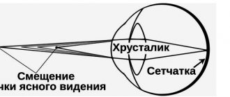
In order for a person to see images clearly, rays of light enter the eye, where the lens changes shape and focuses the rays onto the retina.
This allows you to focus your gaze on objects at different distances.
Presbyopia develops when the lens of the eye gradually loses its ability to change shape under the influence of the ciliary muscle within the ciliary body surrounding the biconvex lens. As you age, it becomes less elastic, making it difficult to focus on close objects.
Presbyopia (age-related farsightedness) is a refractive error. It is considered a natural condition that comes with age. Every person over 40 years old has it to a greater or lesser extent.
Causes
Presbyopia is an age-related change in the visual analyzer. As a person ages, muscles cease to be as elastic as before. This condition is called sarcopenia, and according to scientific research, it begins to develop from the age of 40. This condition is especially noticeable in the arms and legs, requiring more effort to do daily housework and move around.
A similar thing happens with the muscles of the optical system. They lose their former elasticity and firmness. Muscles that are unable to function properly lead to various refractive errors.
At the same time, the lens becomes stiffer and loses its ability to change its shape and curvature. This results in the inability to focus on objects at a certain distance.
Signs of presbyopia are more common among older patients and less common among younger people.
The exact causes of the disease are unknown. Current research confirms that the reason lies in the aging of the body. The following conditions contribute to the development of age-related farsightedness:
- insufficient intake of vitamins;
- external factors (overvoltage, work in insufficiently environmentally friendly areas);
- changes in the normal anatomy of the eyeball.
It is almost impossible to avoid the development of presbyopia, but it will be possible to slow down the progression.
Difference between presbyopia and farsightedness
Age-related farsightedness and hyperopia are identical diseases of the optical system that develop for different reasons. Don't confuse them.
Hypermetropia means that the eyeball is too short. Light passing through it is not focused on the back of the retina. Distant objects become blurry.
Hypermetropia is the cause of an eyeball shortened in the anteroposterior direction and a reduced refractive power of the optical media of the eye. Presbyopia is an age-related change in vision and is a type of farsightedness.
Causes of presbyopia
The causes of presbyopia are the natural aging processes of the lens, leading to weakening of accommodation.
IMPORTANT!
Accommodation is the unique ability of the organ of vision to instantly adapt to the vision of objects located at different distances in relation to each other.
The main pathogenic link in the aging of the lens is phacosclerosis, which is characterized by dehydration of the lens and loss of plasticity. Dystrophic changes occur especially actively in the ciliary muscle, the main function of which is to hold the lens.
Due to developing dystrophy, new muscle fibers of the ciliary muscle cease to form and are replaced by connective tissue. As a result, the contractility of the muscle is significantly reduced.
The described process is related to metabolic disorders through the retinal vessels. The disease develops especially quickly in people suffering from atherosclerosis, hypertension, diabetes mellitus and some other pathologies. A person’s professional activity can also lead to the development of presbyopia if it is associated with increased visual load at close range. The risk group primarily includes people who work at computers, jewelers, laboratory assistants, engravers, and writers.
Symptoms
The near point of accommodation gradually moves outward from 20 cm at the age of 40 years, to 30 cm at the age of 45 years and 40 cm at the age of 50 years.
Presbyopia, like other focus defects, becomes less noticeable in bright sunlight. This is not the result of any mysterious "healing effect", but simply a consequence of closing the iris to the pinhole, so that the depth of focus, regardless of the actual focusing ability, is greatly increased, as in a pinhole camera which produces images without a lens.
Presbyopia develops in both eyes (ou). Causes the following symptoms:
- words are blurry when reading;
- to see clear letters, you need to remove the book some distance from your eyes;
- headaches and dizziness occur due to muscle tension;
- double vision;
- drowsiness and rapid fatigue.
In some patients, cataracts and glaucoma occur together with age-related farsightedness. Itching, burning, increased sensitivity to light, redness of the conjunctiva, and lacrimation are added.
Presbyopia is characterized by pain in the eyes and in the area of the eyebrow, extending to the bridge of the nose. They are sharp or pressing in nature.
Signs of presbyopia
The first signs of presbyopia appear during prolonged work (reading small print, knitting, etc.) at close range or in poor lighting. The patient feels rapid visual fatigue (asthenopia) and tension, headaches, dull pain in the eyeballs, bridge of the nose and brow ridges, photophobia. With the development of age-related farsightedness, a person sees objects that are nearby as fuzzy and blurry. Pathological changes in the visual apparatus progress until 65–70 years of age.
Article on the topic: Albendazole - instructions for use for children and adults, composition, release form and price
In patients with myopia (myopia), presbyopia occurs almost unnoticed, due to the fact that age-related changes in accommodation are compensated for a long time, and the symptoms of age-related farsightedness develop much later (by 50–60 years). People with myopia of more than 3-5 diopters often do not need vision correction.
Diagnostics
Many people over 40 self-diagnose presbyopia based on their inability to see letters clearly at distances that used to be natural and comfortable.
Because the condition develops gradually over many years, most people do not notice small changes in vision and do not seek professional help until problems with focusing cause problems in daily life.
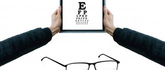
For diagnosis, contact an ophthalmologist. The doctor checks vision using the Sivtsev table. If presbyopia is diagnosed, appropriate studies are performed. These include:
- visometry;
- automatic refractometry;
- ultrasound;
- computer keratotopography;
- biomicroscopy.
Presbyopia treatment
Treatment includes the selection of glasses for near vision (approximate table - an emmetrope at 40 years old needs to have a collective lens of 1.0 D for near vision, at 50 years old - 2.0 D, at 50 years old - 3.0 D).
Essential drugs
There are contraindications. Specialist consultation is required.
- Quinax (has an activating effect on proteolytic enzymes contained in the aqueous humor of the anterior chamber of the eye). Dosage regimen: the drug is instilled 1 drop into the conjunctival sac of each eye 3 times a day.
- Taufon (stimulates reparative processes in eye tissues). Dosage regimen: the drug is instilled 1 drop into the conjunctival sac of each eye 3 times a day.
Treatment
Drug therapy
Drug treatment helps improve the stimulation of metabolic processes, restore damaged tissue and normalize the functioning of eye cells. Prescribed drops activate the capabilities of the lens to fragment protein structures.
The best drugs:
- Taufon;
- Quinax;
- Vita-iodurol;
- Oftan katachrome.
Glasses or lenses
Age-related farsightedness is treated with corrective lenses. Glasses are the easiest way to restore your ability to see objects close up clearly.
If the patient already wears corrective lenses for nearsightedness or farsightedness, two sets of glasses may be needed—one for hyperopia and one for myopia. Glasses with bifocal lenses are used, in which the upper part of the glass corrects long distance, and the lower part corrects myopia.
If the patient has not previously used glasses or glasses, they can only be worn when reading or when there is a need to look at objects up close. Vision correction devices will need to be changed in the future, since presbyopia worsens with age, and lenses with a different optical power are required.
Treatment of presbyopia with surgery
Surgery is performed to change the shape or structure of a patient's eye.
One of the most common surgical procedures, LASIK, involves removing a predetermined amount of corneal tissue.
You may need glasses after the procedure. LASIK does not prevent presbyopia, but it does help improve visual perception.
The following types of refractive surgery can be used in ophthalmology:
- keratoplasty;
- laser surgery;
- subepithelial laser exposure;
- photorefractive procedure.
The operation will permanently relieve vision problems.
Cataract surgery for presbyopia
Presbyopia affects almost everyone who has cataracts. Previously, patients who had surgery to remove it were required to wear CLs, bifocals, or reading glasses after surgery to see things at intermediate or close distances.
Lens replacement surgery allows you to give up glasses and contact lenses forever.
Presbyopia begins due to the natural loss of elasticity of the lens inside the eye. Thus, age-related farsightedness is a disease of the biconvex lens, not the cornea, and therefore it is the lens that is replaced.
Doctors suggest implanting intraocular lenses. To achieve better results, patients first undergo a thorough examination, then the ophthalmologist decides whether to undergo lens replacement surgery or not.
Presbyopia
- Causes of presbyopia
- Pathogenesis of presbyopia
- Symptoms of presbyopia
- Diagnostics
- Treatment
- Surgical treatment of presbyopia
Presbyopia, or senile farsightedness, is an age-related insufficiency of eye accommodation, manifested by a slowly progressive deterioration of uncorrected vision when working at close range.
Such a weakening of accommodation - presbyopia, or senile farsightedness - has long caused the need to use biconvex, collective glasses, and therefore until recently it was not completely separated, or was insufficiently separated from hyperopia, and both of these eye conditions were called in one word: farsightedness.
The Dutch ophthalmologist Donders established the difference between these two eye conditions: refractive error and weakening of accommodation, reserving the word presbyopia only to denote an age-related decrease in accommodation. Donders considers the beginning of the appearance of such presbyopia in a normal eye to be the moment when the nearest point of clear vision is removed by more than 20 cm.
In the presence of emmetropic refraction, presbyopia occurs at the age of 40-46 years, with myopic refraction - later, with hypermetropic refraction - much earlier, often accompanied by deterioration of distance vision.
The diagnosis is established on the basis of characteristic asthenopic complaints, clarification of the patient’s age, determination of visual acuity and refraction; sometimes the position of the nearest point of clear vision for each eye and the volume of accommodation are additionally examined.
Causes of presbyopia
The reason is a weakening of accommodation caused by age-related physiological changes in the lens, which consist of progressive dehydration of the lens tissue, an increase in the concentration of albuminoid, an increase in the yellowish tint, compaction of the nucleus and capsule of the lens and, consequently, a decrease in its elasticity while maintaining transparency (phacosclerosis).
Also an important role is played by the phenomena of involutional dystrophy of the ciliary muscle (cessation of the formation of new muscle fibers, their replacement with connective tissue and fatty degeneration), as a result of which its contractility weakens.
Pathogenesis of presbyopia
The leading role belongs to the compaction of the substance of the lens, as a result of which it ceases to change its refractive power when the gaze moves to a finite distance. This is the oldest theory in the historical sense, but has not lost its relevance to this day.
Despite the evidence of the process of phacosclerosis, this is not the only factor in the pathogenesis of presbyopia. An age-related change in the elasticity of the lens capsule plays a certain role: by the age of 60-75 the capsule becomes thicker, then thins out, its elasticity sharply decreases with age, which prevents the lens from changing its shape.
A number of authors point to the role of age-related changes in the ligamentous apparatus of the lens. Due to an increase in the size of the lens, the zone of attachment of the ligaments of Zinn to the equator of the lens moves forward, the angle between the capsule and the ligaments in the attachment zone decreases. This leads to the fact that during the process of disaccommodation, the tension created by the ligaments on the lens capsule becomes insufficient to flatten it, the lens remains convex and seems to be accommodating all the time.
Involutional changes in the human eye also affect the ciliary muscle. It was found that from 30 to 85 years, the ciliary muscle shortens by 1.5 times; the area of the radial portion decreases, the area of the circular portion increases, the amount of connective tissue in the meridional portion increases, the apex of the muscle approaches the scleral spur, taking on the appearance of an accommodating muscle of a young man. In addition, in the ciliary body, the number of lysosomes in myocytes decreases, myelination of nerve endings is disrupted, and the elasticity of collagen fibers decreases, which leads to a decrease in muscle contractility.
Presbyopia is a physiological condition of the eye, however, an age-related increase in the size of the lens and disruption of the processes of accommodation and disaccommodation can play a significant role in the pathogenesis of glaucoma. Presbyopia itself, while not being a cause of glaucoma, in eyes with an anatomical and biochemical predisposition can lead to changes that cause an increase in intraocular pressure. In small eyes with a narrow anterior chamber angle, angle blockade and angle-closure glaucoma may develop. Most often, these eyes have hypermetropic refraction. In eyes with a wide anterior chamber angle, changes of a different nature may occur. An increase in the size and compaction of the lens leads to a decrease in the amplitude of excursions of the ciliary body, which in turn reduces the volume of fluid displaced from the anterior chamber. This leads to a state of hypoperfusion of the ocular drainage system. Normally, in the trabecular apparatus there is a balance between the processes of synthesis and leaching of glycosaminoglycans. Hypoperfusion of the drainage system leads to an increase in the content of sulfated glycosaminoglycans in it and, as a consequence, to a decrease in its permeability and the development of open-angle glaucoma.
Presbyopia invariably develops in all people, regardless of refraction, and usually manifests itself at the age of 40-50 years.
Symptoms of presbyopia
- Slowly progressive deterioration of near vision, especially in low light conditions.
- Characteristic is rapid, after 10-15 minutes of visual work, fatigue of the ciliary muscle (asthenopia), expressed in the merging of letters and lines;
- Blurring near and momentary blurred vision when looking between close and far objects.
- A feeling of tension and dull pain in the upper halves of the eyeballs, eyebrows, bridge of the nose, and less often in the temples (sometimes to the point of nausea).
- Mild photophobia and lacrimation
- In extreme cases of presbyopia, many complain that their arms have become “too short” to hold material at a comfortable distance.
- Symptoms of presbyopia, like other vision defects, become less pronounced in bright sunlight due to the use of a smaller diameter iris.
Age-related changes occur differently in people with different refractive errors. For example, presbyopia in people with congenital farsightedness often manifests itself in decreased vision, both for reading and for distance. Thus, presbyopia aggravates congenital farsightedness and such patients will require glasses with a large “plus”
Patients' complaints boil down to a decrease in near visual acuity, including with conventional glasses. Obviously, myopes of 2.0-4.0 diopters suffer least from presbyopia - their near visual acuity without correction remains high. Correction of presbyopia comes down to the selection of an additional correction for near - addition (ADD, Add), which gradually increases with age-related weakening of the ability to accommodate and the severity of presbyopia symptoms. The approximate amount of addition can be determined by the age of the patient. Most Russian ophthalmologists know the formula A = (B – 30)/10, where A is the addition value; B is the patient’s age. This formula only applies to a working distance of 33 cm.
Yu.Z. Rosenblum et al. (2003) proposes adding a correction factor of 0.8 (A = 0.8 (B – 30)/10) to this formula, which makes it more consistent with the optical needs of a modern presbyope, however, such a calculation can only serve as a guideline, since when choosing additions take into account not so much age as the usual working distance and the amount of residual accommodation.
Diagnostics
When diagnosing presbyopia, age characteristics, asthenopic complaints, as well as objective diagnostic data are taken into account.
To identify and evaluate presbyopia, visual acuity is checked with a refraction test, refraction (skiascopy, computer refractometry) and the volume of accommodation are determined, and the closest point of clear vision is determined for each eye.
Additionally, the structures of the eye are examined using ophthalmoscopy and biomicroscopy under magnification. To exclude concomitant presbyopia glaucoma, gonioscopy and tonometry are performed.
During the diagnostic appointment, the ophthalmologist, if necessary, selects glasses or contact lenses to correct presbyopia.
Treatment
Correction of presbyopia involves adding positive spherical lenses for working at close range to lenses that correct ametropia (nearsightedness or farsightedness). However, spectacle correction requires a strictly individual approach to each patient in accordance with his initial clinical refraction and age.
The criterion for the correct selection of lenses is the feeling of visual comfort when reading text in glasses corresponding to font No. 5 of the Sivtsev table for working near at a distance of 30-35 cm. With age, it is not vision that changes, but accommodation, and only the illusion is created that myopes see better in old age .
Reading glasses are the simplest and most common way to correct presbyopia and are only used when working at close range.
Glasses with bifocal or progressive lenses are a more modern option for spectacle correction of presbyopia.
Bifocals have two focal points: the main part of the lens is for distance vision, and the lower part is for near vision.
Progressive lenses are similar to bifocal lenses, but have an undeniable advantage - a smooth transition between zones without a visible boundary and allow you to see well at all distances, including medium distances.
If you wear contact lenses, your eye doctor may prescribe reading glasses for you to wear without removing your lenses. A better option would be to choose reading glasses.
The modern contact correction industry today offers gas-permeable or soft multifocal contact lenses, the principle of which is similar to multifocal glasses. The central and peripheral zones of such lenses are responsible for clarity of vision at different distances.
Another option for using contact lenses for presbyopia is called monovision. In this case, one eye is corrected for good distance vision, and the other near, and the brain itself selects the clear image needed at the moment. However, not every patient is able to get used to this method of correcting presbyopia.
Changes in the eye will continue until approximately 60–65 years of age. This means that the degree of presbyopia will change and, as a rule, every 5 years it will increase by 1 diopter. Therefore, periodic replacement of glasses or contact lenses with stronger ones is required.
Surgical treatment of presbyopia
Treatment of presbyopia with surgical methods is also possible and involves several options.
Laser thermokeratoplasty uses radio waves to change the curvature of the cornea in one eye, modulating temporary monovision.
Multifocal LASIK is a new treatment for presbyopia but is still in clinical trials. This innovative procedure uses an excimer laser to create different optical strength zones in the patient's cornea for different distances.
Replacing clear lenses is a more radical way to correct age-related farsightedness, but is associated with a certain operational risk.
If presbyopic age coincides with the onset of cataracts, then this method will be the optimal solution to problems with vision correction.


