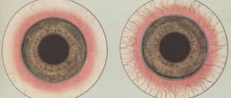
The medical term hemianopsia refers to a condition in which there is an area of blindness in half of the visual field. The visual field is the area of space that a person sees without moving his eyes or turning his head. Hemianopsia is a bilateral “partial blindness”, that is, loss of half the field of vision in each eye.
There are various types of this kind of pathology of visual perception. It is possible to distinguish heteronymous hemianopsia (loss of opposite halves of the visual fields), there is also partial, quarter (upper or lower) hemianopsia. “Darkening” of both temporal halves of the field is called bitemporal hemianopia. The loss of parts of the visible space on the side of the nose is binasal hemianopsia. A “blind spot” in the central part of the visual field is called a hemianopic scotoma.
Homonymous hemianopsia is a condition characterized by loss of the same halves of the visual fields. It can be both right or both left halves at the same time: a person can see only one part of the space falling into the field of view (left or right). The boundary between the visible part and the “blind spot” in homonymous hemianopsia is the central vertical meridian.
Types and classifications
As already noted, a distinction is made between right-sided and left-sided homonymous hemianopia.
Right-sided homonymous hemianopsia occurs in cases where the left optic tract is damaged. Left-sided is caused by damage to the right optic tract.
This pathological condition has another classification. Experts highlight:
- Complete homonymous hemianopsia. In this case, the “blind spot” fills half of the visible space, reaching the periphery of the visual field.
- Quadrant homonymous hemianopsia: the visual defect occupies only the upper or lower quarter (quadrant).
- Partial homonymous hemianopsia. This means "darkening" a more local portion of the visual field.
- Scotoma, in which the center of visible space falls out.
Quadrant homonymous hemianopsia can be upper quadrant or lower quadrant, quadrant partial or quadrant complete.
There is also a distinction between congenital homonymous hemianopia and that which developed as a consequence of a number of diseases.
Treatment of hemianopsia
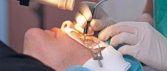
Despite the fact that the disease is associated with a disturbance in the functioning of the organ of vision, treatment of hemianopsia should be carried out by a neurologist and not an ophthalmologist. If the examination results revealed hemianopsia, the patient will be referred to a neurologist. This doctor must not only eliminate the manifestations of the disease, but also cope with its causes.
In rare cases, after visiting a doctor, the patient is referred for examination to a neurosurgeon or oncologist.
These specialists help identify diseases in the form of a benign or malignant tumor. Hemianopsia is treated in the following ways:
- chemotherapy;
- X-ray therapy;
- surgical intervention.
It will not be possible to cope with the pathology using conservative methods. Even with prolonged therapy, the desired results cannot be achieved. If it is not possible to have surgery, you should follow certain recommendations that relate to conservative methods.
Following these tips helps improve your vision and sometimes even increase your viewing angle.
So, ophthalmologists and neurologists advise following the following recommendations:
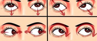
- When reading books, try not to turn your head. It must be stationary. At the same time, you need to try to move your eyes. The larger the range that can be covered, the better.
- When reading, you need to abandon the usual way of perceiving information. You need to read not horizontally, but vertically. In this case, the book or newspaper should be rotated 90 degrees.
- Most often, hemianopia develops in one eye. Therefore, when walking with a sick person, you need to hold his hand on the side on which the anomaly is located.
Recently, special programs have been used to combat this pathology.
Sometimes they help expand your visual range. In this case, the main treatment tactics are aimed at eliminating the root cause of the disease:
- In most cases, traumatic brain injuries require surgery.
- Malignant tumors are eliminated by chemotherapy or radiation. Surgery may also be necessary.
- Migraines require the administration of special nasal sprays that contain sumatriptans.
- For ischemic stroke, thrombolysis is indicated. After this, it is necessary to use nootropic drugs and drugs to reduce blood viscosity. After a stroke, it is imperative to carry out rehabilitation therapy.
- Brain swelling can be eliminated with the help of diuretics.
{banner_gorizontalnyy3}
Most of the pathologies that provoke the appearance of hemianopsia are treated surgically. Conservative treatment methods, as a rule, do not produce results. If the problem cannot be dealt with, the doctor selects a rehabilitation scheme. It helps the patient adapt to external conditions. In each specific case, tactics are selected individually.
Causes
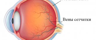
The appearance of this visual defect is explained by pathology in the area of the optic tract, as well as the cerebral cortex.
Homonymous hemianopsia is a consequence of damage to the areas of the brain that need to receive visual signals. Signals from the right and left eyes are transmitted along the optic nerves in such a way that problems in the left hemisphere of the brain cause “darkening” of the right half of the visual field in each eye; destructive processes in the right hemisphere are a factor causing loss of the left half of the visual fields.
The problem may be explained by a congenital pathology. But it can also be caused by various diseases that affect the brain.
The causes of homonymous hemianopia are most often the consequences of a stroke. But this condition can also be explained by the presence of a tumor, abscess, inflammation, trauma or injury to the head area. Find out about the causes of optic nerve atrophy in the article.
Homonymous hemianopia is caused by an aneurysm of the artery of the base of the brain, circulatory problems in the region of the posterior and middle cerebral arteries, basal meningitis, and meningoencephalitis.
All these conditions can cause the development of an inflammatory process, compression and disruption of the blood supply to the optic nerve fibers. Toxins also have a pathogenic effect on nerve fibers. After some time, under the influence of these factors, the visual tract is gradually destroyed.
Depending on what type of homonymous hemianopsia is detected in the patient, the specialist determines the specific location of the lesion. For example, inferior quadrant homonymous hemianopsia develops in case of problems that arise in the parietal lobe of the brain. The disease, localized in the temporal lobe, causes manifestations of complete upper quadrant hemianopsia.
Classification
According to the topic of the lesion, it happens:
- Tractus hemianopsia. Develops due to damage to the visual pathways. It is characterized by atrophy of the optic nerves, lack of pupillary response to light and asymmetry of visual field loss.
- Central hemianopsia. Develops when the central parts of the brain are damaged. The pathology is not accompanied by atrophy of the optic nerves; they continue to respond to light stimuli. The central variant is accompanied by a symmetrical loss of visual fields.
Symptoms
Homonymous hemianopsia may sometimes not be noticeable to the patient. Homonymous hemianopia has no effect on visual acuity. Visual defects in some cases are detected only during visual field examination. Manifestations of this type of hemianopsia are loss of the temporal part of the visual field on one side, and the nasal part on the other. Sometimes half of the visual field disappears completely (complete hemianopia). In some cases, the “darkening” does not reach the periphery of the visual field (incomplete hemianopsia). You can learn about loss of visual fields here.
Cases have been observed in which the remaining half of the visual field slightly “overlaps” the blind side.
Homonymous hemianopsia can make it difficult for a person to move around. People, in some cases, encounter obstacles located on the side of the “dropped out” half of the field of vision. It is almost impossible for the patient to drive a car or distinguish objects located in the “blind spot” of the table. Visual defects make reading difficult: the reader cannot find a new line in a book. Conditions of this kind may not be permanent.
Sometimes a symptom of homonymous hemianopia can be a visual hallucination. This disorder manifests itself especially often in cases where loss of part of the visual field develops after a stroke.
General information
With hemianopsia, a person cannot see on one side of the eye—right or left. The visual picture partially falls out of sight, as if visibility was closed or blocked. Sometimes the disease is accompanied by hallucinations. This condition occurs when the visual structures of the brain, which controls all functions and systems of the body, are damaged. Therefore, pathology is classified as a neurological disease and does not belong to ophthalmology.
Hemianopsia occurs:
- congenital;
- acquired.
The acquired form most often develops in women after 30 years of age. Symptomatic treatment against the background of the underlying disease is indicated.
The congenital form develops against the background of lesions of the central nervous system:
- underdevelopment of the brain and cranium;
- exit of the meninges outside the cranium;
- increased intracranial pressure due to the influence of cerebrospinal fluid;
- dropsy of the brain.
In multiple sclerosis, the nerve membranes are affected, including the visual apparatus.
From a clinical point of view, two forms of pathology are distinguished:
- homonymous (symmetrical);
- heteronymous.
Homonymous hemianopsia is characterized by loss of the left/right part of the visual field in both eyes. Heteronymous pathology has 2 forms:
- bitemporal;
- binasal.
Bitemporal hemianopsia is characterized by loss of the temporal halves of the visual field in both eyes. The names of the pathology with the prefix “bi” indicate that blind areas of the visual field appear on both sides.
There is also a binasal form of hemianopsia, which appears due to the “empty sella” syndrome or chiasmatic arachnoiditis. Binasal pathology is loss of visual fields on the side of the nose. Such a clinical picture is rare; it accompanies oncology, hydrocephalus, inflammatory foci in the arachnoid membrane, and diseases of the central nervous system.
There are cases of partial damage to the visual system in one eye and complete blindness in the other.
Depending on the volume of the affected area, hemianopia occurs:
- full;
- partial;
- square;
- scotoma.
In case of complete hemianopsia, half of the visual field is affected, in case of partial hemianopsia, part of half of the visual field is affected. With a square one, only a quarter of the field; with a scotoma, a round-shaped spot appears in the visual field.
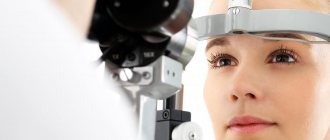
Diagnostics
The violation can be determined after studying the state of three important features of the functioning of the visual organs:
- visual acuity;
- fields of view;
- ophthalmoscopy.
The defect can be detected by visual field testing. The neurological technique includes an indicative test of the visual fields, which the doctor conducts during the examination. A specialist can detect smaller problems of visual perception using conventional or computer perimetry.
To diagnose visual field defects in ophthalmology, visual field analyzer devices (for example, campimetry) are used. Their use makes it possible to study the light and color sensitivity of the visual system. Instruments are also used to study the flicker sensitivity of the visual analyzer.
The doctor can make a final diagnosis based on obtaining a complete picture of the fundus, as well as accompanying neurological symptoms.
To determine the exact causes of a visual disorder, it is important to do an X-ray and CT scan of the brain. The doctor may also prescribe a more complete examination and a series of tests that will help make a diagnosis.
How is the disease detected?
Diagnosis of visual impairment is carried out using visual field testing. The purpose of the express method is to identify field pathologies. In neurological practice, a special hand hammer is used. A neurologist sits down opposite a person and asks him to close one eye with his palm. The gaze of the open eye is fixed on the doctor's nose. The physician slowly moves the instrument from behind the patient's head from the periphery to the center from all sides. The patient must be told immediately when he sees the hammer.
Among instrumental methods, perimetry has the greatest diagnostic value. The procedure is carried out using a perimeter. As a result of the procedure, the doctor receives information about the boundaries of the visual fields for both eyes. Other visual defects are also detected. Computer campimetry is also used. It identifies disturbances in color perception in various areas of the retina and helps determine the level of disturbance.
To identify the cause of the disorder, computer and ultrasound diagnostics are used: computer and magnetic resonance imaging, Doppler ultrasound of cerebral vessels. Computer diagnostics helps in choosing treatment tactics, as it allows you to visualize the source of hemorrhage, tumor or abscess.
Treatment
To get rid of problems of visual perception, it is necessary to completely eliminate the consequences of the diseases that caused homonymous hemianopia. If treatment of the underlying disease is ineffective and untimely, hemianopia can lead to complete loss of vision.
To eliminate neurological pathologies, surgical treatment, chemotherapy, and drug therapy are used, depending on the type and degree of complexity of the underlying problem.
Loss of visual fields with hemionopsia
Hemianopsia practically cannot be cured, however, after eliminating the factor that causes it, a number of methods can be used to improve the patient’s quality of life. For example, a person suffering from homonymous hemianopsia can learn to read by holding text at a 90° angle and viewing it vertically.
There are also a number of recommendations to make moving in space easier.
Diagnosis of hemianopsia
To diagnose hemianopsia, you must consult a doctor promptly.
The specialist performs the following procedures:
- analysis of the visual fields of each eye;
- determination of visual acuity;
- fundus ophthalmoscopy.
The latter procedure may not give the required results. At the initial stages of pathology development, changes in the structure of the fundus may be absent or symptoms may be mild. Subsequently, the manifestations become more noticeable.
Pronounced destructive changes can be seen only after a year of identifying the pathology. The difficulty of diagnosing the disease in the initial stages of its development lies in the fact that it does not cause visual impairment. Such consequences appear only after a few months in the absence of adequate therapy.{banner_gorizontalnyy2}
After a preliminary diagnosis, a more detailed examination is prescribed.
It helps determine the stage and degree of development of the disease. Such diagnostics involve the use of tools. The following procedures apply at this stage:
- tomography;
- angiography;
- radiography;
- ultrasonography;
- application of electromagnetic waves.
To objectively determine the degree of loss of specific areas from the visual field, perimetry is performed.
This procedure helps to project the boundaries onto a spherical plane. If you suspect the presence of a disease, the examination should be carried out in the following order:
- The doctor performs a visual examination of the eyes. This is done one by one. Moreover, one eye is covered with the palm of the hand, and the second is examined from a distance of 1 m.
- During the examination, the ophthalmologist asks the patient to concentrate on his finger. After this, he gradually moves his finger from the areas to the periphery and in the opposite direction. This helps identify the area where the patient is losing vision.
- Provided that the doctor does not have vision pathologies, he and the patient must correctly establish boundaries, beyond which the person cannot see his own finger. This experimental method helps to quickly identify hemianopsia.
conclusions
Hemianopsia is a defect of visual perception in which loss of half the visual field develops in each eye. This condition is caused by factors associated with damage to the areas of the brain that receive visual signals. Read more about hemianopia in this material.
Treatment of hemianopsia is carried out depending on the nature of the underlying disease.
Hemianopsia can be temporary or permanent. This is explained by the severity of the damage to the visual pathway.
If untimely or poor-quality treatment is used, pathological processes can lead to complete loss of vision and, consequently, disability.
Causes
Impaired blood circulation in the brain leads to the development of a pathological condition. This occurs due to a malignant or benign neoplasm, head injury, or cerebral hemorrhage.
Less common causes of the disease include:
- infections;
- seizures or migraines;
- exposure to toxins;
- neurodegenerative disorders;
- non-ketotic hyperglycemia.
Common causes include stroke, tumors or complications from surgery. Also associated with high blood pressure or epileptic seizures.
The disease is caused by aneurysms. In most cases, the cause is a consequence of organic damage to the nerves of the optic pathway.
Complications and prognosis
By itself, bilateral hemianopsia is not life-threatening. But some diseases that cause it can be fatal - traumatic brain injury, severe stroke.
The prognosis for disability depends on the causative factor of the disorder, the degree of damage, and the amount of vision loss. With a slight loss of visual fields and comprehensive rehabilitation, a person with a homonymous disorder can lead a normal lifestyle.
Complications of hemianopsia include possible traumatization of a person due to insufficient perception of the surrounding world. As the causative disease progresses, complete loss of vision is possible.
Heteronymous hemianopsia
The heteronymous variety of this disease occurs when both temporal or nasal halves of the visual fields are affected. In this case, binasal or bitemporal heteronymous hemianopsia develops.
The causes of this particular condition are certain focal changes. This may be ischemia, hemorrhage, tumor, inflammation. They should occur in the area of the pituitary gland, near the base of the brain. In such situations, bitemporal hemianopia is observed, which is a type of partial blindness. It is characterized by loss of perception of the temporal half of the right and left visual fields.
In fact, it is a type of hemianopsia, which is characterized by loss of nasal visual fields, both internal and medial. Bitemporal hemianopsia is observed when uncrossed fibers are affected, against the background of inflammation of the arachnoid membrane of the brain.
If not half of the object, but only a smaller part of it, falls out of the field of view, then they speak of quadrant hemianopia. In this case, only a quarter of the image becomes invisible.
Topical diagnosis of homonymous hemianopsia
Description
Homonymous hemianopsia
can be caused by a disease of both the optic tracts and the parts of the central neuron of the visual tract and the occipital lobe cortex. In this regard, in each individual case it is necessary to resolve the issue of localization of the outbreak. Topical diagnosis of homonymous hemianopsia is of great importance, as it often contributes to the correct recognition of brain disease. This diagnosis is of particular importance in cases where it should help accurately localize the pathological process in the brain.
We distinguish between tractus and central hemianopsia. Under Tractus Hemianopsia
refers to homonymous hemianopsia caused by diseases of the optic tracts; central hemianopsia refers to homonymous hemianopsia caused by diseases of the Graziole bundle or the occipital lobe cortex. In this section we will outline only the differential diagnosis of tractus and central hemianopsia. Topical diagnosis of hemianopsia within the central neuron will be presented in the section on homonymous hemianopsia in diseases of the central neuron of the optic pathway and the occipital lobe cortex.
For the differential diagnosis of tractus and central hemianopsia, it was proposed to use the condition of the fundus, pupillary symptoms and some features of changes in the visual field. Let's consider the meaning of each of these symptoms separately.
Any disease in the optic tract (if it is long enough and the changes are intense enough) leads to atrophy of nerve fibers. From this focus, ascending and descending atrophy spreads. Ascending atrophy reaches the external geniculate bodies, descending - to the nipples of the optic nerves. Thus, tractus hemianopsia
ultimately always leads to the development of simple atrophy of the optic nerves. The situation is different with central homonymous hemianopia. Here, ascending and descending atrophies spread only within the central neuron of the visual pathway and reach, on the one hand, the cortex of the occipital lobe, and on the other hand, to the external geniculate bodies. Therefore, central hemianopsia never leads to the development of simple atrophy of the optic nerves. Therefore, the combination of homonymous hemianopsia with simple atrophy of the optic nerves indicates that the focus of the disease that caused the hemianopsia is localized in the optic tract. From this, however, it does not follow that homonymous hemianopsia with a normal fundus is caused by damage only to the central neuron above the optic tracts. And tractus hemianopia can occur for a long time in the absence of changes in the optic nerves.
We observed several patients with chiasmal disease, in whom a period of 5-8 months turned out to be insufficient for the manifestation of optic nerve atrophy in the fundus. For descending atrophy to descend from the optic tracts to the fundus of the eye, it takes an even longer period than in diseases of the chiasm; It must be assumed that atrophy of the optic nerves with tractus hemianopsia will appear in the fundus in about a year. Thus, with homonymous hemianopsia with a normal fundus, it is possible to confidently exclude the localization of the lesion in the optic tract only if this hemianopsia developed more than a year ago.
It is necessary to take into account that for the topical diagnosis of homonymous hemianopsia, only primary atrophy of the optic nerves
, since secondary atrophy can be a consequence of congestive nipples due to brain tumors and diseases of the central neuron of the visual pathway.
One of the most reliable symptoms is hemianopic reaction of the pupils to light
.
The reaction of the pupils to light is a reflex. The sensitive part of the arc of this reflex passes in the peripheral neuron of the visual analyzer, the motor part of the arc is the oculomotor nerve. In the peripheral neuron, reflex arc fibers pass through the optic nerve, chiasm, and optic tracts. In the posterior part of the optic tract, somewhat anterior to the external geniculate bodies, these fibers leave the optic tract and go to the nucleus of the oculomotor nerve. (These fibers do not reach directly the nucleus of the oculomotor nerve, and the connection is carried out using one or more intercalary neurons. For this question, this detail is not important.) From the nucleus of the oculomotor nerve, the nerve fibers go as part of the trunk of this nerve to the sphincter of the iris.
Therefore, a pathological focus affecting the optic tract turns off not only the peripheral neuron of the visual tract, but also interrupts the arc of the pupillary reflex. In contrast, a pathological focus that interrupts the central neuron of the visual pathway on one side does not affect the arc of the pupillary reflex, since it is located above it.
Hemianopic pupil reaction
observed in tractus hemianopsia and not observed in central hemianopsia. It consists of the following. If a patient with Tractus hemianopia is placed in the remaining half of the visual field, a normal reaction of the pupil to light is caused, but when a light source is placed in the missing half of the visual field, there is no reaction of the pupil to light. In contrast, with central hemianopsia, a normal pupil reaction to light is obtained when the light source is positioned in both the preserved and the lost half of the visual field.
When studying hemianopic pupil reaction to light, a number of conditions must be met
, without which it is impossible to detect her disorders. First of all, it is necessary that when sequentially illuminating the temporal and nasal halves of the retina (the study of the hemianopic reaction is always carried out on one eye), zones equally spaced from the area of the macula are illuminated. Diffuse illumination of the retina should be avoided. Certain other conditions must also be met. Therefore, the hemianopic reaction of the pupil cannot be examined using an ophthalmoscope or by sequentially placing a light source in the right and left halves of the patient’s field of vision. The hemianopic reaction of the pupils is studied using special devices - hemicinesimeters. For this purpose we use a Hess hemicinesimeter.
The hemianopic reaction of the pupils was described by Wernicke in 1883. Subsequently, all authors who dealt with this issue noted its great importance for the topical diagnosis of hemianopsia. Jess studied the hemianopic reaction of the pupils in 5 patients with central hemianopsia, and in 1 with tractus hemianopsia. Only with tractus hemianopsia did he detect a hemianopic pupil reaction. Bunge studied 12 patients with tractus hemianopsia of various etiologies, and in all of them he revealed a hemianopic reaction of the pupils.
We examined the hemianopic reaction of the pupils in 6 patients with tractus hemianopsia and in 9 patients with central hemianopsia. In all cases of tractus hemianopsia
A hemianopic reaction of the pupils was detected, but in central hemianopsia it was absent. Thus, based on our own observations, we can confirm the great importance of this pupillary reaction for the topical diagnosis of hemianopsia.
The need to use special equipment makes it difficult to widely introduce the study of hemianopic reaction into practice. This also explains the small number of observations published in the literature on this issue.
For the topical diagnosis of hemianopsia, Baer attaches great importance to anisocoria
. According to him, with Tractus hemianopsia, the pupil is always wider on the hemianopsia side, for example, with left-sided hemianopsia, the pupil is wider on the left eye. With central hemianopia, the pupils are mostly the same width. If there is anisocoria, then the pupil is wider on the side opposite to hemianopsia. Anisocoria observed with tractus hemianopsia, according to Baer, is explained by the fact that a pathological focus in the optic tract on one side interrupts the arc of the pupillary reflex to light. When both eyes are illuminated, excitation along the fibers of the reflex arc on one side completely reaches the nucleus of the oculomotor nerve, on the other side this occurs only to a small extent and (according to Baer) only due to the double innervation of the macula. Uneven excitation of both nuclei of the oculomotor nerve leads to the development of anisocoria.
Bunge could partially confirm the meaning of this symptom. Among 12 patients with tractus hemianopsia, he found anisocoria in 6 with a wider pupil on the hemianopsia side. Kestenbaum considers this symptom unreliable. Walsh has observed similar anisocoria in several cases of tractus hemianopsia.
We tested the value of anisocoria for the topical diagnosis of homonymous hemianopia on a large material. The data we obtained is presented in table. 33.
Table 33.
Pupils with homonymous hemianopia
These data show that pupil dilation on the hemianopsia side is by no means characteristic of tractus hemianopsia. The significance of such anisocoria for topical diagnosis is further reduced in connection with the data that were obtained with central hemianopsia. Anisocoria with a wider pupil on the side of hemianopsia is often observed with central hemianopsia. Based on our observations, we believe that anisocoria is not important for the topical diagnosis of hemianopsia.
Clinical significance of preservation of the macula area
a number of works are devoted, and many authors attach great importance to this symptom for the topical diagnosis of homonymous hemianopia. The point of view expressed by Lenz (1909) is widely used. When studying 64 cases of homonymous hemianopia collected from literary sources, in which, along with the clinical picture, there were also data from a pathological examination of the brain, Lenz came to the following conclusions. In the vast majority of cases, preservation of the macula area is observed only with lesions of the posterior two-thirds of the central neuron (the border passes through the middle third of the parietal lobe). When the optic tracts and the initial section of the Graziole fascicle are affected, in most cases the dividing line between the preserved and lost halves of the visual field passes through the fixation point, i.e., the area of the macula is not preserved. Therefore, the preservation of the macula area in homonymous hemianopsia indicates a high localization of the lesion, namely, between the cortex of the occipital lobe and the middle of the parietal lobe. In homonymous hemianopia without preservation of the macula area, the lesion lies between the middle of the parietal lobe and the chiasm.
In this regard, many authors believe that tractus hemianopia is characterized by a lack of preservation of the function of the macula area
, and for central hemianopsia - preservation of this area. This point of view on the significance of the symptom of preservation of the macula area for the topical diagnosis of hemianopsia is set out in all manuals on eye diseases (Bellarminov and Merz, Odintsov, Aksenfeld). In this regard, it is widely used in the practice of ophthalmologists, neurologists and neurosurgeons.
Currently, a lot of data have accumulated that challenge the significance of the symptom of preservation of the macula area for the topical diagnosis of hemianopsia. Soon after Lenz's work, a number of observations were published that contradicted his data. Thus, Wilbrand, Genschen, Baer and Bunge described a number of cases of tractus hemianopsia with preservation of the macula area. Utgof, Veve, Wilbrand and Bunge observed hemianopsia due to disease of the occipital lobe, without preservation of the macula area. Baer does not consider it possible to rely on the absence or preservation of the macula area for topical diagnosis of damage to the visual pathways. Kestenbaum also believes that in the topical diagnosis of diseases of the visual pathways, this symptom is much less important than is commonly believed. The unreliability of the symptom of preservation of the macula area for the topical diagnosis of hemianopsia is also noted by Trackuer.
Back in 1945, based on our observations of hemianopsia due to gunshot wounds during the Great Patriotic War, we pointed out the extreme unreliability of this symptom. Currently, we can provide more extensive observations about hemianopsia of various etiologies and localizations.
Before proceeding to the presentation of the relevant data, it is necessary to somewhat clarify the very concept of the preservation of the function of the macula area. The accuracy of perimetry is relative, and, like any subjective research method, it has its own errors. Due to the high sensitivity of the retina in the central zone, the study of visual field defects near the point of fixation is possible with greater accuracy than determining the boundaries of the visual field. However, not every case of hemianopsia, when the dividing line between the lost and preserved portion of the visual field goes around the point of fixation, can be considered as hemianopsia with preservation of the macula area. With conventional perimetry methods, even when examining the central part of the visual field, it is impossible to achieve accuracy within 1-2°. This is especially hampered by those small involuntary movements of the eye, as a result of which it is not possible to accurately maintain the correct fixation of the perimeter arc object. Therefore, we, like some other authors, adhere to the point of view that we can talk about the preservation of the macula area only if the dividing line goes around the fixation point by at least 3°
. In this regard, we include in the group of hemianopsia without preservation of the macula also those cases when the dividing line went around the fixation point by 1-2°. In addition to complete homonymous hemianopsia, cases of quadrant hemianopsia and hemianopic scotomas were also used. In relation to the latter, the group of hemianopsia without preservation of the macula area included those cases when the scotoma in at least one of the sectors reached the fixation point.
The data we obtained on the nature of the dividing line in homonymous hemianopia are given in Table. 34.
Table 34.
The nature of the dividing lines of the visual field in homonymous hemianopia
As can be seen from table. 34, preservation of the macula area in tractus hemianopsia is not uncommon. It should be noted that the disease of the optic tracts in 5 patients with homonymous hemianopia and preservation of the macula area did not raise any doubts, since all had simple atrophy of the optic nerves and in 1, when examined with a Hess hemikinesimeter, a hemianopic reaction of the pupils to light was obtained.
Even more often, the preservation of the macula area in tractus hemianopsia was observed by Bunge. Among 14 patients with tractus hemianopsia, the macula area was preserved in 9. Moreover, 6 of them had atrophy of the optic nerves, and 8 had a hemianopic pupil reaction.
Here is our observation.
Observation 46.
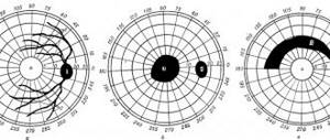
Patient V-va, 42 years old, went to the clinic of the Leningrad Institute of Eye Diseases. A month and a half ago I noticed deterioration in vision and loss of the left halves of the visual field. 2 years ago she suffered from infectious encephalitis. When examined, the pupils are uniform; the reaction to light and installation near is satisfactory; in both eyes there is blanching of the temporal half of the optic nerve nipple; the boundaries of the nipples are distinct, the arteries are narrowed; atrophy phenomena are more pronounced in the left eye; visual acuity of both eyes is 0.7. In the field of view (Fig. 64)
Fig. 64
There is complete left-sided hemianopsia. The area of the macula in both eyes is preserved by 5° white and 3° red. The left halves of the visual field are not narrowed. When examined with a Hess hemicinesimeter, a distinct hemianopic reaction of the pupil is noted. The neuropathologist identified residual effects of encephalitis.
In this case, the localization of the process in the right optic tract is established on the basis of simple atrophy of the optic nerves and hemianopic reaction of the pupils. The macula area is clearly preserved in both eyes. Taking into account that the patient suffered infectious encephalitis, after which, according to the neurologist, residual effects still remained, it should be considered that there is a focus of softening in the right optic tract.
From a comparison of the data given in table. 34, it is clear that with tractus hemianopsia, preservation of the macula area is often observed
. Along with this, with central hemianopsia, the macula area is also very often not preserved. Therefore, the preservation of the macula area or the loss of this part of the visual field does not matter for the topical diagnosis of hemianopsia.
If homonymous hemianopsia is not complete, then the degree of symmetry of visual field defects is essential for topical diagnosis. A pronounced asymmetry of visual field defects is characteristic of tractus hemianopia and is not observed with central hemianopsia.
—-
Article from the book: Diseases of the visual pathway | E. J. Tron
Treatment methods
Treatment of bilateral hemianopia should first be aimed at eliminating the underlying cause:
- Traumatic brain injuries are predominantly treated with surgery.
- Oncological processes require chemical or radiation therapy, surgical intervention.
- Migraines are treated by using nasal sprays containing sumatriptans (Imigrant).
- For ischemic stroke, thrombolysis is performed, after which nootropic drugs and drugs that reduce blood viscosity are prescribed. Rehabilitation treatment after a stroke is mandatory.
- Cerebral edema is eliminated by taking diuretics.
Most of the diseases that provoke the appearance of hemianopsia are treated surgically. Conservative therapy is ineffective in most cases. In cases where it is impossible to eliminate the problem, a rehabilitation plan is drawn up to allow the patient to adapt to the external environment. Recovery tactics are selected individually.
Eye gymnastics, various computer programs and spectacle correction are also prescribed, with the help of which you can increase the field of vision in case of hemianopsia.