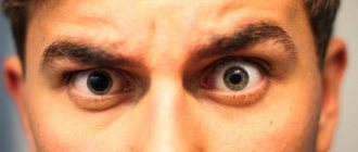
Third eyelid
In addition to fish, reptiles, amphibians, birds and sharks, some mammals such as camels, polar bears and seals have a fully functional membrane known as the "third eyelid". It protects the eyes of crocodiles and beavers from water.
In humans it is present only as a small fold known as the plica semilunaris, connected to the tissue covering the whites of the eyes and the insides of the eyelids, which no longer serves its original purpose but to aid in the drainage of tears. In addition, without it, the conjunctiva would be attached directly to the eyeball, limiting the mobility of the latter.
Why does pathology develop?
The third eyelid is a fold of the conjunctiva located in the inner corner of the eye, which, in turn, consists of the nictitating membrane - a thin mucous membrane lining the surface of the eyeball and the inside of the eyelid. This membrane moisturizes the cornea with tear fluid and cleans it of debris.
We can say that it resembles a cleaning brush on car windows, performing a similar function. In addition, thanks to the lacrimal gland located at the base of the third eyelid, the cornea is reliably protected from pathogenic microorganisms.
If a cat has a slight covering of the eye with the third eyelid, there is no redness, inflammation, or swelling, then this does not always mean blindness. Perhaps the organ of vision is sunken due to a reduction in fat caused by weight loss, or the cat flu is developing in your pet.
The causes of third eyelid prolapse are varied. The most common ones include:
- Viral, fungal, bacterial infections.
- Pathologies of internal organs - kidneys, liver, gastrointestinal tract.
- Antibiotic therapy.
- Malfunction of the central nervous or hormonal systems.
- Inflammatory processes in the ears.
- Weak ligaments responsible for the activity of the eyelids.
- Consequence of head injuries.
- Worm infestation.
- A foreign body that has entered the eye and that the tear fluid cannot wash off on its own.
- Any form of conjunctivitis.
- Adenoma (benign tumor of the third eyelid).
- Atrophy of the eyeball.
Prolapse of the third eyelid can be diagnosed in absolutely any pet, but representatives of the Persian cat breed are most susceptible to this pathology.
Pointed part of the ear
It was first described by Charles Darwin in his book The Descent of Man and Sexual Selection. The scientist named it "Wolner's tip" after the British sculptor. The tubercle occurs in 58% of Swedish children, 40% of adults in India and 10.4% of adults in Spain. It is usually present on both ears, but may be present on only one.
The structure of the onion will help you chop it quickly and easily: the chef's method
Do-it-yourself: how to make a decoration for a children's room from wooden branches
Which shirt to wear with trousers this winter: models for men and women
Symptoms
A condition is considered pathological when the third eyelid does not fold and almost completely covers the organ of vision. Experiencing discomfort, the four-legged furry dog begins to rub its face, blink frequently, squint and hide from the irritating light in a secluded place. His eyes turn red, tears flow from them, mucus and pus are released.
Subsequently, a voluminous dense formation is formed in the corners. The temperature often rises.
If one eye is covered with film, then most likely a foreign body has entered it. When there is drooping of the eyelid and narrowing of the pupil, they speak of a pathology of a neurological nature. But if changes are noticed in both eyes, then this is already a serious systemic disease.
Ear movement
Macaques and other monkeys have more developed auricular muscles, used to move their ears, than humans, chimpanzees, or orangutans. In our country, these muscles, although quite large, do not function.
However, some people can move their ears at will, and anyone can learn to do this. The inability to move the ears in humans and other animals is compensated by the ability to turn the head horizontally, which most monkeys cannot.
Treatment, prognosis
To eliminate infectious pathologies, therapy is practiced, which is based on antiviral and antifungal drugs. Their task is to suppress the development of pathogenic microorganisms.
The animal is also prescribed antipyretics, painkillers, and immunostimulants and vitamin-mineral complexes to strengthen the immune system. Intravenous infusions of pharmacological solutions are advisable.
If prolapse of the third eyelid is associated with an allergic reaction, then the allergen is eliminated and antihistamines are prescribed. For general improvement of the condition, hormonal agents are indicated (in advanced stages).
Eye injuries are treated by applying anesthetic drops and rinsing to remove foreign bodies and debris. Surgery is performed only in severe cases.
If the prolapse of the third eyelid is caused by an adenoma, but the neoplasm does not grow and does not bother the animal, then removal of the benign tumor is not carried out. The operation may have unpleasant consequences, such as dry eye syndrome. As a rule, veterinarians limit themselves to supportive therapy.
Grasp reflex
This ability is formed in the 11-15th week of pregnancy. This is one of the most important reflexes that a newborn must exhibit in order to be considered neurologically healthy.
The reflex can be tested by placing a finger on the child's palm. When trying to remove the finger, the child will grab it tightly enough to support his own weight. The latch can be unpredictable as the baby may let go without warning.
Silence helps you hear yourself: several differences between emotionally developed people
With the help of 60,000 bees and his skill, the artist sculpted a bust of Nefertiti
Lyudmila Artemyeva wants to sue Instagram over verification of a fake page
The reflex disappears after 2-3 months, maximum after six months, when the child gains control over his body. If this does not happen, you may have difficulty holding objects. The reflex can also be observed on the legs.
When to urgently contact a veterinarian
If the appearance of a cat’s third eyelid is unusual in any way, the animal must be taken to a veterinarian for examination as quickly as possible, even if at the moment this is the only manifestation of trouble. It is easier to identify the problem at an early stage, when the vast majority of diseases can be cured. This will keep the cat healthy and reduce the budget spent on its treatment.
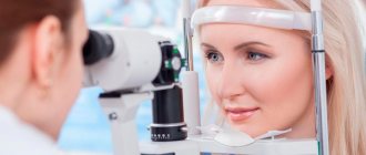
Only a veterinarian in a clinic can conduct a full diagnosis, including specialized ophthalmological diagnostics. The examination usually includes:
- taking an anamnesis - asking the owner what preceded the painful manifestations and how they developed over time;
- examination of the cat and its eyes;
- general blood analysis;
- blood chemistry;
- to clarify the nature of the causative agent of inflammation, material is taken from the conjunctiva of the eye for bacteriological examination or PCR;
- Ultrasound of internal organs in cases of bilateral prolapse of the nictitating membrane;
- Ultrasound of the eyeball;
- CT, MRI - to clarify the nature of the lesion, it is possible to conduct an X-ray of the skull.
Ophthalmological examination:
- examination of the cornea and pupils with fluorescein staining;
- measurement of intraocular pressure;
- examination using special optics of the internal structures of the eye.
Inadmissible actions in case of pathology of the nictitating membrane
In case of pathology of the nictitating membrane, the following are unacceptable:
- attempts at self-diagnosis and self-medication. The diagnosis can only be made by a veterinarian, often after a specialized examination. Self-medication can be dangerous and result in worsening the disease and worsening its prognosis.
- attempts to independently “straighten” the nictitating membrane. They can lead to irreversible injury to the eyeball and necessitate its removal.
Jacobson organ
The vomeronasal organ was first discovered by the Danish anatomist Ludwig Levin Jacobson in 1811. It is located at the base of the nasal cavity just above the palate. The organ contains receptors that help detect certain types of non-volatile or liquid, organic compounds that may come from prey, predators or potential mates.
Unlike the olfactory bulb, which sends signals directly to the olfactory cortex, the Jacobson's organ sends signals to the hypothalamus, which is responsible for regulating hormone production and body temperature.
Jacobson's organ is present in all snakes and lizards, as well as in many mammals.
In humans, it develops in the embryo, but after that it regresses and the nerve connections disappear. The cavities persist in some adults, but without a functional nervous system the organ simply does not function.
Epicanthus
The epicanthus is a fold of skin that hangs over a person's upper eyelid. It is believed that epicanthus is characteristic of the Mongoloid race, and this rudiment is due to the weather conditions in which the ancestors of modern people lived. Harsh winters, dry desert air, scorching sun - epicanthus was additional protection for the eyes. Now we have sunglasses, and the need for skin folds has disappeared over time, although it still occurs in some people.
Goose pimples
Skin bumps appear when tiny muscles at the base of the hair contract in an emergency or due to irritants such as a gust of cold wind. In animals with fur, this helps trap air between the hairs, which creates a layer of insulation and prevents hypothermia.
Unusual dwarf giraffes found in Namibia and Uganda
I combined rice with eggs and baked it: I didn’t expect the dessert to be so delicious
11 tests for covid did not give an unambiguous result: the story of Angelina Dubrovskaya
People experience goose bumps from screeching, metal scraping, music, feelings or strong emotions, watching horror movies, or experiencing exciting events such as winning a sporting event. Some are able to deliberately cause goose bumps in themselves.
Diet after removal of atavism in humans
Postoperative diet
- Efficacy: therapeutic effect after 21 days
- Terms: 1-6 months
- Cost of products: 1300-1500 rubles. in Week
After surgical interventions, patients are recommended to follow a gentle, balanced and fairly lean diet for 6-12 months. depending on your health condition. The diet requires strict control of the consumption of simple carbohydrates, salt and spices. The diet should consist of such dishes as:
- pureed vegetable soups;
- fruit and berry compotes;
- boiled porridge;
- lean varieties of fish and meat;
- low-fat lactic acid drinks;
- boiled eggs and steam omelettes.
It is recommended to completely exclude fresh baked goods, soda, coffee, fatty meats, fish, cheeses, fried and smoked foods, as well as canned food.
Hiccups
According to the hypothesis, this reflex is necessary for tadpoles before their lungs are fully formed. It forces you to swallow air and water through your gills and is similar to the hiccups of mammals.
Hiccups are normal for babies in the last three months of pregnancy and are part of the fetal development process. It is also seen in premature babies, and researchers believe it is "an evolutionary precursor to modern pulmonary breathing."
Tail vertebrae (coccyx)
At one stage of embryonic development, a tail can be seen in a human embryo. The tail is a group of rudimentary vertebrae fused together that are located at the base of the spine. Animals need a tail to maintain balance when moving, but in our country the need for caudal vertebrae has disappeared since people straightened up and began to walk on two legs.
However, there are still known cases of babies being born with a small fatty appendage, which is the ancestor of a full-fledged tail, characteristic of many vertebrates.
Wisdom teeth
Our ancestors compensated for their lack of ability to properly digest plant foods by chewing them thoroughly. But with the advent of agriculture, the human diet changed, and as a result, the jaws began to move forward less, leaving less space for wisdom teeth. Because of this, in some people they do not fully grow in because they are blocked by other teeth and must be removed.
Causes
The origins of man have always aroused keen interest among philosophers and scientists, but over the past century and a half, human atavisms have been more thoroughly studied. Thanks to DNA decoding, it was possible to explain the origin of atavisms. After sequencing and studying the human genome, it was possible to better understand the mechanisms of development of traits and the structure of the genes responsible for them. It turned out that atavism is a manifestation of genes preserved in DNA and responsible for this trait (tail-shaped appendage, additional pair of mammary glands, etc.). Normally, they do not function because they are suppressed by the influence of other genes.
The external manifestation of characteristics may be completely lost, but fragments of genetic “programs” preserved in the genome in special circumstances can “work” and become an impetus for the development of atavism. These are most often mutations in regulatory genes responsible for reduction. They arise under the extreme influence of various chemical and physical factors in the developing embryo.
Why do some people develop atavisms?
Atavisms are congenital malformations. They appear during reexpression - the awakening of dormant genes as a result of hybrid dysgenesis, heredity, exposure to mutagens (ionizing radiation, radiation, alkaloids, pesticides, etc.) and stressful environmental factors. However, most often atavisms are the result of multifactorial influence and their etiogenesis is not fully understood.
Treatment of third eyelid prolapse in cats
Treatment of nictitating membrane protrusion is primarily associated with identifying and eliminating the cause of this condition.
If the cause of prolapse of the third eyelid in a cat is an eye disease, for example, inflammation - conjunctivitis, then during treatment all attention should be focused on it. The same applies to two other groups of causes of protrusion: neurological disorders and systemic diseases. Symptomatic therapy is also used.
In itself, prolapse of the third eyelid is not dangerous, but it may indicate the onset of a serious illness. Therefore, a cat with such a symptom should be shown to a specialist as soon as possible.
Third century inversion
Inversion is the inversion of the third eyelid cartilage with or without prolapse of the lacrimal gland. This pathology is rare in cats. The reasons for the third century inversion are currently unknown.
If you notice that your cat has a rolled-up third eyelid, you need to take her to the veterinary clinic. An inverted third eyelid irritates your pet's cornea, rubbing it, and if left untreated can lead to corneal inflammation and ulceration. You can rid your cat of this problem with surgical correction.
Lymphoid hyperplasia of the third eyelid
This pathology consists of the proliferation of lymphoid tissue on the inner side of the third eyelid. This usually occurs as a result of chronic conjunctival irritation and/or infection. Treatment is symptomatic: hormonal therapy is used in combination with surgical compression of the lymphoid follicles.