Rules for passing vision tests
You should start testing only if you feel well. If you have any discomfort: the temperature has risen, headaches have appeared, you feel weak throughout the body, then it is better to refuse the procedure for now. You should not get checked after prolonged eye strain, or after taking certain medications.
In order to begin the examination, it is necessary to create certain conditions:
- the room where the subject is located should be very light;
- Under no circumstances should you tilt your head;
- when looking at the table, there is no need to squint your eyes;
- The monitor should be level.
Only by following all the rules can you get the most accurate results.
Sivtsev table
The Sivtsev vision test table has black letters printed on a white background of various sizes, the purpose of which is to determine visual acuity. The table contains various options of 7 letters of the same height and width, but having different sizes. According to the benchmarks, 2 equidistant points with an angular resolution of 1/60 degree are well visualized.
Sivtsev’s table helps determine the severity in the interval from 0.1 to 0.5 at a distance of five meters. The rows are presented as follows:
- the first ten rows differ in increments of 0.1 (V = 0.1 – 1.0);
- two rows have a difference of 0.5 (V = 3.0 – 5.0);
- three rows in 0.1. (V = 3.0 – 5.0).
There is additional information. The left column shows the distance, expressed in meters, from which a patient with 100% vision can see, and the right column shows the reference values when viewing from 5 m.
Test rules
To get the most accurate results, you need to follow certain rules. The table cloth is placed five meters from the patient’s eyes. It must be illuminated by 2 fluorescent lamps. The light flux is directed onto the canvas, and not into the eyes of the subject.
Each eye should be checked separately, and they must be covered with the palm of your hand or taken an object for this. There is no need to put too much pressure on your closed eye, nor should you close your eyes. It is permissible to make no more than two or three mistakes. No more than three seconds are allotted for everything. The digital expression is in the last line where errors were made that exceeded the norm. The ability to see all ten lines from a distance of 10 meters cannot be called an indicator of farsightedness; in this case, they speak of a significant excess of the norm, the so-called “eagle vision”.
{banner_horizontalnyy}
How to decipher readings
At the bottom there is a special column starting with “D = ...”, in this area they indicate the distance, expressed in meters, from which a person with ideal visual acuity is able to distinguish elements. Another column “V = …” numerical indicators when reading from five meters.
Visual acuity test
Each of us has already had an eye exam at an ophthalmologist's office, but what if you want to monitor your visual acuity every day? Is it possible to test your eyesight at home? Yes, it is possible, and completely free. There are two options.
First option:
Download the vision measurement chart and print it out. We offer you 3 types of tables:
Table for measuring visual acuity Sivtsev download
Table for measuring visual acuity Golovin download
Table for measuring vision for children Orlova download
Instructions “how to measure vision using downloaded and printed tables”
1.Print the table on A4 sheet.
2. Position the sheet so that the 10th line (the line above the gray stripe) is at eye level. The distance between the eyes and the table is 2.5 meters.
3. Turn on good lighting in the room, the table should be illuminated evenly.
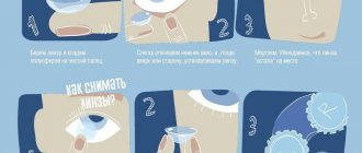
4. Cover one eye with your palm or something opaque (do not close it under any circumstances).
5. Mark the line that you see with each eye separately (to the right of each line is the value V, this is the value of your vision. For example, you see the 4th line from the top - that means your vision is 0.4).
Second option:
Online visual acuity test. We offer a unique algorithm for measuring visual acuity online, in which you do not need to know the resolution, diagonal size and proportions of your monitor. In this case, you will get a result that is as close as possible to the real one (however, visual acuity can be measured most accurately in an ophthalmology office) by synchronizing the dimensions of the monitor with the dimensions of the table. Also, a very important factor for the reliability of measuring visual acuity is the absence of distractions, therefore, when checking vision online on our website, you will see a table on a white background, without banners or advertising. All you need for an online vision test is to have a banknote of the appropriate denomination or a ruler with you.
Instructions “how to measure vision online on the website infoglaza.ru”
1.Attach a sample (bill, card or ruler) to the monitor and mark it as shown in the figure. Select the sample you took (what you measured). Set the desired distance from the monitor. Select a table to measure visual acuity. Click on the “start vision test” button
2.Move away from the monitor so that there is the distance you set between your eyes and the monitor.
3. Cover one eye with your palm or something opaque (do not close it under any circumstances).
4. Mark the line that you see with each eye separately (to the right of each line is the value V, this is the value of your vision. For example, you see the 4th line from the top - that means your vision is 0.4)
Golovin table
Golovin's vision test table consists of several optotypes, with the help of which the quality of vision is determined. It contains four rings of equal height and width with a gap. In accordance with the established norm, a patient with 100% vision visualizes 2 separate lying points located at an equidistant distance with an angular resolution of one minute, which is 1/60 of a degree.
Optotypes are placed as follows:
- 10 rows have a difference of 0.1 steps;
- two rows – 0.5 steps;
- three additional – 0.1 steps.
To decipher the results, additional columns are used: “D =” and “V =”. The first column suggests the distance, expressed in meters, from which a person with perfect vision can see, and the second column gives diagnostic results corresponding to the ability to visualize objects located up to five meters away.
The table will need to be illuminated using two fluorescent lamps with an illumination of 700 lux. Under no circumstances should the light beam be directed at the subject's face.
Reading results
Golovin's technique is used to determine visual acuity from a five-meter distance: from 0.1 in the top row to 2.0 in the bottom. If the patient does not see the signs from the established distance, then the study is carried out from a closer and further distance, in this case the formula should be used in the calculations: V = d/D, while V is visual acuity, d is the distance from the patient to the vision table , D – the distance from which the ordinary eye visualizes the presented series.
With ideal vision quality, all characters in each line will be named accurately. If the sharpness is incomplete, it is permissible to make a mistake once in the rows from 0.3 to 0.6 and twice in the rows from 0.7 to 1.0. Visual acuity of 1.0 is considered normal. To determine visual function within less than 0.1, the patient must be gradually moved closer to the table every half meter and marks made on the floor. This is done until a person can clearly recognize all the signs that are in the first row. Then, using the formula, all the necessary calculations are made. The most convenient option for studying eye acuity 0.1 would be Polyak optotypes.
Vision training using tables for Norbekov's eyes
Both methods are suitable for training, but using a table according to Norbekov, this activity will be much more exciting. For the eyes of small patients, other types of tables are created, on which various animals are drawn.
Based on Norbekov’s book, you need to train according to this scheme.
- If you have farsightedness, the table should be placed at a distance of no more than fifteen centimeters from the eyes.
- For myopia, the table is placed at arm's length, but it should not be less than thirty centimeters.
- If problems with visual function are observed in only one eye, then the healthy eye should be covered with a bandage or hand.
- If one eye is farsighted and the other suffers from myopia, then training should be done alternately.
- After this, you need to take the table in your hands and check your posture. It’s worth looking at it as if fleetingly, without trying to look at it.
- When looking through the table, reading starts from the top line and then down to determine which line will become working. The working line is the one after which the rest of the letters become blurry. Once the patient has found the right line, it is necessary to lift his mood a little, after which the patient will notice glimpses of the next line. So, each time we move down.
Snellen Chart
The most common method for diagnosing vision is the Snellen vision test table, which was compiled in 1862 by the Dutch ophthalmologist Hermann Snellen. It looks like this: letters are placed in several lines, and their size decreases with each subsequent line, starting from the top.
The largest letters are located at the top. A person with perfect vision can easily read them from a 60-meter distance. The lower lines are read from 36, 24, 18, 12, 9, 6, 5 meters.
How to conduct an examination
If it is planned to conduct the examination on a computer, then the subject is seated six meters from the screen. One eye is closed without putting pressure on it, and the signs begin to be read in order with the other. If a person managed to count the very last row, this indicates an excellent level of vision. Normally, you need to read one of the last lines while being at a distance of six meters (6/6) from it, some manage to do this from five meters. In the case where the patient is able to recognize only lines that are above the row that is normally read from a 12-meter distance, visual acuity is noted to be 6/12.
Recommendations for parents
A vision test in children at an appointment with an ophthalmologist has the most correct result if several rules are followed.
The diagram should be fixed so that the tenth line is at the child’s eye level. The room should be well lit. Before the procedure, it is extremely important to show the child the images and agree on names for them. If the images are printed on an A4 sheet, it is recommended to test from a distance of 2.5 meters. It is forbidden to squint during testing. The time to respond should be no more than five seconds.
Interesting to know! As children learn the alphabet, testing opportunities expand. The Sivtsev method is the most accurate diagnostic method.
During the test, ask your baby to close one eye with his palm or other opaque object. Start testing from the top row, gradually working your way down. It is enough to point to one or two images from each row to avoid overwork before the end of the study.
A mistake is not a death sentence
If an error is made on a certain line, there is an opportunity to correct it. If the error repeats, you need to go to the line above.
The result is determined by the line in which the child named all the images (one mistake is considered acceptable if it was corrected independently). Next, you need to repeat the procedure for the second eye.
Orlova table
Orlova's table is used to determine visual acuity in children under seven years of age. The lines contain pictures that decrease from line to line, starting from the top. On the left and right sides indicators of the norm are indicated: D – distance, expressed in meters, from which a child with perfect vision can visualize all lines, the readings are taken as the standard: 50 m for the first line and 2.5 m for the last; V - expressed in conventional units, it shows visual acuity from a distance of five meters, the assessment is made as follows: 0.1 - if only the first row is clearly visible from 5 meters, 2.0 - if the last row is visualized.
Ideal vision is fixed at 1.0 - this means that a preschooler can clearly distinguish the 10th line from five meters. If the child cannot see the first row from a standard distance, he should be placed closer to the table every half meter until he is able to see the items printed at the very top.
Differences in children's vision testing
Before starting the procedure, the baby needs to be brought to a sign and asked to name several images. You should make sure that the child understands what is drawn in the pictures, at the same time, he must understand exactly what is required of him, only in this case can the study begin. {banner_horizontalnyy2}
The younger the child, the faster he will get tired, so you should not show every picture. It is enough to invite him to name two or three images in each line. But if the baby in one of the rows did not name the picture, he is asked to look and try to name all the objects that are in this line. The problem series will indicate the quality of vision.
History of origin and principles of creation
Golovin-Sivtsev table
Tables for testing visual acuity as such appeared relatively recently. The first prototype in this area is considered to be the table of Hermann Snellen, a Dutch ophthalmologist who worked in the second half of the 19th century.
He suggested making a table of capital letters of different sizes. In the rows of the table, the number of letters varied: one letter in the top row, two in the next, etc. The number of lines was not limited, but the top 7-8 lines were mainly used.
The letters were chosen in such a way that their width and length were the same. These letters were called optotypes to highlight their suitability for use in vision charts.
In each row of this scheme, the size of the letters was the same, and from the top row to the bottom it decreased. The principle of using the Snellen scheme was that a person with healthy eyes could clearly distinguish:
- the letter of the top row from a distance of 60 m;
- letters of the second row - from 36 m;
- letters of the third row - from 24 m;
- letters of the fourth row - from 18 m;
- letters of the fifth row - from 12 m;
- letters of the sixth row - from 9 m;
- letters of the seventh row - from 6 m;
- letters of the eighth row - from 5 m.
To test vision, a person was shown this table from a distance of 6 m. The study was carried out separately for each eye, the second eye had to be closed to ensure the purity of the results.
Checking visual acuity is a preventive diagnostic measure
If the patient clearly distinguished letters only up to the row that was clearly visible with healthy eyes from a distance of 12 m, his visual acuity was assessed as 6/12.
The proportions of the letters decreased from line to line in such a way that the characters in the 7th-8th lines were clearly visible from a distance of 6 m for a person with normal vision. This indicator was taken as one (the result of dividing 6/6).
The Snellen chart is still used in ophthalmology in English-speaking countries. Based on this vision testing technique, various scientists subsequently developed their own versions of tables (diagrams), trying to improve the existing one or introduce something new.
Soviet ophthalmologists were no exception. The corresponding table was created at the beginning of the 20th century by S.S. Golovin, and then (with modification) by his student and follower D.A. Sivtsev. They were guided by the same principles as G. Snellen.
Soviet diagrams also consisted of several rows of letters, decreasing in size from the top row to the bottom. And visual acuity was tested on them in the same way.
The only difference was that in Golovin’s scheme, Landolt rings were used as optotypes - rings with a cut-out part located in different quarters of a two-dimensional plane, and in Sivtsev’s scheme - letters of the Russian alphabet with equal width and height.
Subsequently, these two tables were combined. The resulting diagram was called the “Golovin-Sivtsev table.” On the left side of this diagram, opposite each row of symbols, the distance in meters (D) is indicated at which they are clearly visible to a healthy eye.
For the top line this figure is 50 m, for the bottom line - 2.5 m. In total, the table contains 12 lines, and seven letters are used as optotypes: Ш, Б, М, Н, К, У, И.
On the right side of the diagram, opposite each row, the corresponding visual acuity (V), established experimentally, is indicated. It is measured in conventional units and is determined by the ratio of the distance at which the diagnosis is carried out to the distance at which a person with healthy vision distinguishes the symbols of a given series.
Siemens star
The Siemens Star is made in the form of a figure with a diameter of 10 cm, consisting of 54 rays that stand out against a white background. The rays are directed from the edge to the center.
A person with good vision is located at a distance of five meters from the image and begins to trace the merging of the rays into a single whole at half the length, this is 2.5 cm from the center. The size of one ray is 5 cm. If you move further away, the rays will unite into a single gray spot. This occurs because the retina has a limited resolution function.
If there are refractive errors, the rays will blur and overlap each other. When approaching, they may even merge with the general background, but in the center they will be clearly visualized. The general appearance of the picture will begin to resemble a negative, that is, where the black beam lies, a light background will appear, and vice versa, a black stripe of the beam will appear in place of the background. This process can be observed in several areas, especially if the figure is brought very close to the face.
Tables for diagnosing specific vision
Rabkin color table
Rabkin's color tables for testing vision allow you to determine the presence of a disease called color blindness. This means that a person cannot adequately perceive colors and makes mistakes in their definition. Using this table, the ophthalmologist identifies this diagnosis, and also divides patients into three types:
- trichromant is an indicator of the norm;
- protoanope – inability to accurately perceive the color red;
- Deuteranope – inability to accurately perceive the color green.
Before the examination, you need to prepare, the patient must be in a good mood. Poor health can affect and distort the results. One image must be viewed for 10 seconds. Then they begin to examine another picture. The pictures show numbers on a polychromatic background, which makes it difficult to visualize the numbers. If the patient has problems distinguishing colors, he may not see some elements. At the end of the procedure, the specialist counts the correct answers, and based on the data obtained, he analyzes the patient’s degree of color perception.
Bailey-Lovey system
The technique looks like this: Latin letters are arranged in a table in a geometric progression. There are different intervals between the signs, which complicates perception. In the first row, the letters are large in size, they are located at a great distance from each other. In the next row, the size of the letters decreases, and the spaces between them become smaller. This diagnostic option is often used to certify pilots and drivers.
RORBA table
This technique is very similar to Sivtsev’s table, with the difference that it contains not 7, but 11 letter icons. They use not only the Cyrillic alphabet, but also the Latin alphabet. The system is placed on two sheets, on one - four rows, with capital letters, on the second - 12 lines. Thus, visual acuity can be detected in the range from 0.25 to 2.0.
There are a huge number of developments and programs on the Internet that can be used to test your vision. After undergoing an examination and discovering a deviation from the norm, you can consult a doctor, but this method should not replace examination by a specialist. It is not always possible to identify deviations from the norm in this way, which means missing the onset of the disease. To prevent this from happening, you need to regularly visit an ophthalmologist for preventive purposes.
Schulte table test
The Schulte table test helps to see how well developed a person’s peripheral vision, memory, concentration and attention are.
Other exercises with a similar effect:
- Reading by sighting
- Memory games
- Games to develop attention
- Thinking games
Try to go through the complicated version of the Schulte Table presented in the picture below. There, instead of 25, as many as 90 values are distributed over an uneven grid and have a complex, confusing shape:
Schulte table results
The results of the Schulte table will allow you to read faster, find the necessary material faster, better navigate the text and also contribute to the development of memory, because when finding the right numbers, one way or another you have to remember the location of the next or previous ones in order to better navigate them and complete this task faster.
Increase reading speed by 3 times Develop super memory and attention
More Schulte tables:
- Letter tables
- Red-black Gorbov-Schulte tables
How to test drivers' eyesight
A vision test for drivers is a mandatory procedure; it is done in order to issue a medical certificate to the driver. Only with such a certificate can you obtain a driving license. For diagnostics, they offer three tabular options: the Sivtsev and Golovin methods and the color perception test for drivers according to the Rabkin table. Usually, one of the color vision testing options is sufficient; others are offered when additional information is needed. The main diagnostic method is the Rabkin table.
The procedure is standard. The driver is asked to look at the signs alternately with one eye and the other. The doctor then interprets the results.
In cases where a deviation from the norm has been recorded, the driver will have to take measures to improve the quality of vision. You will need to undergo a course of vision correction, or you will need to purchase contact lenses or glasses, and possibly undergo surgery - this will be decided by the doctor.
When the problem is resolved, a re-check will follow, and if the indicators are good, the driver will be issued a certificate. If you have poor eyesight, you will not be given a certificate; such a driver is a danger on the road and can cause an accident.
{banner_horizontalnyy3}
Vision testing for the visually impaired
In cases where the subject cannot accurately visualize the optotypes while being two meters away from them, the study is carried out by moving the fingers in front of the patient's face. The symbol for the meter is taken to be 0.2. The person is asked to name what he sees. If he cannot do this, they suggest determining whether a movement is being made to the right or left of him, while the ophthalmologist waves his arms alternately on each side. If the patient fails to do this, then use a light source. When a person does not react to light, they are diagnosed with zero vision.
Treatment methods
Laser correction. It is worth knowing that this is not an operation. Laser exposure does not cause harm and can restore vision after several manipulations. The method is effective for eliminating astigmatism, hypermetropia and myopia. Within a couple of hours after the procedure, vision returns in full, and after 3-5 days the eyes can be loaded as if they were in a healthy state.
Photostimulation. The procedure involves applying rhythmic signals of different colors to the retina. Stimulation raises the level of brain reaction and develops the visual apparatus as a whole. The method is used to eliminate serious diseases such as glaucoma, as well as combat other pathologies of the optic nerve. It is also used to eliminate myopia and astigmatism.
Magnetotherapy. Helps restore damaged or atrophied tissue by exposure to a magnetic field. Doctors use this method in postoperative rehabilitation, for patients with a damaged structure of the connective tissue of the eye organs, when eliminating the consequences of inflammatory processes.
Electrical stimulation. The electrical impulse tones the muscles and also restores the nerve endings. The method is successfully used for strabismus, atrophy and myopathy. It is usually prescribed as a course, and not as a one-time procedure.
Glasses. Optical devices at this age are often corrective in nature, with the prospect of reducing diopters and removing glasses altogether. The attending physician selects lenses so that the number of pros or cons does not harm the baby, fixing the minimum value in the prescription.
Lenses. They are used for aesthetic purposes, but in adolescence this aspect is most important. To ensure the stability of the teenager’s psycho-emotional state, parents rush to the ophthalmologist for advice on when they can put lenses on their child.
| Previously, lenses were allowed to be used from the age of 14, but with progress in this area and the development of science in general, soft lenses are currently used in treatment at the age of 8-9. |
But it is important to remember that the child must be able to cope with hand hygiene, maintaining lenses and applying them to the eyes on his own, although he knows that you are always ready to help. If he is old enough for this, it is worth discussing the issue with a doctor
Visual gymnastics. The doctor provides a number of exercises, explaining in detail each movement and the expected effect from it. Training is performed in the first half of the day, and before going to bed it is recommended to carry out palpation (massage with fingers) in the eye area. Pediatricians and ophthalmologists from all over the world offer their methods of performing gymnastics: Zhdanov, Norbekov, Komarovsky and others.
Is it possible to memorize vision test tables?
Some people who have insufficiently good eyesight want to memorize the data from vision testing tables in order to undergo diagnostics and receive a certificate authorizing this or that activity. All the methods that doctors use in their practice are freely available on the Internet, they have a standard form. Anyone can easily learn and remember letters or table symbols and use them at their own discretion.
Using online tables, a person has the opportunity to independently undergo a vision test on a computer or print the tables in A4 format. Still, self-diagnosis is not a substitute for visiting a doctor. The most accurate results can be obtained only by contacting an ophthalmologist.
Of course, you can learn all the tables for testing vision, undergo diagnostics and get a certificate, but this approach is not recommended, since it provides false results. Moreover, if no measures are taken in time, vision will begin to decline and the disease will drag on. If a driver with a visual impairment obtains a driving license fraudulently, he can create an emergency situation on the road, as a result of which he and the people around him will suffer. It is highly undesirable to resort to deception when wanting to obtain a certificate. This option is dangerous. It is necessary to detect the problem in a timely manner and take measures to eliminate it.
The sooner treatment is started, the higher the chance that the quality of vision will return to normal, then it will be possible to obtain a certificate in an honest way.
Tips and tricks
Experts recommend undergoing a timely preventive examination by an ophthalmologist to avoid many problems. This measure will allow a person to keep his health under control and, if necessary, begin treatment in a timely manner.
Even if there is no deterioration in vision, you need to visit an ophthalmologist:
- after birth;
- at 6 months of age;
- at 3 years old;
- in preparation for entering school;
- annually during school periods;
- between the ages of 20 and 65, you should visit a doctor once a year.
Such measures are necessary in order not to miss the onset of the disease and to monitor the condition of the eye organs in case of injury. People who experience increased eye strain due to their work activities can visit a doctor twice a year and take measures to eliminate the negative consequences. Often, a certificate from an ophthalmologist is necessary to issue permission to engage in a particular type of activity.
Just looking at the tables will not cure and restore vision; in this case, even the most progressive developments will not help. This is just a diagnostic method that will help document the presence of pathology. Based on the information received, the specialist must prescribe a course of treatment to avoid further progression of the disease. It is for this purpose that you should regularly visit an ophthalmologist for a preventive examination.
Checking for visual impairment using the duochrome test
The duochrome test allows you to suspect the presence of nearsightedness (myopia) or farsightedness (hyperopia) using a two-color picture. This check can be carried out online, on a computer or phone. If you wear glasses or contacts, remain wearing them during the test - the test can also show whether your correction device is correctly selected.
Sign up for a free vision test
The distance to the monitor during a vision test should be 50-70 cm. Determine the desired distance and look at the picture: if the letters on a green background seem clearer to you, this may indicate the presence of farsightedness. And vice versa - clearer letters on a red background may indicate the presence of myopia.
Let us clarify once again: a home vision test in front of a computer will not replace a full examination by an ophthalmologist, so even if you have passed the test and suspect any disorder, the final word will be with a specialist.
How to prepare for the test?
Give your eyes a little rest - relax, close your eyelids and sit like that for at least 5-7 minutes. If you are using correction equipment, please remain wearing it during the test.
The test only makes sense if you are in good health. Any ailment - headache, fever or illness - can affect the results.
How often should I test myopia/farsightedness at home?
This verification method does not require frequent repetitions. It is enough to take the test a couple of times a year. You can also take the test if you change your correction device, but only after your vision has adapted to the new glasses or contact lenses.