Uveitis in a child is a collective term for infectious and inflammatory pathologies of the vascular structures of the eye. Clinicians identify several causes of pathology: autoimmune diseases, infections, negative endogenous or exogenous effects on the choroid (choroid). The clinical picture depends on the form and localization of the inflammatory process.
For the purpose of diagnosis, a whole range of measures are carried out aimed at clarifying the diagnosis and causes of uveitis. Treatment of the disease is complex. If conservative therapy is ineffective, surgical intervention is indicated.
Causes
The choroid of the eye is the choroid of the eyeball, which is a massive conglomerate of capillaries with slow blood flow. Slow blood flow is a favorable factor for the sedimentation of infectious pathogens, allergens, immune complexes, and toxic compounds. The close location of the choroid to the orbital artery is important in the development of the infectious process, which increases the risk of inflammation transferring from the maxillofacial area and the dental system.
If infectious uveitis occurs according to the general rules of the inflammatory process, then non-infectious uveitis is more often caused by the characteristics of the immune system and hereditary burden.
Uveitis in children is a polyetiological disease. But it is not always possible to clarify the true cause of the pathology within the framework of ophthalmological diagnostics.
The reasons are conventionally classified into several groups:
- Infections. Most often, the causative agent of uveitis in children is herpes viruses (herpes simplex, cytomegalovirus), less often - pathogenic microorganisms. Uveitis is often a complication of syphilis, tuberculosis, and Lyme disease. It is extremely rare that the disease is caused by Candida fungus.
- Autoimmune pathologies. According to medical statistics, in 25-30% of cases, uveitis in a child is considered a complication of the childhood form of idiopathic arthritis. Sometimes the pathology is associated with systemic lupus erythematosus, Behcet's disease, dermatomyositis, and arthropathy.
- Regional infectious and inflammatory processes. Given the proximity to the ophthalmic artery, the infection easily spreads from distant foci of the maxillofacial system. The disease develops as a complication of sinusitis, pulpitis, acute otitis, alveolitis and others.
- Toxic effects. Uveitis can develop with long-term drug therapy with drugs from the group of bisphosphonates, antibiotics for the treatment of tuberculosis, sulfonamides, and checkpoint inhibitors for correcting immune function. Poisoning and severe intestinal infections contribute to the pathology.
The prevalence of uveitis in children is 15-21 cases per 100,000 people. The pathological process accounts for 15-17% of all cases of ophthalmological diseases.
The peak detection of clinical cases in pediatrics occurs during the child's puberty. The nonspecificity of symptoms, polyetiology, and the absence of gender differences allow us to speak of uveitis as a dangerous pathological condition in ophthalmology with severe complications.
Causes
The causes of the development of the disease are associated with eye injuries of varying degrees , resulting from mechanical or chemical effects, allergic reactions. The disease can be triggered by food allergies, blood transfusions, and vaccinations.
mainly occurs in children with impaired metabolism .
The risk group includes children with diabetes mellitus, liver and kidney failure, prone to manifestations of vegetative-vascular dystonia, and obesity.
Uveitis is also more common as a concomitant disease with rheumatoid arthritis, polyarthritis, rheumatism, psoriasis, syphilis, herpes, tuberculosis, histoplasmosis and toxoplasmosis.
Kinds
The types and modern classification of pathology make it possible to clarify the nature of the course of the disease and prescribe treatment tactics. Uveitis in children is classified according to clinical course, form and stage of development.
Clinicians distinguish three main forms:
- acute for up to 3 months;
- chronic with exacerbations;
- recurrent, when the symptoms practically do not subside.
According to the etiology, uveitis in a child is provoked by endogenous or endogenous factors: toxic, allergic, infectious and inflammatory triggers. The greatest clinical significance in systematizing the disease is classification according to the type and location of the pathological process. In this regard, the following are highlighted:
- anterior (35-60%) – the most common form of pathology with inflammation in the iris (iritis), often combined with damage to the ciliary body;
- median (4-5%) – the pathology involves the tissues of the posterior parts of the ciliary body, the distant part of the retina and the choroids that unite them;
- posterior (12-35%) – damage to the choroid (development of choroiditis) and retinal tissue (chorioretinitis), often involving the optic nerve in the pathological process;
- extensive (in 7-30%) – develops as a complication of other types of uveitis. It is characterized by total damage to all vascular membranes of the eye and the spread of inflammation to nearby anatomical structures.
Uveitis is classified according to the type of inflammatory changes into:
- serous;
- fibrinous;
- purulent;
- hemorrhagic.
These same types of inflammation determine the stage of development of the infectious process.
Uveitis in children
Uveitis refers to a group of inflammatory diseases that cause swelling of the middle layer of the eye and destroy tissue.
These disorders cause permanent damage to the eyes, causing vision loss or, in rare cases, blindness.
The eye consists of three parts - the iris, the ciliary body and the choroid. The choroid (uveal tract) contains many blood vessels, veins, arteries and capillaries.
When the eye becomes inflamed, various structures can be damaged, including the lens (cataract), retina, or optic nerve (high blood pressure or inflammation).
Uveitis is known as iritis or iridocyclitis, depending on which part of the visual organ is affected by inflammation.
In a population-based study, the annual incidence of uveitis was 4.3 per 100,000 children and 27.2 per 100,000 adults; Prevalence rates were 27.9 and 93.1 per 100,000.
Cause of childhood uveitis
Uveitis can be caused by infection, injury, or an autoimmune or inflammatory disease. However, in many cases the exact cause cannot be determined. This type is known as idiopathic, non-infectious uveitis.
Uveitis is caused by inflammatory reactions within the visual organ. Inflammation is the body's natural response to germs, toxins, or tissue damage. The disease develops against the background of:
- Lyme disease or tuberculosis;
- viral infections - herpes or Epstein-Barr virus;
- in rare cases, fungal and parasitic infections;
- juvenile idiopathic arthritis, vasculitis or inflammatory bowel disease;
- trauma to the organs of vision, in some cases one eye may become inflamed due to severe trauma to the opposite eye (sympathetic uveitis).
In approximately half of the cases, the cause of the inflammatory process remains unknown.
Risk group
All patients with juvenile arthritis are at risk of developing uveitis and should be screened for its complications. Children with tuberculosis, eye injuries, and single lesions of the joints of the lower extremities are susceptible to developing the disease.
A chronic form of the inflammatory process develops in half of sick children by the age of 6.
Symptoms
Symptoms of uveitis may not be obvious. Sometimes children complain of increased sensitivity to light or blurred vision. The child's vision appears red or cloudy. These symptoms usually develop slowly and are characteristic of anterior uveitis.
Iridocyclitis is characterized by the formation of exudative and cellular deposits, which contain epithelial cells known as Koeppe's nodes. Intraocular pressure at an early stage of development is normal or slightly higher.
Acute iridocyclitis is manifested by sudden pain in the head and eye on the affected side. Palpation causes severe pain. The following signs appear:
In acute iridocyclitis, the acuity of visual perception does not change or decrease. The injection of the eyeball is deep. Precipitates and exudation in the humor of the anterior chamber are detected in the media. The iris is swollen and changes color. The child’s general condition does not change, but if such symptoms are present, he becomes irritable, moody and sleeps poorly.
With the viral form of the disease, rashes appear and body temperature rises to 39–40 degrees. The child experiences severe redness and swelling of the iris.
The tuberculous form of the pathological condition is characterized by lethargy and frequent relapses. The disease provokes the appearance of large sebaceous precipitates on the posterior surface of the cornea, and new vessels appear on the iris. This form is characterized by clouding of the vitreous body and the formation of tubercles of a yellowish-gray hue.
Diagnostics
An eye examination performed by an ophthalmologist is painless and lasts a few minutes. Be sure to tell the doctor about the medications your child is taking.
Before examining the visual organ, the ophthalmologist applies drops to dilate the pupils. Dilation helps the doctor see the inside of the eyes.
To detect inflammation, the ophthalmologist uses a special microscope called a slit lamp. Shines a thin beam of light into one eye.
To detect changes in vision, a visual field examination and the following tests are performed:
- skin test;
- X-ray examination;
- optical coherence tomography.
If necessary, the child is referred to a rheumatologist. The doctor diagnoses autoimmune and inflammatory disorders associated with joints.
These doctors know a lot about the immunosuppressive drugs that can be used to treat the disease and monitor potential side effects.
The role of the rheumatologist is important in the care of patients with inflammatory eye diseases, so regular monitoring is necessary.
Medicines
If uveitis is diagnosed, different types of drops are prescribed. Eye drops to dilate the pupils are prescribed to prevent scarring.
Excluding an infectious cause of the inflammatory process is of paramount importance before prescribing nonspecific anti-inflammatory and immunomodulatory treatment. Corticosteroids remain the first line of treatment for noninfectious uveitis in children.
Topical corticosteroids are initially used to treat anterior segment inflammation. Periocular or sub-Tenon's corticosteroid injections are used to treat intermediate or posterior uveitis, especially in unilateral cases or to treat cystoid macular edema.
Long-term use of topical corticosteroids is associated with a higher risk of ocular complications in children. Increased intraocular pressure and steroid-induced glaucoma occur more rapidly and may be refractory to treatment.
Systemic corticosteroids are used only for short-term treatment because of the significant systemic side effects associated with long-term use, including growth retardation, weight gain, hypertension, osteoporosis, gastrointestinal disorders, psychosis, and electrolyte imbalance.
Methotrexate is the most widely used first-line immunomodulatory agent in children with uveitis. Anti-TNF drugs are used in patients who do not respond to conventional immunosuppressive therapy and are at high risk of vision loss.
Infliximab and Adalimumab have been successfully used to treat resistant childhood uveitis.
To cure a disease of the leptospirosis type, you will need to take gamma globulins, doxycycline, and sulfones for 2 years or more.
Surgery
Surgery or laser treatment is prescribed if complications develop. Indications:
- retinal detachment;
- glaucoma;
- cataract.
Recurrent forms of the disease are treated with extracorporeal methods. Used for complex forms of pathology. More often they turn to the use of ultraviolet or ultrasound irradiation of blood, plasmapheresis and hemosorption.
After treatment, the ophthalmologist registers the child and schedules a visit for a preventive examination. This will allow a relapse of the disease to be detected in a timely manner and appropriate measures taken.
Complications
If inflammation is detected late, serious visual impairment may occur. Glaucoma, cataracts, and blindness are all complications that can result from severe uveitis.
Among the most dangerous consequences are retinal dystrophy and detachment.
Forecast
Uveitis is a serious disease. If left untreated, it leads to serious and severe complications. With early diagnosis and treatment—along with ongoing monitoring—the child will have fewer complications and improved vision.
Prevention
Carefully following the doctor's recommendations and attending all scheduled appointments with the rheumatologist and ophthalmologist, your child's vision problems should be under control - the main preventive measures.
It is recommended to visit doctors regularly (every 3–6 months) to ensure that there are no changes in the condition of the optical system.
Prevention is as follows:
- proper hygiene;
- minimizing injury;
- strengthening the immune system with drugs and hardening;
- minimizing exposure to toxic substances.
Poor vision significantly worsens the quality of life and makes it impossible to see the world as it is.
Not to mention the progression of pathologies and complete blindness.
MNTK "Eye Microsurgery" published an article on non-surgical restoration of vision up to 90%, this became possible thanks to...
Read more Was this article helpful?
Source: https://proglazki.ru/bolezni/uveit-u-detej/
Symptoms
Clinically, the symptoms of uveitis in children depend on the stage of the disease, the location of the infection, and the type of pathology. The insidiousness of the disease lies in its long asymptomatic course. The younger the child, the more difficult it is to diagnose the disease. That is why the peak diagnosis of uveitis occurs at conscious age.
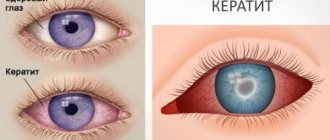
Red eyes are the main sign of uveitis in children at different stages of the disease.
Main signs, according to the type and location of uveitis:
- Front. The increased sensitivity of the eye, such as chronic conjunctivitis, redness of the whites, tearing, and decreased visual acuity come to the fore.
- Rear. The appearance of light flashes, “spots” before the eyes, decreased visual function, distortion of objects in the field of vision, swelling. There are practically no symptoms at an early stage.
- Spicy. An exacerbation of the disease is accompanied by sudden pressing pain in the eyes, which intensifies at night or when the light intensity changes. Acute uveitis is characterized by swelling of the eyelids, photophobia, and lacrimation. When gentle pressure is applied to closed eyelids, tension occurs in the area of the eyeball. This can characterize both an acute process and the addition of inflammation of the trigeminal nerve.
- Chronic. In the absence of significant symptoms and discomfort, epithelial cells begin to be deposited on the iris. Vision decreases and focus clarity decreases.
- Sluggish process. The intensity of pain in the eye varies greatly. The disease is accompanied by spontaneous exacerbations and self-healing.
- Necrosis. With retinal necrosis, children complain of a sharp decrease in visual acuity, acute pain, increased intraocular pressure and chronic headaches. After 2-3 months, the dangerous process of retinal detachment begins. Inflammation with necrosis first occurs as a one-sided process, and then spreads to the second eye.
- Tuberculous. Secondary pathology against the background of pulmonary tuberculosis and its complications. It is accompanied by the accumulation of subcutaneous fat and fat on the posterior membranes of the cornea, and the formation of a new vascular component. The pupil becomes covered with inflammatory nodes with new vessels, along its edge there are clusters of cells that look like small pieces of cotton wool. Discomfort increases, quality of life deteriorates sharply.
- Syphilitic. Uveitis against the background of syphilis is characterized by keratitis, neuritis of the nerves of the eye or maxillofacial apparatus, and damage to the retina. Syphilitic sore throat and papules on the skin and membranes of the oral cavity are often simultaneously diagnosed.
Patients complain of discomfort when blinking, a feeling of a foreign body in the eyes, and constant lacrimation. Clarity of vision disappears, cloudiness increases. During an ophthalmological examination of the fundus, the venous and arterial structures are sharply changed.
Complications and prevention
This disease can be prevented. To do this, you must follow the following recommendations:
- promptly treat infectious diseases;
- Wear safety glasses when performing work that is hazardous to the eyes;
- exclude injuries;
- prevent eye burns;
- visit an ophthalmologist periodically;
- monitor hormonal levels;
- do not contact with allergens;
- lead a healthy lifestyle.
The most common causes of uveitis are infection, trauma, and systemic disease. They need to be prevented or treated in the early stages. Most often, uveitis is a complication of another pathology. Prevention should be carried out from a young age. To protect children from this pathology, it is necessary to prevent bacterial and viral infections.
If uveitis does develop, the goal is to prevent complications. To do this, you need to visit your doctor in a timely manner and follow all his prescriptions. Self-medication can lead to dangerous complications, including loss of an eye. Thus, uveitis is a very common ophthalmological pathology.
Diagnostics
Timely diagnosis of uveitis in children is difficult due to the specificity of symptomatic manifestations. Most often, uveitis is diagnosed accidentally during an examination of the fundus or examination by a doctor regarding the patient’s complaints of visual impairment. A wide range of diagnostic measures are required to differentiate uveitis from other ophthalmological pathologies.
Diagnosis begins with an examination by an ophthalmologist, assessing the condition of the fundus and visual acuity.
Considering that the main reason for the progression of the pathology is the infectious-inflammatory process, a blood and urine test is required. To exclude pulmonary tuberculosis, fluorography is indicated.
Uveitis in children: how to treat posterior urolithiasis in a child, symptoms and treatment
Uveitis is a fairly common eye disease where the inflammatory process begins in the choroid.
Its structure consists of the iris, choroid, and ciliary body. Since the disease can cause blindness, timely diagnosis and treatment are very important.
Causes
Code according to the international classification of diseases ICD-10:
- H20 Iridocyclitis;
- H21 Other diseases of the iris and ciliary body;
- H30 Chorioretinal inflammation.
The disease is characterized by severe redness of the conjunctiva and deformation of the pupil.
Many different factors can cause the disease. Below we list the main ones:
- allergic reaction, diseases of the autoimmune system;
- eye injuries;
- impaired metabolism, as well as hormonal levels;
- viruses and infections that cause inflammation;
- the presence of fungal microorganisms and parasites;
- sepsis;
- diabetes;
- chronic inflammatory diseases;
- intoxication of the body;
- the presence of a genetic predisposition.
The disease can occur in people of different ages. In the elderly and children, infectious uveitis is observed in most cases.
Let's make life easier - learn about the causes and treatment of pain in the eyes from the article.
Diabetes mellitus often provokes the development of the disease
In almost half of cases, the disease is caused by pathogenic microorganisms:
- fungi;
- streptococcus;
- toxoplasma;
- treponema pallidum;
- herperviruses.
The disease may be a consequence of a progressive pathology in the body. Most often, the disease is diagnosed in patients with:
- ulcerative colitis;
- psoriasis;
- spondyloarthrosis;
- rheumatoid arthritis;
- multiple sclerosis;
- reactive arthritis;
- autoimmune thyroiditis.
Uveitis is also caused by ophthalmological diseases: conjunctivitis, blepharitis and scleritis.
Psoriasis can also cause uveitis
An effective medication for the treatment of bacterial infections in ophthalmology - instructions for Fucithalmic eye drops.
Kinds
The disease can be classified according to various criteria. Depending on where the disease is located, uveitis is divided into:
- Front. The most common form. It can be divided into cyclitis and iritis. In the first case, the inflammation affects the cicatricial body, in the second - the iris. If both of these elements are affected, then this pathology is called iridocyclitis. During the cold season, the disease may recur.
- Peripheral. In this form, both eyes become infected. Diagnosis is quite difficult, since inflammation affects an area that is difficult to study using standard methods. Namely the central region, retina, choroid, vitreous body.
- Rear. This form develops over a long period of time and appears quite late, since the symptoms are mild and do not worsen the general condition of the patient. Posterior uveitis is complicated by macular ischemia, macular edema, retinal detachment, and retinal vasculitis.
- Diffuse. It is less common than other types and is the most dangerous, since inflammation affects the entire choroid of the eye.
Only an ophthalmologist can accurately diagnose the disease and only using special diagnostic methods (visometry, ophthalmoscopy, visual examination).
When the prescription of Hilabak eye drops is indicated, read here.
Based on the nature of the course, uveitis is divided into:
- Spicy. Occurs for the first time. The symptoms are quite pronounced.
- Chronic. The disease can develop into this form if symptoms persist for a long time and there is no treatment. There is a change in periods of time during which the symptoms worsen and subside.
- Recurrent. In this case, the disease often worsens, and the patient again develops quite pronounced symptoms.
- Sluggish. In this case, the symptoms are mild and may last for more than 2 months.
Let us separately highlight rheumatoid serous uveitis. As a rule, it has a chronic course, the clinical picture is quite blurred. It is a rare form.
During the disease, corneal precipitates and posterior adhesions of the iris are formed, the ciliary body is destroyed, and the lens darkens. It lasts quite a long time and is difficult to treat.
May be complicated by the development of similar eye diseases.
There are many types of uveitis. In addition to those listed, they are also distinguished according to the nature of inflammation: purulent, exudative, fibrinous-lamellar, mixed type.
First of all, treatment is aimed at avoiding complications that can lead to vision loss.
A broad-spectrum antibiotic from the aminoglycoside group - instructions for Tobropt eye drops.
Why daily eye tonometry is performed, find out at the link.
Prognosis and prevention
Preventive measures include:
- Compliance with personal hygiene rules (daily eye care, use of a personal towel, etc.)
- Avoiding hypothermia;
- Prevention of eye injuries;
- Timely treatment of various diseases, especially ophthalmological, as well as allergic reactions.
If the treatment was comprehensive and timely, the recovery process takes no more than 6 weeks. The prognosis is less favorable if a complication of uveitis occurs, namely, cataracts, glaucoma, heart attack, retinal detachment or dystrophy develop, in which case blindness may occur.
Chronic uveitis is prone to relapses due to a general decrease in immunity.
GCS for topical use in ophthalmologyMost often the reason lies in the uncontrolled proliferation of streptococcus
Uveitis in children
In children, the disease can be caused by food allergies, vaccinations, and also occur through blood transfusions. Most often, infectious-allergic inflammation of the choroid is observed in children with impaired metabolism.
Treatment is similar to therapy in adults; it is necessary to carefully select drugs according to age and the form of the disease.
Metabolic disorders in babies can manifest themselves in the development of uveitis
Even the slightest redness of the eyes, itching, decreased vision, etc. should arouse suspicion. In this case, it is necessary to immediately consult a doctor, because the sooner treatment is started, the higher the prognosis for a favorable outcome.
Source: https://ProZrenie.online/zabolevaniya/vospalenie/simptomy-i-metody-lecheniya-uveitov.html
Treatment tactics
Treatment of uveitis in children can be conservative or surgical. Drug therapy is aimed at suppressing the inflammatory process, preventing the replacement of membranes with fibrous tissue, and eliminating the provoking factor.
Therapy is often prescribed in collaboration with a pediatrician, rheumatologist, pediatric allergist, infectious disease specialist and other doctors. For treatment purposes, the following medications are prescribed:
- Mydriatics. Eye drops with M-anticholinergics help dilate the pupil and prevent fibrotic complications. Effective in exacerbation of the disease.
- Non-steroidal anti-inflammatory drugs, glucocorticosteroids. They are used as symptomatic therapy to relieve pain, inflammation, and swelling. In addition, NSAIDs enhance the therapeutic effect of treating underlying diseases.
- Immunosuppressants. Used when hormonal therapy is ineffective. The administration of cytostatics is advisable in cases of autoimmune nature of the pathology and changes in connective tissue.
- Fibrinolytic agents. Required for the prevention of fibrinous plastic processes and other complications. Doctors prescribe enzyme preparations in the form of solutions for subconjunctival injections. Popular products in this group are Collalysin, Lidaza, Wobenzym.
- Preparations for detoxifying the body: saline or glucose-containing solutions, antihistamines, sorbents.
- Antibiotic therapy. Etiotropic antibiotic therapy is performed when uveitis is of a bacterial nature. First, broad-spectrum antibiotics are prescribed, and after analyzing the sensitivity of the pathogen, targeted ones are prescribed.
If etiotropic and symptomatic therapy is ineffective, the child’s quality of life does not improve, and the disease progresses, the question arises about the need for surgical treatment. In case of complicated uveitis for children, the following is performed:
- laser coagulation;
- laser synechiotomy;
- iridotomy;
- vitrectomy;
- evisceration of the eyeball.
Some methods can complement each other.
Treatment
Treatment of various forms of the disease uses pharmaceuticals, as well as surgical, laser, and extracorporeal therapy.
Drugs
Corticosteroids are used as anti-inflammatory drugs . If anterior uveitis, local hormonal corticosteroid therapy is sufficient. Treatment is supported by mydriatics: atropine, phenylephrine. Posterior uveitis will require corticosteroid injections into the skin of the lower eyelid. If the inflammatory process occurs with complications, the drug is used systematically.
When anti-inflammatory therapy has no effect, cytostatic treatment with Cyclosporine is used. Sometimes this drug is used in combination with Prednisolone.
Depending on the causative agent of the disease, the following drugs are used:
- Drugs of the tetracycline, sulfonamide group and aminoglycoside series are used to treat bruzcellous uveitis. Treatment is carried out quite quickly, within 4 weeks.
- To cure leptospirous uveitis, it will take several years of use of gamma globulins, doxycycline, amoxicillin, sulfones, and rimpamycin.
- In the treatment of syphilitic uveitis, benzylpenicillin compounds with benzathine, novocaine and sodium salts are used. If the child does not tolerate benzylpenicillin, the drug can be replaced with doxycycline, tetracycline, or erythromycin.
- If the disease has developed due to the activity of parasites - toxocariasis, treatment will consist of thiabenzene and mebentazole.
- Uveitis caused by toxoplasmosis is treated with the drugs pyrimethamine and sulfadimezine, or you can use a combination drug - Fansidar. Folic acid is prescribed simultaneously with pyrimethamine. An analogue of pyrimethamine is aminoquinol.
- To treat tuberculous uveitis, you will need to take isoniazid and rifampicin for about 2-3 months. Then rifampicin is replaced with ethionamide, therapy continues for another 3 months
- If uveitis is caused by a herpes virus, the disease should be treated with antiviral drugs: acyclovir, valacyclovir.
Operation
Surgical and laser therapy is used in cases of secondary glaucoma, complicated cataracts, and retinal detachment . Similar complications occur with traumatic uveitis.
Prevention
Carefully following the doctor's recommendations and attending all scheduled appointments with the rheumatologist and ophthalmologist, your child's vision problems should be under control - the main preventive measures.
It is recommended to visit doctors regularly (every 3–6 months) to ensure that there are no changes in the condition of the optical system.
Prevention is as follows:
- proper hygiene;
- minimizing injury;
- strengthening the immune system with drugs and hardening;
- minimizing exposure to toxic substances.
Cause of childhood uveitis
Uveitis can be caused by infection, injury, or an autoimmune or inflammatory disease. However, in many cases the exact cause cannot be determined. This type is known as idiopathic, non-infectious uveitis.
Uveitis is caused by inflammatory reactions within the visual organ. Inflammation is the body's natural response to germs, toxins, or tissue damage. The disease develops against the background of:
- Lyme disease or tuberculosis;
- viral infections - herpes or Epstein-Barr virus;
- in rare cases, fungal and parasitic infections;
- juvenile idiopathic arthritis, vasculitis or inflammatory bowel disease;
- trauma to the organs of vision, in some cases one eye may become inflamed due to severe trauma to the opposite eye (sympathetic uveitis).
In approximately half of the cases, the cause of the inflammatory process remains unknown.
Rheumatic eye diseases (Part 2)
Description
Sclerosing keratitis
a rare, but severe eye disorder, the cause of which, along with others, can be rheumatic disease. The process is predominantly bilateral, occurs more often in elderly and old people, begins with limited swelling of the sclera or with annular scleritis, in which inflammatory infiltration of the sclera surrounds the cornea in a ring.
The infiltrate forms a gelatinous shaft up to 1
cm, starting with a steep rise slightly away from the limbus and gradually flattening towards the arches of the conjunctival sac. The conjunctiva covering the shaft is pale or slightly hyperemic, the eye is moderately irritated.
Massive infiltration of predominantly deep corneal layers often spreads from the shaft into the cornea in separate wide “tongues”. The initially grayish areas of infiltration eventually turn white and become similar to sclera. This impression is reinforced by the individual blood vessels running through the cloudiness. Sometimes the infiltration and subsequent opacification surrounds all or most of the periphery of the cornea, but the center remains transparent and appears in the opacification ring as a microcorneal contact lens, which is why this pattern is called a “contact lens” cornea.
Patients are concerned about not intense, but constant eye pain, moderate photophobia, lacrimation, blurred vision due to reactive edema of the epithelium of the center of the cornea. A similar, but more severe lesion of the sclera, complicated by iridocyclitis, retinal detachment, and sometimes secondary glaucoma, was previously described under the name “progressive scleroperikeratitis”.
In cases of scleral swelling in a limited area (or areas) in the cornea, one, sometimes two or three deep limbal infiltrates also appear with separate vessels, less often without them. Turning into persistent opacities, their “tongues” make the contour of the cornea scalloped.
No matter how rheumatic sclerosing keratitis begins, it is usually accompanied by anterior uveitis or panuveitis expressed to varying degrees, and proceeds cyclically, with long-term remissions and exacerbations, often associated with colds, tonsillitis, etc.
In addition to the forms described, diseases of the sclera in rheumatism can manifest themselves as diffuse granulomatous scleritis, and in the most severe cases - in the form of perforating scleromalacia. The latter is manifested by the appearance in some area of the anterior part of the eyeball of a softening of the sclera, initially grayish-gelatinous, then, as the sclera thins, dark in color. Eye irritation and pain can be expressed to varying degrees. Despite the most vigorous measures, including scleroplasty, softening, having captured a fairly large area, steadily spreads into the depths and after some time perforates the wall of the eye. The disease ends with its atrophy.
Along with the anterior part, rheumatoid scleritis can affect the posterior pole of the eyeball. For example, malignant scleritis is well known. Developing close to the optic disc, it often mimics an intraocular tumor and is recognized only histologically after enucleation of the eye. Despite the diagnostic error, removal of the eyeball in such patients is justified, since the disease is incurable and fraught with serious consequences. However, such scleritis is observed very rarely.
Of much greater practical and scientific interest may be sluggish and imperceptible posterior rheumatic scleritis, which, however, causes weakening of the sclera and its stretching with the progression of myopia. This possibility was pointed out back in 1953 by N.M. Pavlov and L.A. Molchanova, who established a more frequent development and progression of myopia in children suffering from rheumatism. E. S. Avetisov draws attention to the importance of common diseases for weakening the sclera in mions.
All forms of scleritis in patients with rheumatism are considered as a single disease with differences only in the depth of the lesion, localization, extent along the surface of the eye, the severity of subjective and other symptoms, they are considered a manifestation of the true rheumatic process in the episclera rich in vessels and mesenchyme, as well as in scleral tissue and therefore All these diseases are combined into a single concept “rheumatoid scleritis”.
The authors attach the leading importance in its development to allergic-hyperergic reactions such as infectious allergies. Successful therapy, mainly with glucocorticoids, in the majority of patients with rheumatoid scleritis confirms the validity of this view. Nevertheless, the previous, more differentiated, isolation of sclerites is more convenient for the clinic.
In the treatment of episcleritis and scleritis, together with glucocorticoids, other antirheumatic and symptomatic therapy recommended above for tenonitis and myositis may be useful. Of course, uveitis and others, which complicate this entire pathology, require special measures.
Uveitis and chorioretinitis.
Rheumatism of the vascular tract is of utmost importance in ophthalmic-rheumatology and occupies a prominent place in all uveal pathology of the eye. According to domestic authors, this etiology is detected in 19,3%
from all uveitis, and according to foreign statistics - in
8-50%
. Rheumatic uveitis is primarily characterized by the diversity of its clinical forms and manifestations. The disease may involve predominantly the anterior part of the uveal tract, the flat part of the ciliary body, the choroid, or all parts of the vascular tract may be affected simultaneously.
Anterior, peripheral, posterior uveitis and panuveitis develop accordingly. In any case, the process can be acute or chronic, occur against the background of general rheumatic symptoms or without them, and be predominantly exudative or transudative (serous).
Like the rheumatic diseases of the eye described above, uveitis is more often observed in young and middle-aged patients with rheumatism, they are prone to relapses, which, like primary eye diseases, more often occur in spring and autumn, and are often associated with colds, sore throat, etc. Among patients with rheumatic uveitis women predominate.
In the nosological diagnosis of these sufferings, biomicroscopy of the anterior and posterior parts of the eyeball, examination of the vitreous body, and ophthalmoscopy are of great importance. Etiological diagnosis, as with any other rheumatic eye disease, is helped by anamnesis, general clinical and instrumental examination, laboratory data, consultations with rheumatologists, cardiologists, pediatricians, therapists, otolaryngologists, etc.
In unclear cases, it is necessary to exclude another etiology; treatment is quite legitimate. However, even if all possibilities are used, the rheumatic nature of uveal pathology may remain presumptive or unrecognized. Particular difficulties in etiological diagnosis are presented by the monosymptomatic course of rheumatism, when its only manifestation is uveitis.
Due to such patients, as well as the incomplete use of diagnostic methods, the rheumatic nature of uveal processes is not always recognized and some of them join the category of uveitis with an unclear etiology. This can, apparently, explain the significant discrepancy in statistical data in the literature.
Rheumatic plastic anterior uveitis.
It begins acutely or with conjunctivitis. In the first case, all the symptoms of the disease increase rapidly. When conjunctivitis precedes uveitis, the patient is bothered for several days by the sensation of a foreign body in the eye, pain, and serous discharge, but these phenomena are moderate.
If the patient consults a doctor in this phase of the disease, which rarely happens, then examination will reveal hyperemia and looseness of the conjunctiva of the eyelids, conjunctival injection and unmotivated mild mydriasis. All these phenomena persist 2—5
days, then the conjunctival injection turns into ciliary, mydriasis gives way to miosis and plastic anterior uveitis quickly develops, proceeding further as rapidly as during its development without conjunctivitis.
Already on the 2-3rd
the day after the first signs of uveitis appear, all its symptoms are completely different (Fig. 20). The patient is bothered by severe aching pain in the eyes, lacrimation, and photophobia. The eyelids may be somewhat swollen, the eye is very irritated, and the conjunctiva is also swollen to the point of chemosis. Ciliary pain may be so severe that the patient does not allow the eyes to be touched.
When examined with lateral lighting, swelling of the cornea and banding of its posterior layers are revealed. Biomicroscopically it is discovered that the stripes are nothing more than folds of Descemet's membrane. Intersecting each other, these folds form a kind of lattice, which is considered to some extent characteristic of rheumatic uveitis (lattice symptom).
If the eye gets sick for the first time, then its cornea retains tactile sensitivity well, which makes it possible to exclude the viral nature of the disease.
Changes in the iris with the described uveitis are so significant that they differ even with a pronounced violation of the transparency of the cornea. Due to edema, hyperemia and predominantly superficial exudation, the color of the iris changes, its pattern is blurred, and cone-shaped and wider pigmented posterior synechiae appear along the pupillary edge.
Their strength in the first days is small, but if there is a delay for any reason in prescribing mydriatics, the synechiae become strong.
In patients with a more transparent cornea, opalescence of the moisture of the anterior chamber of the eye is detected, grayish-white or grayish-yellow, sometimes jelly-like exudate on the anterior surface of the iris, and where there is no exudate, the blood-filled vessels of the iris are often clearly visible.
If the environment allows, then using a slit lamp, changes in the vitreous are detected: increased Tyndall phenomenon, absence of retrolental space and anterior limiting membrane, abundant inflammatory suspension behind the lens. These changes, as well as the pronounced ciliary soreness noted above, together with hypotension, indicate inflammation of not only the iris, but also the ciliary body.
Thus, the process manifests itself as iridocyclitis, and not iritis, although the latter is mentioned very often in the literature. Ophthalmoscopically, such patients sometimes reveal moderate hyperemia of the optic nerve head. The process is often one-sided, and if both eyes are affected, one eye may first become ill, and a little later the other eye.
Without treatment, the acute period of anterior plastic rheuveitis can last several weeks, then the inflammation gradually disappears, but powerful posterior synechia remains, up to the fusion and fusion of the pupil, cataracts, secondary glaucoma and other complications characteristic of severe uveitis can develop. Repeated, often repeated attacks (relapses) of rheumoveitis that occur weeks or months later have a negative impact on the condition of the eye and its functions.
They occur in the same or other forms of uveitis and can be protracted. Their irreparable consequences are often observed. Timely active treatment, especially of the primary disease, is very effective. Using appropriate means and methods, it is possible to quickly relieve pain in the eye, reduce its irritation, break up existing ones and stop the formation of new posterior synechiae, etc. Complete relief of the disease often requires hospital treatment for 1—17
g month When a patient is discharged for outpatient follow-up treatment and observation, it cannot be guaranteed that after some time he will not be admitted again with a relapse.
Hemorrhagic rheumatic uveitis
the main manifestations are similar to the previous form, differing from it only in more massive exudation into the anterior chamber of the eye, and blood is mixed with the exudate. Symptoms of uveitis in such patients, in particular pain in the eye, its irritation, ciliary tenderness, are sharply expressed, the cornea is swollen, there is a “grid” symptom, and in the anterior chamber of the eye exudate is found that is dirty red in some areas and grayer in other areas .
Along the entire circumference or over its large extent, exudate fills the corner of the anterior chamber, and closer to the pupil, loose masses cover the iris and the pupil area, which cannot be seen, especially if the pupil is narrow. As the exudate resolves, posterior synechiae are detected, often powerful, even circular. However, the formation of such synechiae can be prevented by timely vigorous atropinization. Despite its very severe course, hemorrhagic rheuveitis responds well to therapy.
Serosoplastic anterior rheumatic uveitis
is observed less frequently than previous forms, and differs from them in a less acute, but often longer course, the formation, along with synechia, of precipitates on the posterior surface of the cornea, the absence of clear signs of exudation of the iris, overflow of its vessels with blood, etc.
Only in severe cases of the disease, along with precipitates, signs of the “grid symptom” can be observed, a noticeable blurring of the iris pattern, a change in its color, and an increased tendency for it to fuse with the lens.
Usually the disease begins with mild aching pain in the eye, slight photophobia, and the appearance of a pericorneal injection around the limbus. Gradually, these phenomena increase; when examined with a slit lamp, precipitates are detected, mostly small, grayish, at the beginning of the disease, round in shape, with clear boundaries, protruding in clumps into the anterior chamber of the eye.
Every day there are more and more “fresh” precipitates. From the Türkic line they spread in both directions, first in a triangle with the base downwards, then they occupy the entire lower half of the cornea, and some of them rise above its horizontal meridian. After a few days, the precipitates that appeared first begin to flatten, their contours become scalloped, and the surface is inhomogeneous, which indicates the aging of the precipitates and their resorption that has begun.
However, it proceeds slowly; instead of disappearing precipitates, new ones appear, and in general they can last for months. Precipitates clearly predominate in the picture of the disease, since the phenomena of exudation and formation of posterior synechiae in such patients are often moderately expressed, and during treatment the synechiae rupture and quickly cease to form again. In patients with this form of rheumoveitis, the vitreous body is almost always available for study, in which significant changes are revealed using a slit lamp.
In the Vogt section (macroscopic examination of the optical section), a diffuse suspension may be visible behind the lens, enhancing the glow of the light beam of the lamp illuminator, and sometimes posterior, less often anterior vitreal detachments are revealed. Using a slit lamp, instead of moiré of the anterior limiting membrane and filamentous skeleton, pronounced granular destruction of the vitreous body is revealed.
An abundant suspension of small grayish round particles occupies the anterior and central vitreal sections. Suspension particles move with eye movements, many of them settle on the remaining fibers or, sticking together, form coarser opacities in the form of lumps and flakes. With lens attachment. Coarse suspension, although not so intense, is often found in the preretinal vitreal layers.
Vitreous detachment in such patients is a consequence of its long-term destruction. However, much more often, even a very thick suspension disappears over time, especially under the influence of resorption therapy, which indicates its predominantly inflammatory nature. While this suspension is contained in the vitreous body, the uveal process cannot be considered complete.
Serous-plastic uveitis is the most common form of rheumatic uveitis in children. In childhood, especially in early and preschool age, such uveitis occurs without irritation of the eye, although small grayish precipitates, swelling of the corneal endothelium, posterior synechiae, and often translucent gelatinous exudate in the anterior chamber of the eye are always found. The process is well controlled by complex therapy.
Serous anterior rheumatic uveitis
It is rarely observed and differs very little from similar uveitis of other etiologies. The rheumatic nature of the disease can only be established with a general and special examination. The main manifestations of the disease are corneal precipitates and granular destruction of the vitreous body, which, when examined with a slit lamp, look similar to the form of rheumatic uveitis described above, but differ from it in that they occur in a quiet eye and without noticeable signs of exudation.
The disease is characterized by a chronic, multi-month course, is less responsive to therapy, can be complicated by secondary glaucoma, cataracts, sometimes develops into serous-plastic and plastic uveitis, but more often with persistent treatment it still ends without significant consequences. The tendency to relapse is very high.
Nodular and rheumatic uveitis
Currently, it is very rare, mainly in patients with rheumatoid arthritis. Inflammatory changes in the eye are usually moderately expressed: mild pericorneal injection, mild hyperemia and blurred pattern of the iris are observed, in the tissue of which vaguely capitulated, grayish-pink color, the size of a poppy seed to a match head, nodules are visible.
In varying quantities, rarely abundant, they are scattered throughout the entire circumference of the pupillary girdle of the iris. If the irritation of the eye is more pronounced and the swelling of the iris is more massive, then the nodules are barely noticeable with a slit lamp. Through 1—2
weeks they disappear, leaving behind atrophic spots. New nodules appear to replace those that have resolved, posterior synechiae appear, and relapses of the disease in one form or another lead to
To secondary glaucoma and other complications. The nodules are rheumatic granulomas. An unfavorable outcome can be prevented by treatment, which gives good results.
Rheumatic panuveitis
can proceed according to the type of any of the above forms of anterior rheumatic uveitis, differing from them primarily in the participation in choroidal disease. Along with inflammatory changes in the anterior parts of the uveal tract of a serous or serous-plastic nature, in such patients with transparent eye media, fresh or older choroidal foci are detected very early and after clearing of the media in cases of clouding.
More often they are in small quantities (2—3
) are located in the middle periphery or in the posterior pole of the fundus and do not exceed
1/4-1/3
PD in size. Fresh lesions have the appearance of loose formations, with unclear boundaries, yellowish in color, slightly protruding into the vitreous body. Old lesions are well defined, white, partially pigmented, and around them there may be an area of depigmentation of the pigment epithelium or pigment sputtering.
Along with this, at the beginning and height of the disease, as a rule, hyperemia of the optic nerve head is observed. However, similar types of lesions and disc hyperemia occur with choroidal pathology of other very different etiologies. It is not the lesions themselves that are suspicious of rheumatic lesions, but their detection against the background of anterior uveitis and, to some extent, their small number and small size.
Rheumatism is even more evidenced by the phenomena of retinovasculitis and, especially, vasculitis of the choroidal vessels that often accompany chorioretinitis. Sometimes the participation of the vascular network in panuveitis in rheumatism is manifested by diffuse choroiditis, characterized by effusion, which, permeating the choroid and retina for a considerable extent, also penetrates the vitreous body and causes clouding of its preretinal layers, granular destruction, formation of films, etc.
During ophthalmoscopy, the fundus of such patients is visible behind the “fog.” If it is possible to conduct a biomicroscopic examination with an attachment lens, then a thickening of the optical section of the retina due to its edema is revealed. All these changes can last for months after the symptoms of anterior uveitis disappear, then the vitreous body clears up, although signs of its destruction remain, and macular depigmentation, cysts, and exudative retinal detachment that does not require surgical intervention may be detected in the fundus.
In the form of panuveitis, ocular rheumatism can occur in school-age children, but this form is observed less frequently than anterior uveitis.
Peripheral rheumatic uveitis
is a kind of transitional form between anterior and posterior uveitis, since the disease develops in the flat part of the ciliary body. The process can occur without any features characteristic of rheumatism or in the form of annular exudative pseudomembranous diclitis. This disease occurs in patients with rheumatism in one or both eyes, also starting with inflammation of the flat part of the ciliary body.
In the absence of signs of uveitis, the patient is bothered by blurred vision and “flying spots”. Upon examination, pupil rigidity, an inflammatory suspension in the vitreous body, and at the extreme periphery of the fundus there is a low, circular ciliary detachment with scalloped contours are revealed.
The grayish-brown blisters of this detachment are tightly stretched, translucent during diaphanoscopy, remain stable for months, but immediately disappear after the release of subchoroidal fluid, and glucocorticoids prevent their reappearance. A process not recognized in a timely manner leads to the constant development of flat retinal detachment mainly in the lower half of the eye, and choroidal foci appear throughout the fundus of the eye, posterior synechiae may appear, and therefore the disease becomes severe.
Posterior rheumatic uveitis
during systematic observation of patients with rheumatism, rheumatism is diagnosed more often than lesions of the anterior part of the vascular tract. The forms of manifestation of this pathology are also varied, the course is often severe, and the clinical data are not much different from those for a similar pathology of another genesis.
If the nosological diagnosis of these lesions is made on the basis of ophthalmological, bioophthalmomicroscopic, perimetric, electrophysiological and other studies of the eye, then the rheumatic nature of the process, as with all manifestations of ocular rheumatism, is established through a general examination of the patient, exclusion of other etiologies, and treatment.
Rheumatic disseminated chorioretinitis
It often develops in one eye, proceeds sluggishly and for a long time and, without affecting the optical zone, often remains unrecognized, or many years later old choroidal foci and sclerosis of the choroidal vessels (choroidosis) are discovered. Ophthalmoscopically, fresh and old lesions in such patients look the same as in rheumatic panuveitis. The predominance of small foci (no more than 1/2
PD) indicates that rheumatism primarily affects the zone of the middle vessels and the choriocapillaris layer of the choroid.
Secondarily, the retina is involved in the process, damage to which leads to depigmentation of the pigment epithelium in some areas and accumulations of pigment in other areas. O.I. Shershevskaya describes the sclerosis of choroidal vessels often observed in such patients in areas of depigmentation, the development here of “the mooring of proliferative choroiditis in the form of delicate banded cords, films, in places covering the choroidal vessels.”
She also draws attention to the accompanying stripes identified biomicroscopically in these patients along the medium and large choroidal vessels in cases where such vessels are accessible for inspection. The author considers these greyish-colored, wide, vaguely contoured stripes, appearing as a result of increased permeability of the vascular wall, to be plasmorrhagia and, in accordance with the concept of A.I. Nesterov and Ya.I. Sigidin about rheumatic lesions of the capillaries, evaluates them as a characteristic sign of rheumatic lesions of the eye. It is no coincidence that with choroiditis, perivasculitis of the choroidal vessels is detected.
Rheumatic diffuse chorioretinitis
is in many ways similar to panuveitis, in which the choroid is diffusely affected, but occurs without anterior uveitis. Swelling of the choroid and retina and effusion into the vitreous in such patients impair visual functions, which forces them to consult a doctor.
As long as the opacification of the preretinal vitreal layers remains and the fundus of the eye is unclear, no features of the disease that would indicate its rheumatic etiology can be identified.
After resorption of vitreous opacities, which lasts for months, the same pathology is found in the fundus as in focal rheumatic chorioretinitis, excluding, however, chorioretinal and choroidal foci. Areas of retinal depigmentation after such chorioretinitis can be extensive, and sclerosis of the choroidal and retinal vessels, inadequate for age and refraction, is noticeably pronounced. Secondary degenerative processes in the retina and resulting detachments are possible.
Jensen's rheumatic chorioretinitis
begins with the appearance at the edge of the optic nerve disc on either side of a white, raised above the fundus, loose, similar to a piece of cotton wool, size 7g-11/g
RO.
The nerve disc is slightly swollen, with a ring of 17g-2
RB around it.
there is an edematous retina, in the field of view there is a fan-shaped or sectoral Jensen's scotoma. This condition lasts 7-10
days, then the lesion begins to settle and decreases somewhat. Its boundaries become clearer.
Pigment appears around and in the lesion itself, and swelling of the disc and retina disappears. A choroidal lesion remains near the optic nerve head, the size of 74—1
RO. Sometimes, accordingly, Jensen's scotoma or paracentral scotoma is preserved without breaking through to the periphery.
Since similar chorioretinitis of tuberculous and other nature proceeds similarly to that described, concomitant changes, primarily in the radical and choroidal vessels, which often respond to the disease by disrupting the permeability of their walls, vasculitis, etc., help to suspect its rheumatic origin.
Rheumatic central chorioretinitis also does not have noticeable differences from similar processes of other etiologies. Initially, swelling of the retina of the center of the posterior pole of the eye, including the macular area, occurs.
The affected area acquires a grayish color and a disc up to 3-4 RB in size protrudes above the surrounding retina, simulating to some extent its detachment. Vision decreases to hundredths, a central relative (depression), and sometimes even absolute (defect) scotoma is found in the field of vision.
The optical section of the retina is greatly thickened, its transparency is impaired. During 2—3
During the week, the swelling increases, “subretinal precipitates” begin to appear through the clouded retina as white dots, and small choroidal foci can be visible.
The process continues for 11/2-2
months, then the edematous disc begins to settle and decrease in size, the retina takes on an almost normal appearance, and vision is restored. At the site of edema, a slight spray of pigment is visible, sometimes small foci of choroidosis.
The outcome is favorable, but the risk of relapse is very high. The rheumatic etiology of the process is established primarily on the basis of the general signs of rheumatism and by excluding other etiologies.
The reaction of eye vessels to rheumatism is not limited to chorioretinovasculitis. Much more often than such, patients with rheumatism experience circulatory disorders with corresponding changes in blood vessels, in particular the conjunctiva and fundus. Thus, when examining with a slit lamp in patients with rheumatism, a narrowing of the adductor and expansion of the abducent limb of the vascular loops at the limbus are often detected with a slowdown in blood flow in them. Corkscrew-shaped tortuosity of individual vessels, their expansion in the form of ampoules, and pinpoint hemorrhages are also revealed.
In the fundus, vascular disorders caused by rheumatism can be of the nature of functional angiopathy of the retina and choroid. Like angiopathy of any other etiology, they are manifested by narrowing, less often dilatation of the retinal arteries, and the appearance of transudate stripes along them. The same changes occur in the choroid and, when examined with a lens attachment, can be observed in areas of the choroid accessible to inspection.
Even more often, microcirculatory disorders in the fundus of the eye, which are in the nature of microangiopathies, are detected bioophthalmomicroscopically. This is the name for the expansion, tortuosity, uneven caliber of the perimacular capillaries of the retina, the appearance of yellowish-white pinpoint spots around them, and the sputtering of pigment. V.P. Zavgorodiya observed these changes in 52%
examined patients with rheumatism. She considers them so specific for rheumatism that she even suggests using them for the purpose of diagnosing it.
Diseases of the optic nerve of rheumatic origin are rare. The literature describes only isolated cases of retrobulbar neuritis, papillitis, and optic nerve atrophy caused by rheumatism. Much more often, patients with rheumatism experience blanching of the optic discs with preservation of full visual acuity, but a narrowing of its field by 10—30
°. O.I. Shershevskaya (1965) explains this blanching by some disturbance in the nutrition of the disc due to the narrowing of the vessels supplying it with blood. The reversibility of blanching indicates its predominantly functional nature.
Due to intracranial hypertension, congestive discs can develop in rheumatic brain lesions. Cerebral rheumatism can also cause damage to the chiasm, visual pathways and centers that fall under the competence of neurologists and neuro-ophthalmologists.
Article from the book: Therapeutic ophthalmology | Krasnov M.L.; Shulpina N.B..