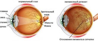
Retinal functions
The retina perceives ambient light and transfers the received information to the brain. For full visual functionality, the presence of receptors is necessary:
Externally, the organ is characterized by the following properties:
- transparency;
- softness;
- inelasticity.
According to anatomy, the retina lines the inside of the eyeball. Due to its functionality, the visual organ provides:
- Central vision. Allows you to clearly see objects, read and drive. Thanks to central vision, a person sees objects at a distance.
- Peripheral vision. Thanks to this type of vision, a person orients himself in space and sees objects located to the side.
- Perceives color. Thanks to the visual organ, the eye distinguishes shades.
- Ability to see at night. The retina perceives surrounding objects even in low light conditions or complete darkness.
Thanks to the organ, the light entering the visual apparatus is converted into a nerve impulse. After which it enters the cerebral cortex and the processing of the received information begins.
Causes
This disease mainly occurs in people who have white skin color. This is a unique fact. People who lead an unhealthy lifestyle and are overweight have an increased risk of encountering pathology. The main reasons that lead to thinning of the retina include:
- diabetes;
- impaired renal and liver function;
- hypertension;
- complications after ARVI, influenza;
- infectious eye diseases;
- the presence of myopia and farsightedness;
- hereditary predisposition.
Each patient has individual reasons for the development of the disease. Their presence and method of treatment can only be determined after a comprehensive examination. If there are representatives in the family who are faced with such a pathology, everyone needs to undergo examinations with a doctor more often. This will help to identify thinning in time and take the necessary measures.
Structure
According to the structural division of the visual organ, several areas are distinguished:
- Optical part. It occupies almost the entire scale of the retina, namely 2 parts out of 3. It is a thin and transparent structure that is filled with nerve fibers.
- The blind part. It is characterized by smaller sizes and consists of a pigmented layer.
The retina is nourished by the vitreous body and vascular formations. The visual organ is not uniform in thickness and consists of 3 parts:
- 0.4 mm is occupied by a thickened area, which is located close to the edge of the optic nerve;
- 0.1 mm in thickness is the retinal zone near the dentate line;
- 0.075 mm constitutes the main area responsible for the perception of the environment.
The cells of the retina of the eye consist of 3 types of neurons:
- photoreceptor neuron;
- association neuron;
- ganglion neuron, reaches brain structures.
Neurons are responsible for transmitting impulses and processing perceived information.
Diagnostics
Diagnosis is carried out by an ophthalmologist. Initially, it is necessary to study the patient's medical history to exclude the presence of predisposing factors. After this, you should listen to the patient's complaints and conduct a visual examination. Often a person can talk about the presence of spots or films before the eyes. They can be located in different areas, depending on the location of the thinning. To establish an accurate diagnosis, the doctor uses the following diagnostic methods:
- ophthalmoscopy – allows you to study the condition of the fundus of the eye;
- measurement of intraocular pressure;
- visual acuity test;
- determination of the state of visual fields;
- tests for the perception and discrimination of colors and shades;
- Ultrasound;
- MRI and CT.
After a thorough diagnosis, the doctor can determine the cause and type of disease. This is very important, because the method and success of therapy will depend on it. It is also important to consult a doctor in a timely manner and not try to get rid of unpleasant symptoms on your own. Lack of treatment can lead to severe consequences. Doctors especially do not recommend self-medication with folk remedies.
Layering
According to its structure, the retina consists of about 10 layers, which respond and perform various functions:
- Pigment epithelial cells. They absorb light rays and prevent them from scattering.
- Receptor layer. Consists of tentacles that extend from the epithelial structure. Prevents light from scattering between receptors. The zone is characterized by the absence of vascular formations.
- External limiting membrane. The region consists of Müller cells, which contain segments of photoreceptors. Under a microscope, the accumulation of cellular structures has a homogeneous structure.
- Nuclear layer. The nuclei of cones and rods are located in this zone.
- Outer mesh. Thanks to this area, contacts are formed with the postsynaptic elements of neurons.
- The inner layer is nuclear. Consists of several types of interneurons, namely bipolar, amacrine, horizontal and interplexiform structures.
- Mesh inner. Contains presynaptic and postsynaptic elements.
- Ganglion cells. Responsible for transmitting information to the brain.
- Optic fiber. The area consists of ganglion cell axons that are adjacent to the vitreous body.
- Internal limiting membrane. Represents processes of Müllerian cells.
Symptoms
Each type of this disease is accompanied by severe symptoms. In the peripheral form, the initial stage does not cause pain. There are also no visual defects observed. The first symptoms begin to appear when the retina ruptures. Often patients who are faced with this form of the disease begin to complain of the appearance of black spots before their eyes. This makes it difficult to lead a normal lifestyle.
When the form is wet, visual distortion of objects begins. A narrowing of the visual fields may also occur. Sometimes this causes the visual fields to begin to fall out. The main symptoms of retinal thinning include:
- decreased visual acuity;
- disorientation in a dimly lit room;
- dysfunction of the receptors that are responsible for twilight vision;
- color vision impairment;
- the appearance of a film and blurred vision.
Each patient experiences symptoms differently. If the disease appeared in childhood, it may develop slowly and not cause suspicion among parents. A small child will not be able to talk about what is bothering him on his own. Therefore, in order to avoid such consequences, it is necessary to undergo a scheduled examination by a doctor in a timely manner.
How the retina works
The mechanism for receiving and transmitting information to the brain by the retina is based on the following stages:
- the light signal affects the membranes of cones and rods, which reduces their permeability;
- a current of ions is obtained, which is characterized by a certain retinal potential;
- the potential spreads through ganglion structures that create nerve impulses.
The visual organ evaluates the environment according to the following parameters:
- image spectrum;
- illumination level;
- contrast.
If there is a pathological condition in any part of the retina, the mechanism of its operation is disrupted, which is why corresponding symptoms arise.
Complications
The main complication is irreversible processes in the retina. This leads to impaired visual acuity. Sometimes images and objects may become distorted. Typically, such complications occur in the absence of proper and timely treatment. With proper treatment, retinal thinning rarely has serious consequences. With the help of laser coagulation you can get rid of this pathology.
It is important to consider that in old age, ophthalmological diseases are difficult to treat. Therefore, often against such a background, irreversible processes occur in the form of a decrease in the quality of vision. The congenital form of the disease is also difficult to treat. A person always has to take vitamin complexes and supporting medications for the visual organs. During prolonged exercise, it is recommended to use drops to prevent the development of dry eye syndrome.
Symptoms of retinal diseases
Symptomatically, pathologies of the visual organ are characterized by the following manifestations:
- decreased visual acuity;
- narrowing of the field of vision or the appearance of blind spots;
- deterioration of adaptation to darkness;
- the appearance of darkened areas;
- vagueness of letters when reading;
- development of color perception anomalies;
- deterioration of orientation in space in poor lighting;
- the appearance of blurred images;
- the formation of sparks in front of the gaze.
With problems with the retina, there is no pronounced pain syndrome, since the nerve fibers in the structure of the visual organ work in a different direction.
Classification
The disease is acquired and hereditary in nature. Ophthalmologists divide the extreme into two types: pigmented (occurs due to dysfunction of certain receptors), dotted white (usually occurs in childhood and is characterized by slow development).
The acquired form is often associated with age-related changes in the body. It is mainly diagnosed in older people. The disease is accompanied by the development of cataracts. This type of disease is divided into:
- Peripheral – leads to a decrease in visual acuity and quality. This type of thinning occurs as a result of injury or an inflammatory process.
- Central - the lesion leads to disruption of central vision. Significantly affects daily lifestyle.
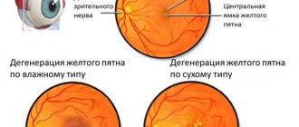
The central view has two distinct forms, which have certain differences:
- wet - leads to disruption of the vascular system in the visual organs, as a result, fluid begins to accumulate under the retina, which circulates in the vessels;
- dry – a disorder of the vascular system, which leads to the accumulation of foods and the breakdown of nutrients.
Only an ophthalmologist can determine the type of retinal thinning after a thorough examination. For this purpose special equipment is used. First of all, the presence of a genetic predisposition should be excluded. This type of pathology can be detected in young children.
List of retinal diseases
Even in the absence of pain, it is possible to develop pathological conditions in the area of the visual organ:
To carry out diagnostics and timely determine the pathology, consultation with an ophthalmologist is necessary. Predisposed to problems with the visual apparatus:
- elderly category of the population;
- pregnant women;
- patients with sugar pathology;
- people with myopia or farsightedness;
- female category of the population;
- people with hypertension;
- genetically predisposed;
- overweight people.
Pathologies are eliminated using drug therapy and surgical interventions. Treatment of retinal pathologies is not always characterized by positive results. In case of detachment or atrophy, the main goal of treatment is to stop the progression of the disease, since it is impossible to completely cure the pathology.
Symptoms of retinal dystrophy: how to identify the disease
Persons who are predisposed to developing the disease should visit an ophthalmologist 1-2 times a year. The main danger of the anomaly is that in the first stages it is asymptomatic. However, over time, signs of violations begin to appear more and more noticeably.
It is important for the patient to pay attention to the following symptoms:
- Loss of brightness of colors in the perception of the surrounding world. Contrast appears less and less.
- The outlines of objects and contour lines of objects lose clarity. The patient wants to turn on the light or add lighting in order to gain the perceptual features that were inherent to him before.
- While reading a book or looking through a newspaper, a person discovers that he does not see all the letters even with glasses that previously made his vision sharp and clear. The same thing happens with reading texts on the Internet.
Gradually, all these changes lead to loss of quality of vision near and at long distances. Then a spot appears in the visibility zone that interferes with viewing. At first it is slightly cloudy, still transparent, then it grows and becomes completely dark. Over time, a person loses the ability to distinguish faces and see objects in the environment.
Age-related macular degeneration affects both eyes, one of which more rapidly loses the ability to see normally. The second eye may be affected by pathology after 4-7 years. When you detect the first signs of an anomaly, you need to carefully monitor your health.
General symptoms of retinitis
In any form of this ophthalmological disease, all vision functions are affected.
The patient is concerned about the following symptoms:
- decreased visual acuity;
- color vision impairment;
- narrowing of visual fields;
- diplopia;
- the appearance of spots before the eyes, flashing flashes, sparks, lightning;
- night blindness;
- blurred vision in bright light.
There are also signs that are visible only under a microscope: optic nerve atrophy, vascular damage, hemorrhages in the retina. An accurate diagnosis will require a lot of research, as there are so many causes and forms of retinitis. Treatment is determined by these factors.
Kinds
Due to delamination:
- Traumatic;
- Rhegmatogenous;
- Traction;
- Exudative.
The rhegmatogenous form is the most common in ophthalmopathology. The “rupture” allows fluid to penetrate from the cavity of the gel-like substance into the subretinal space, which leads to the separation of the photosensitive membrane.
| If diagnosis and treatment are not timely, a retinal tear occurs. |
Physiology of color perception
- The physical essence of light and color
- Perception of color by the eye
- Color rating
- Color spaces
- Physiology of retinal receptors
Color perception (color sensitivity, color perception) is the ability of vision to perceive and transform light radiation of a certain spectral composition into the sensation of various color shades and tones, forming a holistic subjective sensation (“chromaticity”, “chromaticity”, coloring).
Color is characterized by three qualities:
- color tone, which is the main characteristic of color and depends on the wavelength of light;
- saturation, determined by the proportion of the main tone among impurities of a different color;
- brightness, or lightness, which is manifested by the degree of proximity to white (the degree of dilution with white).
The human eye only notices color changes when the so-called color threshold (the minimum color change noticeable to the eye) is exceeded.
The physical essence of light and color
Visible electromagnetic vibrations are called light or light radiation.
Light emissions are divided into complex and simple.
White sunlight is complex radiation, which consists of simple color components - monochromatic (one-color) radiation. The colors of monochromatic radiation are called spectral.
If a white beam is decomposed into a spectrum using a prism, you can see a series of continuously changing colors: dark blue, blue, cyan, blue-green, yellow-green, yellow, orange, red.
The color of the radiation is determined by the wavelength. The entire visible spectrum of radiation is located in the wavelength range from 380 to 720 nm (1 nm = 10-9 m, i.e. one billionth of a meter).
The entire visible part of the spectrum can be divided into three zones
- Radiation with a wavelength from 380 to 490 nm is called the blue zone of the spectrum;
- from 490 to 570 nm - green;
- from 580 to 720 nm - red.
A person sees different objects painted in different colors because monochromatic radiation is reflected from them in different ways, in different proportions.
All colors are divided into achromatic and chromatic
- Achromatic (colorless) are gray colors of varying lightness, white and black. Achromatic colors are characterized by lightness.
- All other colors are chromatic (colored): blue, green, red, yellow, etc. Chromatic colors are characterized by hue, lightness and saturation.
Color tone is a subjective characteristic of color, which depends not only on the spectral composition of the radiation entering the observer’s eye, but also on the psychological characteristics of individual perception.
Lightness subjectively characterizes the brightness of a color.
Brightness determines the intensity of light emitted or reflected from a unit surface in a direction perpendicular to it (the unit of brightness is candela per meter, cd/m).
Saturation subjectively characterizes the intensity of the sensation of a color tone. Since not only the radiation source and the colored object, but also the eye and brain of the observer are involved in the emergence of the visual sensation of color, some basic information about the physical essence of the process of color vision should be considered.
Perception of color by the eye
It is known that the eye is similar in structure to a camera, in which the retina plays the role of a photosensitive layer. Radiations of various spectral compositions are recorded by retinal nerve cells (receptors).
Receptors that provide color vision are divided into three types. Each type of receptor absorbs radiation differently from the three main zones of the spectrum - blue, green and red, i.e. has different spectral sensitivity. If blue zone radiation hits the retina, it will be perceived by only one type of receptor, which will transmit information about the power of this radiation to the observer’s brain. The result will be a blue sensation. The process will proceed similarly if the retina of the eye is exposed to radiation from the green and red zones of the spectrum. When two or three types of receptors are simultaneously excited, a color sensation will arise, depending on the ratio of the radiation powers of different zones of the spectrum.
With the simultaneous stimulation of receptors that detect radiation, for example, the blue and green zones of the spectrum, a light sensation may arise, from dark blue to yellow-green. The sensation of more blue shades of color will occur in the case of greater radiation power in the blue zone, and green shades - in the case of greater radiation power in the green zone of the spectrum. Equal radiation power from the blue and green zones will cause a sensation of blue color, green and red zones - a sensation of yellow color, red and blue zones - a sensation of purple color. Cyan, magenta and yellow are therefore called dual-zonal colors. Equal radiation power from all three zones of the spectrum causes the sensation of gray color of varying lightness, which turns into white with sufficient radiation power.
Additive light synthesis
This is the process of obtaining different colors by mixing (adding) radiation from the three main zones of the spectrum - blue, green and red.
These colors are called the main or primary radiations of adaptive synthesis.
Different colors can be produced in this way, for example, on a white screen using three projectors with filters of blue (Blue), green (Green) and red (Red). In areas of the screen illuminated simultaneously from different projectors, any colors can be obtained. The color change is achieved by changing the ratio of the power of the main radiations. The addition of radiation occurs outside the observer's eye. This is one of the types of additive synthesis.
Another type of additive synthesis is spatial displacement. Spatial displacement is based on the fact that the eye does not distinguish separately located small multi-colored image elements. Such, for example, as raster dots. But at the same time, small image elements move across the retina of the eye, so the same receptors are successively affected by different radiation from neighboring differently colored raster dots. Due to the fact that the eye does not distinguish between rapid changes in radiation, it perceives them as the color of a mixture.
Subtractive color synthesis
This is the process of obtaining colors by absorbing (subtracting) radiation from white color.
In subtractive synthesis, a new color is obtained using paint layers: cyan (Cyan), magenta (Magenta) and yellow (Yellow). These are the primary or primary colors of subtractive synthesis. Cyan ink absorbs (subtracts from white) red radiation, magenta absorbs green, and yellow absorbs blue.
In order to obtain, for example, red color using a subtractive method, you need to place yellow and magenta light filters in the path of white radiation. They will absorb (subtract) blue and green radiation, respectively. The same result will be obtained if yellow and purple paints are applied to white paper. Then only red radiation will reach the white paper, which is reflected from it and enters the observer’s eye.
- The main colors of additive synthesis are blue, green and red and
- The primary colors of subtractive synthesis—yellow, magenta, and cyan—form pairs of complementary colors.
Complementary colors are the colors of two radiations or two colors that, when mixed, make an achromatic color: F + S, P + Z, G + K.
With additive synthesis, additional colors give gray and white colors, since in total they represent radiation from the entire visible part of the spectrum, and with subtractive synthesis, a mixture of these colors gives gray and black colors, since the layers of these colors absorb radiation from all zones of the spectrum.
The considered principles of color formation also underlie the production of color images in printing. To obtain printed color images, so-called process printing inks are used: cyan, magenta and yellow. These paints are transparent and each of them, as already indicated, subtracts the radiation of one of the spectrum zones.
However, due to the imperfection of the components of subtactive synthesis, a fourth additional black ink is used in the manufacture of printed products.
It can be seen from the diagram that if process paints are applied to white paper in various combinations, then all the basic (primary) colors can be obtained for both additive and subtractive synthesis. This circumstance proves the possibility of obtaining colors with the required characteristics when producing color printed products using process inks.
Changes in the characteristics of the reproduced color occur differently depending on the printing method. In gravure printing, the transition from light areas of the image to dark ones is carried out by changing the thickness of the ink layer, which allows you to adjust the basic characteristics of the reproduced color. In gravure printing, color formation occurs subtractively.
In letterpress and offset printing, the colors of different areas of the image are transmitted by raster elements of various sizes. Here, the characteristics of the reproduced color are regulated by the sizes of the raster elements of different colors. It was already noted earlier that colors in this case are formed by additive synthesis - spatial mixing of the colors of small elements. However, where halftone dots of different colors coincide with each other and colors are superimposed on one another, a new dot color is formed by subtractive synthesis.
Color rating
A standard measurement system is required to measure, transmit and store color information. Human vision may be considered one of the most accurate measuring instruments, but it is not able to assign specific numerical values to colors, nor remember them exactly. Most people don't realize how significant the impact of color is on their daily lives. When it comes to repetition, a color that appears "red" to one person is perceived as "reddish-orange" to another.
Methods by which objective quantitative characterization of color and color differences are carried out are called colorimetric methods.
The three-color theory of vision allows us to explain the occurrence of sensations of different color hue, lightness and saturation.
Color spaces
Color coordinates L (Lightness) - color brightness is measured from 0 to 100%, a - color range on the color wheel from green -120 to red +120, b - color range from blue -120 to yellow +120
In 1931, the International Commission on Illumination - CIE (Commission Internationale de L'Eclairage) proposed a mathematically calculated XYZ color space, in which the entire spectrum visible to the human eye lay within. The system of real colors (red, green and blue) was chosen as the base, and the free conversion of some coordinates to others made it possible to carry out various types of measurements.
The disadvantage of the new space was its uneven contrast. Realizing this, scientists carried out further research, and in 1960, McAdam made some additions and changes to the existing color space, calling it UVW (or CIE-60).
Then in 1964, at the suggestion of G. Vyshetsky, the space U*V*W* (CIE-64) was introduced. Contrary to the expectations of specialists, the proposed system turned out to be insufficiently perfect. In some cases, the formulas used to calculate color coordinates gave satisfactory results (mainly in additive synthesis), while in others (in subtractive synthesis) the errors turned out to be excessive.
This forced the CIE to adopt a new equal-contrast system. In 1976, all differences were resolved and the Luv and Lab spaces were born, based on the same XYZ.
These color spaces are used as the basis for the independent colorimetric systems CIELuv and CIELab. It is believed that the first system is more consistent with the conditions of additive synthesis, and the second - with subtractive synthesis.
Currently, the CIELab color space (CIE-76) serves as an international standard for working with color. The main advantage of space is independence from both color reproduction devices on monitors and information input and output devices. Using CIE standards, all colors that the human eye perceives can be described.
The amount of color being measured is characterized by three numbers showing the relative amounts of mixed radiation. These numbers are called color coordinates. All colorimetric methods are based on three dimensions i.e. on a kind of volumetricity of color.
These methods provide the same reliable quantitative characteristics of color as, for example, temperature or humidity measurements. The difference is only in the number of characterizing values and their relationship. This relationship of the three basic color coordinates is expressed in a coordinated change when the color of the illumination changes. Therefore, “three-color” measurements are carried out under strictly defined conditions under standardized white lighting.
Thus, color in the colorimetric sense is uniquely determined by the spectral composition of the measured radiation, but the color sensation is not uniquely determined by the spectral composition of the radiation, but depends on the observation conditions and, in particular, on the color of the illumination.
Physiology of retinal receptors
Color perception is related to the function of cone cells in the retina. The pigments contained in cones absorb part of the light falling on them and reflect the rest. If some spectral components of visible light are absorbed better than others, then we perceive this object as colored.
The primary discrimination of colors occurs in the retina; in the rods and cones, light causes a primary irritation, which is converted into electrical impulses for the final formation of the perceived shade in the cerebral cortex.
Unlike rods, which contain rhodopsin, cones contain the protein iodopsin. Iodopsin is the general name for cone visual pigments. There are three types of iodopsin:
- chlorolab (“green”, GCP),
- erythrolab (“red”, RCP) and
- cyanolab (“blue”, BCP).
It is now known that the light-sensitive pigment iodopsin, found in all cones of the eye, includes pigments such as chlorolab and erythrolab. Both of these pigments are sensitive to the entire region of the visible spectrum, however, the first of them has an absorption maximum corresponding to the yellow-green (absorption maximum about 540 nm), and the second yellow-red (orange) (absorption maximum about 570 nm) parts of the spectrum. Noteworthy is the fact that their absorption maxima are located nearby. These do not correspond to the accepted "primary" colors and are not consistent with the basic principles of the three-part model.
The third, hypothetical pigment, sensitive to the violet-blue region of the spectrum, previously called cyanolab, has not been found to date.
In addition, it was not possible to find any difference between the cones in the retina, nor was it possible to prove the presence of only one type of pigment in each cone. Moreover, it was recognized that the cones simultaneously contain the pigments chlorolab and erythrolab.
The non-allelic genes chlorolalab (encoded by the OPN1MW and OPN1MW2 genes) and erythrolab (encoded by the OPN1LW gene) are located on the X chromosomes. These genes have long been well isolated and studied. Therefore, the most common forms of color blindness are deuteronopia (impaired formation of chlorolab) (6% of men suffer from this disease) and protanopia (impaired formation of eritolab) (2% of men). At the same time, some people who have impaired perception of shades of red and green perceive shades of other colors, for example, khaki, better than people with normal color perception.
The cyanolabe gene OPN1SW is located on the seventh chromosome, so tritanopia (an autosomal form of color blindness in which the formation of cyanolabe is impaired) is a rare disease. A person with tritanopia sees everything in green and red colors and cannot distinguish objects in the twilight.
Nonlinear two-component theory of vision
According to another model (nonlinear two-component theory of vision by S. Remenko), the third “hypothetical” pigment cyanolab is not needed, the rod serves as a receiver for the blue part of the spectrum. This is explained by the fact that when the lighting brightness is sufficient to distinguish colors, the maximum spectral sensitivity of the rod (due to the fading of the rhodopsin contained in it) shifts from the green region of the spectrum to the blue. According to this theory, the cone should contain only two pigments with adjacent maximum sensitivity: chlorolab (sensitive to the yellow-green part of the spectrum) and erythrolab (sensitive to the yellow-red part of the spectrum). These two pigments have long been found and carefully studied. In this case, the cone is a nonlinear ratio sensor, providing not only information about the ratio of red and green colors, but also highlighting the level of yellow color in this mixture.
Proof that the receiver of the blue part of the spectrum in the eye is the rod can also be the fact that with color anomaly of the third type (tritanopia), the human eye not only does not perceive the blue part of the spectrum, but also does not distinguish objects in the twilight (night blindness), and this indicates precisely the lack of normal operation of the sticks. Supporters of three-component theories explain why the sticks always stop working at the same time the blue receiver stops working, and the sticks still cannot.
In addition, confirmation of this mechanism is the long-known Purkinje Effect, the essence of which is that at dusk, when illumination falls, red colors turn black, and white colors appear bluish. Richard Phillips Feynman o.
At night, when the flow of photons is insufficient for the normal functioning of the eye, vision is provided mainly by rods, so at night a person cannot distinguish colors.
To date, it has not yet been possible to come to a consensus on the principle of color perception by the eye.
Retinitis
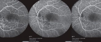
Diagnosis and differential diagnosis of various forms of retinitis are carried out on the basis of ophthalmological tests (visometry, achromatic and color determination of visual fields, computer perimetry, color testing), examination of eye structures (ophthalmoscopy, diaphanoscopy, biomicroscopy of the fundus), optical and radiological studies (OCT, fluorescence angiography). The etiology of retinitis is established based on the patient's epidemiological history.
The greatest importance in diagnosing retinitis is the assessment of the fundus. The ophthalmoscopic picture of tuberculous retinitis is characterized by disseminated retinal damage with the presence of numerous small or several large chorioretinal foci. In syphilitic retinitis, multiple foci of light and dark colors (“salt and pepper”), diffuse edema of the retina and optic disc, and pigmented areas of choroidal atrophy are identified on the periphery of the fundus. Toxoplasma retinitis occurs with damage to other membranes of the eye (iridocyclitis and episcleritis); ophthalmoscopy reveals a central loose focus of yellow-green color with symptoms of perifocal inflammation. With solar retinitis, yellowish-white spots with a gray rim are first identified in the fundus of the eye, which then turn into clearly defined red lesions.
When examining the visual fields, scotomas (peripheral, pericentral, central) and concentric narrowing of the visual fields are detected. Using contrast angiography of retinal vessels, changes in blood vessels are detected: their narrowing or dilation, uneven caliber, formation of couplings, obliteration. Optical coherence tomography for retinitis helps evaluate structural changes in retinal tissue. In order to more thoroughly assess retinal function, electrophysiological studies are performed - electroretinography, determination of the critical frequency of flicker fusion, etc.
To identify a bacterial or viral pathogen, blood culture is performed for sterility, PCR and ELISA testing. If an autoimmune etiology of retinitis is suspected, specific immunological tests are performed.
In addition to the ophthalmologist, other specialized specialists are involved in the diagnosis and treatment of retinitis of a specific etiology: phthisiatrician, venereologist, infectious disease specialist, rheumatologist, endocrinologist-diabetologist, etc.