A baby's eyes begin to develop in the womb, during the third week of pregnancy. Active formation of the visual apparatus continues for several years after birth. During this period, it is important to pay attention to the child’s vision, know the stages of its development, and regularly visit the ophthalmologist for checks. Let's talk about this in more detail.
Correct formation of the visual apparatus is important for the full mental development of the baby, because the eyes are connected to other senses. With poor vision, a child will lag behind in school and experience difficulties with active activities (sports, outdoor games), so good vision not only helps to understand the world around us, but also contributes to the full development of the child. Most vision problems can be eliminated in early childhood: the later the pathology is detected, the more difficult it is to correct and treat. It is very important to keep this aspect under control in the initial period of a baby’s life. Parents of newborn children should know the main stages of formation of the child’s visual apparatus.
Vision in newborns in the first days of life
How does a baby see in the first days of his life? Of course, not at all like an adult. The fact is that his eyeball still has a flattened shape and continues to actively grow, the retina is not fully formed, and the yellow spot, responsible for the acuity of central vision, is completely absent. The baby is not yet able to see clearly and three-dimensionally, focus his gaze on objects, or evaluate visual images. What functions are available to it?
Immediately after birth, the baby is bombarded with a huge amount of visual stimuli: from the darkness he finds himself in the bright world around him. But the vision of newborns in the first days of life is still very imperfect. Children at this time have infantile strabismus, which should go away after a certain time (we will talk about this below). The child's eyes look slightly in different directions or are reduced to the nose, since the ciliary muscles are still weak and their movements are not coordinated. In the first days of life, infants perceive black and white images, and colors are still indistinguishable to them. The first color that a child begins to distinguish is red, since it has the longest wavelength in the spectrum, 620-780 nm. Therefore, it is recommended to buy the first toys and rattles in red.
It is also not recommended to completely turn off the lights in the room where the baby sleeps. It is better to leave the lamp working. The child, upon waking up, will begin to look for a light source with his eyes, which, firstly, will calm him down, and secondly, will contribute to the development of the oculomotor muscles.
If your newborn has trouble opening his eyes
When do newborns' eyes open? Normally, the baby's eyes should open during the first breath. Sometimes this happens 3-5 minutes after birth, when the baby has already been placed on the mother’s stomach. Also, often the baby opens his eyes very wide for the first time when he first goes outside.
A newborn baby laughs in his sleep - reasons and what parents should do
There are situations when a newborn's eyes remain closed for several days. Reasons for this condition:
- Swelling of the soft tissues around the eyes. It can appear due to birth trauma when the facial part of the head is compressed. Or swelling occurs due to the child “standing” for a long time in the pelvis. With swelling, the baby's eyelids enlarge, sometimes very much. Also, the skin may shine and become red if an inflammatory process has begun in the eyelid.
- If a newborn baby's eyes cannot open, it may be due to an infection. Congenital infectious diseases often occur in infants, the symptoms of which include swelling of the tissues around the eyes and forehead, accumulation of pus on the conjunctiva, and sticking together of the upper and lower eyelids. Sometimes the eye is full of purulent discharge.
- The newborn does not open his eyes at all due to prematurity. In babies born prematurely, all organs, including the eyes, are immature, so their opening slows down; the eyelids begin to open 3-7 days after birth.
Note! If the child was already opening his eyelids, but suddenly stopped doing this, his eyes turned red and became very swollen, you need to contact your pediatrician; conjunctivitis may have become infected.
Newborn has swollen eyes
When a newborn child opens his eyes completely, all parents should know, since it is by this sign that one can determine whether his organs of vision are healthy and whether he will see well. If your baby cannot open his eyes, you should consult a doctor. He will conduct an inspection and determine what exactly the problem is.
If one eye of a newborn does not open at all on days 2-5 of life, this may be due to the development of conjunctivitis or a pathology such as ptosis, which requires immediate intervention. An increase in body temperature should also alert parents. This will be a symptom that an infection is developing in the baby’s body. With purulent discharge, the baby needs help, since there is a high probability of developing pathological processes inside the eye.
Possible reasons
- Ptosis. If a newborn has difficulty opening one eye, this may indicate that the child has ptosis. Due to underdevelopment of the muscle that raises the eyelid, or damage to the optic nerve that controls the movements of this muscle, drooping of the upper eyelid occurs. A drooping eyelid prevents light from entering the eye.
- Conjunctivitis. Inflammation of the conjunctiva occurs - the outer transparent mucous membrane that covers the inner surface of the eyelids. In a newborn, this disease is accompanied by redness of the conjunctiva, swelling of the eyelids and the presence of mucopurulent discharge.
- Inflammation of the lacrimal sac (dacryocystitis). The baby has purulent tears flowing from his eyes, and crusts form around the eyelids. The lacrimal sac area swells, becomes tense and red. This happens if the child has preserved the membrane of the nasolacrimal duct, which normally should dissolve after birth, but it happens that it remains intact.
baby crying
How does vision change in children under one year old?
So, what stages of formation does a child’s visual apparatus go through? What can and should a baby distinguish, what should it react to, how does the vision of newborn children change by month?
- 1 month: where is he looking?
At this age, the baby is able to distinguish between light and shadow, the outlines of large objects at a distance of no more than 30 cm. Wise nature designed it so that in the initial days of his life the child more clearly sees the most important thing - the mother’s face, her smile, it is at this distance that it is located during feeding. Everything else doesn't matter to him yet. At a further distance, objects look blurry to him, since visual acuity has not yet been formed. During this period, it is recommended to take the child in your arms more often during his waking moments and talk to him. It is important to check how the pupil reacts to light. The ophthalmologist will do this during your first visit.
- 2-3 months: life in bright colors
In the first 2-3 months of life, the light sensitivity of the retina will increase almost five times and the visual apparatus is actively developing. The extraocular muscles are strengthened. The baby already more confidently fixes his gaze on a horizontally moving object (toy, person) and turns his head behind it, but it is still difficult for him to catch vertical movement. It more clearly distinguishes the outlines of objects, but so far they are visible in two dimensions: length and width. At the same time, the world begins to be filled with more and more diverse colorful shades. At this age, newborns can already distinguish between red and green. Cool (blue, cyan) shades will become available later, since there are fewer photoreceptors in the retina that capture the short-wave part of the spectrum. Hang the mobile above the crib at a distance of 40-50 cm - it helps strengthen the ciliary muscles, as the baby follows objects by moving his eyes.
It is also recommended to carry the child around the apartment, show objects and name them. The baby also recognizes the faces of loved ones that it sees all the time.
- 4-6 months: grab it, or it will run away!
From three months of life to six months, children's vision continues to actively develop. The formation of the macula occurs - the zone of the retina responsible for central vision, visual centers in the cerebral cortex. The child can already clearly distinguish the parents’ faces, facial features, and facial expressions. The most exciting activity for him during this period is playing with his own hands and feet. In addition, along with the visual function, the grasping reflex also develops, since the baby is interested in exploring a clearly visible object. 4-6 months is an important period; parents need to be especially careful. By this time, infant strabismus should have passed, since the extraocular muscles are already working quite well. If this does not happen, then you need to urgently visit an ophthalmologist and conduct a thorough examination. In addition, during the same period, the doctor must examine the condition of the visual organs and finally make sure that there are no obstacles to their further development (congenital glaucoma, retinopathy of prematurity, etc.).
- 7-12 months: exploration of space
This is the period of development of motor activity: the baby actively crawls, moves in a walker, tries to take the first independent steps, holding on to furniture. He begins to understand the difference in the shape of objects (for example, he distinguishes between a cube and rings), strings rings on the axis of a pyramid, in a word, he actively learns about objects and distances (an aspect of assessing the distance of binocular vision). During this period, you should be especially careful, since the child causes many injuries to himself, grabbing everything and trying to put it in his mouth, as well as falling from the sofa or bed.
Vision of a newborn up to 1 year: norm and pathology
Vision is the most informative and at the same time the most fragile external analyzer. It is through the eyes that a person receives most of the information about the world around him. The visual centers are connected and have a strong influence on almost all vital structures of the brain (vision is involved in digestive, motor, vestibular, sexual and other types of body activity). The first year of life is especially important in the formation and development of vision, when the eyes and the child’s body as a whole are easily susceptible to various harmful influences of both internal and external factors.
If at this age the visual organ is damaged, then the child develops impaired coordination of movements, the baby experiences fear of the outside world, which often leads to a significant lag in the child’s development, since the other senses are not able to fully compensate for the lack of information.
In the first year, a child's vision develops very rapidly. The almost complete blindness of a newborn baby (non-directional light perception) within a few months develops into the ability to analyze objects and their movement, evaluate and compare objects according to their various characteristics, including color. Therefore, it is especially important for parents to understand the basic principles of vision development in children in the first year of life and to be aware of the early signs of eye pathology.
Development of the visual system
There are several periods in the development of the organ of vision. The most important of them is the laying and intrauterine formation. At this stage, the action of damaging factors can lead to catastrophic consequences (developmental anomalies - hypoplasia of the optic nerves, congenital cataracts, glaucoma; inflammation of the membranes of the eye, etc.). The next period is from birth to 1 year. At this time, areas of the visual cortex of the brain are actively developing, receiving information about the world around them. Simultaneous eye movement is trained, visual control of hand movements is formed, and the “library” of visual images is filled. If at this stage there is a restriction in the flow of light to the retina (violation of the transparency of the optical media of the eye), a violation of the focusing of objects (the presence of myopia or a high degree of farsightedness) or a deterioration in the perception of visual images (damage to the optic nerves, visual centers of the brain), then vision may stop at the initial stage of development and does not form to a normal level.
Immediately after birth, the child is able to perceive only the presence or absence of a light source. In the first months of life, various objects of the surrounding world appear before the child, as if out of a fog. At first, the baby only fixes his gaze on large objects (first month), then tries to track their movement in space - studies parents passing by, follows moving toys (3-4 months). At this age, you should not hang toys directly in front of your eyes - place them on the sides of the child or on your feet. At 6 months, a child’s visual acuity allows him to observe small objects, visually recognize “his own,” grab and throw toys, while learning the three-dimensionality of space. Place rattles and crinkles in the area where your baby's hands move to make them easier to grasp.
A one-year-old baby is already collecting “small debris” on the floor and is actively moving towards a bright toy. Use distant objects to attract attention. Receiving powerful visual stimuli, the baby begins to strive for objects that interest him, makes attempts to stand on his feet and takes his first steps. Only by the age of 6-7 years does a child’s vision reach the level of an adult (according to special tables, he names the 10th line).
Parental control
Already in the maternity hospital, a visual examination of the newborn can reveal signs of some congenital eye diseases. Cataract is a clouding of the lens that appears as a grayish glow instead of a black pupil. Treatment is most often surgical - removal of the cloudy lens. The long-term existence of interference with the passage of light into the eye will lead to a significant delay in the development of vision (obscuration ambiopia). After such an operation, the child wears special glasses or a contact lens that replaces the lens. Recently, the technique of early implantation of an artificial lens has become widespread. Some types of translucent cataracts cannot be operated on in early childhood. In such cases, periodic courses of stimulating treatment are carried out (exposure to the eye by light and laser radiation, electric and magnetic fields, classes on special computer programs) and delayed surgical intervention is carried out in adulthood.
External manifestations similar to cataracts can be detected with a more dangerous disease - retinoblastoma (retinal tumor). In the early stages, the tumor can be affected using special radiation applicators - plates with a radioactive substance applied to them. They are sutured directly to the sclera at the site of tumor projection, the shadow of which is determined during surgery by shining through the sclera with a diaphanoscope (a device similar to a flashlight). The radioactive material in the applicator destroys the tumor through the sclera. In the later stages, when there is a danger of the tumor spreading beyond the eye, there is only one way - removal of the affected eye.
Congenital glaucoma is an eye disease that is characterized by increased intraocular pressure due to congenital disorders of the formation and outflow of intraocular fluid. As a result, the child's eye stretches and increases in size, moving forward (up to limiting the complete closure of the eyelids). Glaucoma may also cause clouding of the cornea (cataract). Since this disease is associated with structural changes in some parts of the eye, treatment is mainly surgical. The purpose of the operation is to ensure normal outflow of intraocular fluid from the eye cavity. If at the time of surgery the cornea and optic nerve are not affected, then it is possible to preserve and develop full vision.
Inflammatory diseases (conjunctivitis - inflammation of the outer membrane of the eyes, covering the back surface of the eyelids and the front surface of the eyeball to the cornea, dacryocystitis - inflammation of the lacrimal sac, uveitis - inflammation of the choroid, etc.). The main signs of this group of eye diseases are redness of the eye, lacrimation, swelling of the eyelids and conjunctiva, and excessive discharge from the eyes. The means and methods of treatment in such cases should only be determined by an ophthalmologist, since improper treatment can worsen inflammation and complicate the process. Unreasonably prescribed antibiotics often lead to allergization of the mucous membrane of the eye, and their long-term use disrupts the vital activity of normal bacterial flora.
In children in the first months of life, mucous discharge from the eyes, similar to pus, may appear. There is a blockage in the lacrimal drainage system. Often, in order to cope with banal “festering of the eyes”, ordinary hygiene procedures in the form of washing and massaging the area of the lacrimal canaliculi are sufficient. First, use a cotton pad moistened with boiled water to remove mucous films and crusts from the surface of the eye. Then use your little finger to massage the inner corner of the eye towards the nose. After this, pour a puddle of boiled water into the inner corner of the eye (the child should lie on his back) and try to make the child blink. When blinking, the nasolacrimal canaliculi are actively washed, which helps improve the outflow of tear fluid. If necessary, repeat this procedure after each sleep, when the outflow of tears is blocked by tight squeezing of the eyelids.
In case of inflammatory diseases of the eyes, you should not instill breast milk into them - this is an excellent breeding ground for harmful microorganisms, and besides, the fat contained in milk interferes with the outflow of tears.
The most common external manifestations of eye pathology, which can be identified during a non-specialized examination of a newborn, also include:
- nystagmus is a twitching of the eyes in a horizontal or vertical direction, due to which the child does not have gaze fixation and does not develop clear vision (that is, the eye cannot fixate on an object and therefore sees its details as “blurry”). The cause may be various eye diseases (high degree of myopia, damage to the central part of the retina, etc.) or damage to the brain;
- ptosis (drooping) of the upper eyelid - when one or both eyes do not open completely. This happens due to damage to the corresponding nerve or muscle that lifts the upper eyelid (as a result of hemorrhages, birth injuries, etc.). The possibility of vision development in such a situation is determined by the degree of ptosis. If the eyelid covers the pupil, then the child requires special plastic surgery. If such a problem does not interfere with the baby, he is able to look at toys at different distances with this eye and does not develop strabismus, the issue of surgical intervention can be postponed to a later date, since surgical assistance in this case will be needed only for cosmetic purposes. To maintain normal functioning of the eye in this case, it is necessary to carry out special training.
During a medical examination, parents or an ophthalmologist may detect strabismus in the baby (a change in the correct position of one or both eyes in the palpebral fissure). It occurs due to visual impairment in one or both eyes, changes in muscle tone of the oculomotor muscles, damage to the oculomotor nerves, etc. The object in question is focused not on the central part of the retina, but on the adjacent area, where visual sensitivity is significantly lower, which poses a threat to the formation of binocular baby's vision. Binocular vision is vision with two eyes with the combination of images received simultaneously by them, which allows you to localize objects by direction and by their relative distance. In this case, it is necessary to start treatment as early as possible. A “curtain” of gauze napkin is glued onto the non-squinting eye using a patch (in the case of bilateral strabismus, the napkin with the patch is attached alternately to each eye), while training the “problem” eye is carried out. The only exceptions are cases when visual acuity in both eyes is sharply reduced and patching can lead to inhibition of vision development in the eye that sees better.
If the angle of deviation of the eye is large enough, surgical correction of strabismus cannot be avoided. This in no way cancels the implementation of patches and stimulating treatment. With the help of these measures, by the time of the operation (most often it is performed at the age of 4–5 years, so that binocular vision can be formed before school), it is possible to reduce the angle of strabismus and maintain good visual acuity. And this contributes to a smaller volume of surgical intervention, a better postoperative effect and makes it possible to further normalize visual functions.
Specialized examination by an ophthalmologist
When examining a baby in a maternity hospital (and for premature babies - in preterm care units), an ophthalmologist can identify other eye diseases that do not have external manifestations in the early stages. The most dangerous of them today are considered to be retinopathy of prematurity and optic nerve atrophy.
Retinopathy of prematurity is a disease of the retina in which the normal development and growth of its vessels stops, and pathological vessels begin to develop that do not fulfill their function of delivering oxygen to the retina. The vitreous becomes cloudy and thickens, causing tension and detachment of the retina, and if not treated adequately, this can lead to permanent vision loss. Unfortunately, this disease does not manifest itself in any way, and only at the last stage, when it is no longer possible to help the child, does the gray glow of the pupil become noticeable. The disease in its early stages can only be diagnosed by an experienced ophthalmologist. Mild stages of retinopathy may leave behind minor changes that do not significantly affect vision. But when the child reaches the 3rd threshold or 4th stage of the disease, it is necessary to urgently operate.
Optic atrophy is damage to the nerve fibers that carry visual signals from the eye to the visual centers of the cerebral cortex. The main cause is various damage to the structures and ventricular system of the brain. If the optic nerve atrophy is complete (which is rare), then vision may be completely absent. In the case of partial atrophy, visual acuity is determined by the degree and location of damage to the optic nerve. For optic nerve atrophy, stimulating functional treatment with the help of special devices, nootropic (improving metabolic processes in the brain) and vasodilator therapy are used.
Dynamic monitoring of the child
After the maternity hospital, parents need to carefully monitor the development of their baby, paying attention to the formation of visual functions.
It is important that the first examination by an ophthalmologist is carried out in the first 3 months of the baby’s life (during this period, most congenital diseases can be diagnosed in the early stages, which is the key to successful treatment). If there is no pathology at the initial examination, the next visit to the doctor is needed when the child is six months old (maturation of the main structures of the eye responsible for correctly focusing the image on the retina).
The primary examination is carried out according to the following algorithm:
Determination of acuity (at 1 month - by the reaction of fixation on an object, at 2-3 months - by tracking a bright toy 15-20 cm in size on a light plain background, at 4-5 months - by clarity of tracking to a distance of 3-5 m) and fields of view (field of view is the maximum space examined by one eye.). Visual fields are determined approximately - the doctor moves the toy forward from behind the child’s head until the baby reacts to the object. At the same time, an examination of the appendages of the eye is carried out: muscles, lacrimal ducts, eyelids (eye movements in different directions, patency of the lacrimal ducts, completeness of opening and closing of the eyelids), as well as the optical media of the eye and fundus using an ophthalmoscope and a slit lamp (devices that send slit-like or round beam of light through the optical media of the eye).
The ophthalmologist also measures refraction using skiascopy (shadow test with optical rulers) or refractometry (using a special apparatus).
If visual acuity cannot be determined (the baby has an unclear fixation or tracking reaction), then a study of brain impulses in response to visual stimuli is performed (visual evoked potential method). Based on its results, one can judge the presence of functional and structural lesions of the visual analyzer or a delay in its development.
At 6 months of age, during an examination by an ophthalmologist, in addition to the standard examination of the child, the dynamics of the refraction of the eye is monitored, that is, the newly obtained and primary data from this study are compared. In most children at 6 months, the refraction fluctuates between +1–+2.5 D. Sometimes at this age a shift towards minus refraction may appear, which indicates the baby’s predisposition to developing myopia. In this case, it is necessary to limit visual stress - remove small and close-hanging toys, focus on distant and moving objects. If myopia of more than 2 D is detected, especially if, along with this, the child’s visual acuity decreases and strabismus appears, vision correction with glasses is prescribed as early as possible. If necessary, glasses can be prescribed as early as 6 months (sometimes, with large degrees or pronounced asymmetry between the eyes, vision correction with contact lenses is used).
Even if at 6 months a preventive examination did not reveal any pathology of the visual organ, in the future it is necessary to undergo an examination by an ophthalmologist every six months, since during this time the refractive index of the eyes may begin to change (myopia, astigmatism form), some genetic syndromes may appear, occurring with a sharp impairment of visual acuity. In addition, regular monitoring of the baby gives the doctor the opportunity to promptly identify hidden inflammatory processes.
When to see a doctor urgently
The first symptoms of many diseases that you should pay attention to in the first months of life are the absence or slow tracking of the movement of an object, the appearance of strabismus, redness of the tunica albuginea, discharge accumulating in the inner corners of the eyes and on the eyelids. If such signs appear, you should immediately consult a specialist.
It is important to know: even if the doctor has identified a pathology, this is not a reason to panic. In most cases, impaired function of the organ of vision can be restored with adequate treatment.
Be attentive to your children! Do not self-medicate!
Author of the article
: pediatrician, ophthalmologist, candidate of medical sciences, head of the department of ophthalmology, NIIP and DH, Ministry of Health of the Russian Federation, Petr Petrovich Skripets
Binocular vision of newborns
Binocularity is a property of vision in which the image on both retinas is combined into one three-dimensional image. Binocular vision also helps to determine the shape of objects, the distance to them or between them. In its absence for any reason, the child develops pathologies: strabismus, amblyopia, myopia and others.
In the period up to one year, active development of near space occurs - an important stage in the development of binocular vision. At two or three months, the baby still sees objects in two dimensions (width and height), but the sense of touch helps him supplement them with volume (adding depth). The child gets the first idea of the volume of objects.
At 4-5 months of life, the grasping reflex develops, and the direction of movement is determined correctly, but there are still difficulties in assessing the distance to an object. Also, the baby is not able to fully appreciate the volume: he tries to grab the sun's glare or moving shadows with his hands. After six months, the exploration of distant space begins. The tactile reflex replaces active crawling, and after 10 months - walking. At the same time, the child learns to estimate the distance to the object to which he is moving, and there comes an understanding that it is possible to fall from the edge of the bed. Binocular vision in newborns immediately after birth is still absent due to the unformed ocular apparatus. But at 6-8 weeks he should already be able to fix his eyes on an object; at 3-4 months this should become the norm. Parents should pay special attention to this.
Preschool age
In the second year of life, the baby already confidently moves his gaze from one object to another, studying their properties. Of particular importance is eye-hand coordination and the ability to perceive objects distant from each other.
Uncontrolled watching of TV has a very negative effect on the vision of a two-year-old child, because his fragile eye muscles are not able to “keep up” with a television picture that is too bright and moves too quickly. This causes muscle strain and, as a result, reduces visual acuity.
In three-year-old children, the development of visual function is very closely intertwined with speech. It's very simple: a child who has poor vision is not able to exactly repeat the articulation of an adult, which means his speech development is delayed. At this age, the baby tries to talk about everything he sees, so the world around him, filled with vivid images, will stimulate his communication skills.
Photo: Depositphotos
At the age of three years, it becomes possible to check the child’s visual acuity using special Orlova tables. In such tables, various silhouette pictures are arranged in ten rows; their size decreases from top to bottom. They are offered to the young patient sequentially - this way the ophthalmologist has the opportunity to promptly diagnose visual impairments and prescribe treatment (wearing glasses, gymnastics, etc.).
- The age norm for a child 3−4 years old is visual acuity 0.6−0.8.
Color vision in the first year of life
Color vision is formed thanks to cones - special color receptors in the eyes. They perceive spectrum waves of a certain length, and the brain interprets these signals. Babies' brain cells begin translating these signals around two months. At first, children are able to perceive red, yellow, bright green colors - those that have a longer wavelength. Only later, by six months, does the baby recognize the color blue. Disorders associated with the lack of color vision can only be identified when the baby learns to understand the meaning of words and can show with his finger where the color is red and where the green is. In general, children under one year old have four color categories: red, yellow, blue, green. They begin to understand shades such as pink or purple as their speech develops.
What parents can do
It is parents who can create conditions for the full development of vision in the baby. To do this, it is important to follow these recommendations:
- The children's room should have good lighting. This applies not only to electric lighting, but also to natural lighting. Poor lighting in a nursery causes the ability to distinguish colors to be delayed;
- Toys should be large and bright. This will create an incentive for the baby to study them, which contributes to the active development of vision;
- Up to 3 months, it is advisable to place toys above the crib or stroller at a height of 40–50 centimeters from the child’s eyes. This will prevent the development of strabismus;
- Starting from 2 months, it is recommended to lay the awake baby on his stomach. This contributes to the rapid development of not only motor, but also visual skills.
It is important to attend preventive examinations with a pediatric ophthalmologist on time. You need a doctor immediately if:
- There was an eye injury;
- Any chemical has come into contact with the eye;
- There is a foreign body in the eye;
- Redness of the eyes or eyelids occurs;
- Previously normal pupils became different sizes;
- Mucous or purulent discharge appears in the corners of the eyes.
Mandatory consultation with a specialist is necessary if strabismus does not disappear after 3 months.
What needs to be done to help the correct formation of vision in newborns?
During the period of active formation of the visual apparatus up to a year of a child’s life, his children’s room should be properly decorated. You should not glue wallpaper that is too colorful. It is better to place bright objects of different colors and sizes, from different materials, against a neutral background. These can be purchased toys, homemade ones, or some household item; it is important that it does not cause harm to the child.
By exploring objects of various shapes and materials, the baby forms stereoscopic vision, his own idea of the world around him.
You should also not overwhelm your baby with many toys at the same time. Let there be several of them, and when he begins to lose interest in them, replace them with others. With many objects, the child will grab first one or the other, without thoroughly exploring and constantly being distracted.
Stages of development of visual perception of a premature baby
In children born prematurely, the development of the visual apparatus is often observed later. The stages of vision development by month are as follows:
- fixation of gaze on large objects - at 3–4 months;
- recognition of mother's face - from 4 months;
- color difference - from 5 months of age.
Premature babies develop a clearer vision of surrounding objects by the age of 2 years. By this age, babies acquire the ability to recognize different colors. At 3–4 years old, a prematurely born child develops binocular vision (the ability to fully see with both eyes).
At 2 years of age, the visual acuity of a premature baby often does not exceed 50%. In the absence of ophthalmological diseases, it becomes 100% by 4–5 years.
How to determine visual impairment in newborns?
The very first examination of the baby’s eyes is carried out in the maternity hospital for congenital diseases, then it is necessary to carry out preventive examinations at 1 month, 3 months, six months and a year. At the same time, during the growth process you should pay attention to certain factors.
- pupils of different diameters;
- pupils do not constrict in bright light;
- the child does not fixate his gaze on an object after 2 months;
- after 3 months, strabismus does not go away;
- frequent watery eyes or cloudy pupils.
All these are reasons for an urgent unscheduled visit to the ophthalmologist; they can be signs of dangerous pathologies. If there are hereditary eye diseases in the family or the mother suffered any serious illness during pregnancy, then special care must be taken in this case. Parents are unable to recognize some diseases, for example, retinoblastoma (retinal tumor), and when they sense something is wrong, it may be too late.
Congenital vision pathologies
At 1 year of age, a repeat examination is carried out by a pediatric ophthalmologist. It allows you to timely identify problems such as nearsightedness or farsightedness, astigmatism and other problems. It is important to strictly adhere to the doctor’s recommendations and, if necessary, begin vision correction as early as possible.
A child at an appointment with a pediatric ophthalmologist.
In some cases, congenital vision pathologies occur. The cause of their development may be intrauterine infection of the fetus, hereditary pathologies, injuries during pregnancy and other reasons. Congenital pathologies of the visual system include:
- Congenital glaucoma. This is increased intraocular pressure, developing due to impaired outflow of intraocular fluid. For treatment, a pediatric ophthalmologist will prescribe special drops that reduce intraocular pressure. In severe cases, surgery is indicated;
- Congenital cataract. This is a congenital clouding of the lens that prevents light from entering the eye. Treatment is only surgical, since the cloudy lens must be removed. After this, the child is prescribed special glasses, and then special contact lenses that replace the function of the lens;
- Ptosis. Pathological drooping of the upper eyelid, caused by improper development of the muscle that raises the eyelid or the nerve. For children under 3 years of age, treatment consists of fixing the upper eyelid with a plaster. At the age of 3 to 7 years, surgical treatment is indicated;
- Strabismus. This problem is typical in children under 4 months of age, since the extraocular muscles are still underdeveloped. If this problem is persistent, the cause must be eliminated;
- Nystagmus. Expressed in involuntary eye movements, most often horizontal. It does not allow vision to be fixed on specific objects. The child's vision decreases. Its treatment consists of timely vision correction.
If a baby is born prematurely, neonatal retinopathy often develops. This is a retinal disease in which normal blood vessel growth stops. Without timely treatment, retinal detachment occurs. Treatment is only surgical.
Early prevention of proper development of the visual system in children
The formation of a baby's eyes begins long before birth. Weeks 3-12 are the most important period of intrauterine development, at this time the formation of the most important vital organs and systems occurs, and the little man is very susceptible to destructive actions.
Infectious disease of the mother, smoking, alcohol, taking dangerous medications, unhealthy diet can lead to adverse consequences.
Disturbances in the formation of the visual organs can be caused by a lack of vitamin A (blindness), excessive administration of glucose-lowering drugs (underdevelopment of the optic nerve, congenital cataracts). Even taking aspirin can cause eye pathologies. Expectant mothers need to be especially attentive to their health.
Vision is a magical gift given to us by nature. Take care of your child, closely monitor his development, and regularly visit an ophthalmologist to avoid problems that cannot always be solved.
Vision in the first days and weeks after birth
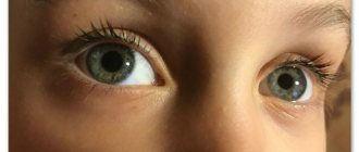
It is worth saying that at this age babies often have cross-eyed eyes, but most often this is not a dangerous phenomenon at all. If a mother notices this in her baby, it is best to see a specialist who will determine whether such a condition is normal or a deviation.
Every mother should pay special attention to the vision of her newborn. The stages of its development will help you understand whether it is developing correctly, and what you should pay special attention to to avoid unwanted and negative changes.
Basic rules for caring for a newborn's eyes
Caring for the eyes of a newborn at home after discharge from the maternity hospital is based not only on washing and instilling eye drops, although this is also an important point. How quickly and correctly a baby’s vision develops depends largely, of course, on the parents. What they should take care of:
- In order for a newborn’s eyes to develop more quickly, they need sufficient exercise. That is, the child should be in a well-lit room, the walls and ceiling of which will also be light. You should not deliberately darken the nursery with curtains and blinds during the day, as many mothers mistakenly do. The more bright light, the better.
- The eyes, not only of a child, but also of adults, are a fragile and sensitive organ. From the very beginning, they need proper hygienic care, and they must also receive all the necessary nutrients, that is, the baby should be provided with adequate nutrition, both breastfeeding and complementary feeding.
- At birth, nature limited the baby’s visual organs from perceiving a large number of visual stimulants. But then, on the contrary, there should be as many of them as possible for proper development. In the nursery you should place as many bright objects of different shapes, sizes and colors as possible. At the same time, it is important to change them periodically so that the load on the eyes also changes and is varied.
In addition to following the rules of hygiene, constant visual stress is necessary for the organs of vision and their successful development; bright multi-colored toys and objects will help with this
The mother and immediate family should play and talk with the child as much as possible, name different colors, and demonstrate them in the outside world. And if any abnormalities or simply suspicious phenomena are suddenly discovered, it is recommended to consult an ophthalmologist as soon as possible.
The world through the eyes of a newborn – what does he see?
The first stage of vision development in a child begins, in fact, long before his birth, in the womb, approximately in the third week of pregnancy. During this period, the formation of the pupillary membrane and optic nerves occurs, and further formation of the eyes occurs until the birth of the baby.
There is a myth that a child is born with black and white inverted vision, but this statement is not entirely true, this is only his perception, and vision is no different from an adult
There is no exact data on how well the fetus sees or whether it sees at all in the womb. During an ultrasound examination, it is easy to notice how an unborn baby opens his eyes slightly, squints, closes and turns away from the light, but this is not confirmation that he is, in fact, already able to see. It is only precisely confirmed that the eyes of even babies born at 28 weeks of pregnancy already react to bright light. Fully term and full-term children are also born with an imperfect visual analyzer. The complete completion of its formation occurs only by the age of 10.
Nowadays, the theory that a newborn's vision is upside down has become widespread. But is this really so? If you think sensibly, based only on confirmed facts, then the situation looks like this. The visual image of every person, including adults, not only babies, is displayed on the retina, indeed, upside down. This is an objective law of optics. But the cerebral cortex, which is responsible for processing the information received through the optic nerves, adapted to this phenomenon and learned to “flip” the picture.
Whether such features of the nerves of the cerebral cortex can be attributed to congenital properties or whether they appear after birth, scientists do not know for sure. This is explained by the fact that a newborn child cannot yet clearly explain how exactly he sees his mother’s face - in a normal position or upside down. Therefore, it cannot be said with certainty that infants have inverted vision. Likewise, one cannot say that they see in black and white. It has only been proven that children begin to react to bright colors only after a few months.
The fact that babies are born half-blind is quite logical, natural and thought out by nature itself. Just imagine how much visual information bombards a newborn as soon as he emerges from the dark womb into a huge, bright world. His still immature visual analyzer is simply not able to cope with the processing of all stimuli. Therefore, nature is limited to the perception of only the most important visual stimulus - the mother’s face bending towards the baby. But he also sees him vaguely and only at a very close distance - about 40–50 cm. It is interesting that it is precisely at this distance that the baby’s face is removed from the mother’s when feeding.
At birth, the child’s visual apparatus is not yet ready to perceive a large number of visual stimulants, but during the first year active adaptation occurs
Item tracking
Vision in newborns develops individually over months. After birth, children are unable to follow objects due to underdeveloped visual muscles. Due to unfocused vision, the child is able to keep his gaze on a stationary object only by 1.5 months.
Gradually by 2 months. The ability to fixate the gaze on a moving object develops. Already at 8-10 weeks, an infant is able to follow the movement of objects at a distance of 60 cm.
Gradually, the child’s horizons increase. By 5-6 months. the child fixes his gaze on objects located at a distance of 2-3 m. Vision trains every day and becomes more clear.
From this period to 1 year, the child develops the ability not only to follow objects at a distance, but also to determine the range of objects. Even from a distance, the baby distinguishes between relatives and friends, tries to remember or recognize different types of objects (car, tree, house).
Gradually, the speed and ability of visual tracking improves and reaches a maximum. All children starting from 12 months. see the picture of the world more clearly and keep track of different objects better than many adults.