How a visible image is created
Each of the 6 human sense organs (analyzers) includes three important links: receptors, nerve pathways, and the brain center. Analyzers belonging to different sense organs work in close “commonwealth” with each other. This allows you to get a complete and accurate picture of the world around you.
The function of vision is provided by a pair of eyes.
Binocular vision
This function of the organ of vision allows you to see with two visual organs, as a result of which the image is assembled into a single picture. For humans, the binocular function of the eye has the following positive qualities:
- increase in the visible border in the horizontal plane;
- increased visual acuity;
- the ability to sense the depth of the incoming picture;
- the ability to estimate the distance to objects.
The functions of the eye and methods for diagnosing them are very important concepts, without which it will be impossible to determine the presence of certain disorders of the visual system in a person. Using the presented techniques, you can give a complete assessment of the state of the optical system and pay attention to those functions that deviate from the norm.
Optical system of the human eye
The human eye has a spherical shape with a diameter of about 2.3 cm. The front part of its outer shell is transparent and is called the cornea. The back part, the sclera, consists of dense protein tissue. Directly behind the protein is the choroid, penetrated by blood vessels. Eye color is determined by the pigment contained in its anterior (iris) part. The iris contains a very important element of the eye - the hole (pupil), which allows light to enter the eye. Behind the pupil is a unique invention of nature - the lens. It is a biological, completely transparent biconvex lens. Its most important property is accommodation. Those. the ability to reflexively change its refractive power when examining objects at different distances from the observer. The convexity of the lens is controlled by a special group of muscles. Behind the lens is a transparent vitreous body.
The cornea, iris, lens and vitreous body form the optical system of the eye.
The coordinated work of this system changes the trajectory of light rays and directs light quanta to the retina. A reduced image of objects appears on it. The retina contains photoreceptors, which are branches of the optic nerve. The light stimulation they receive is sent along the optic nerve to the brain, where a visible image of the object is formed.
However, nature has limited the visible part of the electromagnetic scale to a very small range.
Only electromagnetic waves with a length of 0.4 to 0.78 microns pass through the light-conducting system of the eye.
The retina is also sensitive to the ultraviolet part of the spectrum. But the lens does not transmit aggressive ultraviolet quanta and thereby protects this most delicate layer from destruction.
Mechanism of binocular vision
The fusion reflex is a fundamental mechanism of binocular vision. In this case, the images that were formed in the retinal plane merge into a single picture with stereoscopic characteristics. This fusion occurs at the level of the cerebral cortex.
For the image to become unified, it is necessary to match the images obtained from the right and left retinas. This takes into account the size and shape of the image, as well as the area where it is projected onto corresponding identical areas of the retina. Each point in the retinal plane has its own corresponding point on the opposite side. Asymmetrical areas are called non-identical points, or disparate. When image points fall into these disparate points of the retinal plane, binocular vision becomes impossible. Instead of merging the image, doubling occurs.
In newly born children, coordinated movement of the eyeballs is not possible, and therefore binocular vision is absent. At about one and a half months, babies develop the ability to fix their gaze with two eyes, and at 3-4 months we can already talk about stable binocular fixation. The fusion reflex is formed only by 5-6 months, and full binocular vision - only by 12 years. In this regard, strabismus is more often present in patients of preschool age.
To form normal binocular vision, several conditions must be met:
- Ability to fusion (bifoveal fusion).
- Coherence in the work of all muscle fibers responsible for eye movement. They should ensure a parallel position of the eyeballs while the patient looks into the distance. When the gaze moves to a closer position, a proportional convergence of the visual axes occurs. This process is called convergence. Also, the extraocular muscles must respond to the associated movements of the eyeballs directed towards the object being studied.
- The visual acuity of each eye should not be less than 0.3-0.4, which is sufficient to form a clear image.
- The eyes should be located in the same horizontal and frontal plane. If, as a result of injury, tumor, inflammation or surgery, a change in symmetry occurs, then the merging of images becomes impossible.
- Images projected onto the retinal plane must be of this size (iseikonia). When the size of objects on the retina is different, we are talking about anisometropia, which occurs when the refraction of the two eyeballs is different. In order for binocular vision to be present, the degree of anisometropia should not exceed 2-3 diopters. This must be taken into account when choosing, despite good visual acuity, binocular vision will be absent.
- Also a necessary condition for binocularity is the transparency of the media of the eye (lens, vitreous body, cornea), the absence of pathological changes in the perceptive apparatus (retina) and the conduction system (optic nerve, optic tract, chiasm, cerebral cortex, subcortical centers).
Yellow spot
Opposite the pupil on the retina is the macula macula, where the density of photoreceptors is especially high. Therefore, the image of objects falling into this area is especially clear. Whenever a person moves, it is necessary that the image of the object is kept in the area of the macula. This happens automatically: the brain sends commands to the extraocular muscles, which control eye movement in three planes. In this case, eye movements are always coordinated. Obeying the received commands, the muscles force the eyeballs to turn in the desired direction. This ensures visual acuity.
But even when we look at a moving object, our eyes make very fast movements from side to side, continuously supplying “food for thought” to the brain.
Vision development in children
In this article
- Vision development in children
- What is binocular vision?
- Formation of binocular vision in children
- Testing binocular vision in children
A child's eyes and visual system begin to develop almost from birth. Already in the first seconds after birth, the baby has some unconditioned reflexes of the visual organs in the form of the reaction of the pupils to light, and there are attempts to track moving objects with the eyes. As the child grows and matures, other visual functions develop.
The very first such function is photosensitive. It appears immediately after birth. Photosensitivity becomes the basis for the formation of all other vision functions. When light rays hit the retina of a newborn’s eye, a visual image does not appear, but protective reactions of the pupils are already observed. Light sensitivity gradually increases, and visual functions improve.
At about 2-3 months, central vision appears. It develops and over time the child goes from the ability to find an object with his eyes to the ability to recognize it and distinguish it from other objects. Central vision in children under one year of age develops and improves very quickly. At 4-6 months they react to faces; at the 7th month they recognize the shapes of objects and geometric figures. Visual acuity also gradually increases. In parallel with its development, the formation of color perception is observed. Children begin to recognize colors at 2-6 months. Color perception is finally formed by the age of 4-5 years. Later than all these visual functions, binocular vision develops.
Color and twilight vision
The retina consists of two types of nerve receptors - rods and cones. Rods are responsible for night (black and white) vision, and cones allow you to see the world in all the splendor of colors. The number of rods on the retina can reach 115–120 million, the number of cones is more modest - about 7 million. Rods even react to individual photons. Therefore, even in low light we can distinguish the outlines of objects (twilight vision).
But cones can show their activity only with sufficient lighting. They require more energy to activate because they are less sensitive.
There are three types of light-perceiving receptors corresponding to red, blue and green.
Their combination allows a person to recognize the whole variety of colors and thousands of their shades. And their overlay gives white color. By the way, the same principle is used in color television.
We see the world around us because all objects reflect the light falling on them. Moreover, the wavelengths of reflected light depend on the substance or paint applied to the object. For example, the paint on the surface of a red ball can only reflect wavelengths of 0.78 microns, but green foliage reflects the range from 0.51 - 0.55 microns.
Photons corresponding to these wavelengths, hitting the retina, can affect only the cones of the corresponding group. A red rose illuminated by green light turns into a black flower because it is unable to reflect these waves. Thus, bodies themselves do not have color. And the entire huge palette of colors and shades available to our vision is the result of an amazing property of our brain.
When a light flux corresponding to a certain color falls on a cone, an electrical impulse is formed as a result of a photochemical reaction. The combination of such signals rushes to the visual zone of the cerebral cortex, building an image there. As a result, we see not only the outlines of objects, but also their color.
Light perception and color ability
Taking into account the structure and functions of the eye, concepts such as light perception and color visual function are distinguished.
Light perception is the ability of the visual organ to perceive light flux, as well as recognize its brightness and intensity. Light perception is the most sensitive function of the optical system. It is its pathological changes that are determined before all other changes in other functions.
If light perception is impaired, the following diseases are diagnosed:
- glaucoma;
- damage to the central nervous system;
- liver pathologies;
- lack of vitamins.
When twilight and darkness set in, the last thing a person loses is their sense of light. Each person has their own perception of light. It directly depends on the condition of the retina and the amount of substance in it that is capable of perceiving the flow of light. Light perception is also determined taking into account the general state of the optical system, primarily from the activation of nervous tissue.
Methods for determining the sensation of light involve adaptation of the eye to twilight. Use special devices. A sharp deterioration in the ability to see in twilight conditions is called hemeralopia (night blindness). Most often it occurs with pathologies of the retina, optic nerve, and lack of vitamin A.
Color visual ability is the ability of the eye to recognize objects by their color. Visual functions are very important, as it is possible to recognize the world around us much better.
Color perception plays an important role for the person driving and doctors.
The eye function in question affects the psychological and emotional component of a person.
Our own methods have also been developed for the study of color vision. There are certain tables. They are based on the principle of controlling brightness and saturation. The table contains a set of tests. Each of them assumes the presence of circles of the main and auxiliary colors. Certain tables have round numbers or shapes. They can only be recognized by people who have impaired color perception. This increases the accuracy of the study and also gives it greater objectivity.
Such methods of determining the function of an optical system should only occur under good light conditions. A person is seated with his back to the light stream at a distance of 1 m from the tables located. The doctor shows him the test tables one by one and waits for the person being examined to answer the visible signs. Each test exposure has its own duration, but it does not exceed 10 seconds. The first two tests are answered by people with normal or impaired color perception. Their role is to control and explain to a person his tasks. Research methods allow us to make a diagnosis of color blindness. Spectral methods for determining disorders of the color function of the eye include anomaloscopy.
Visual acuity
One of the most important properties of vision is its acuity. That is, his ability to perceive two closely located points separately. For normal vision, the angular distance corresponding to these points is 1 minute. Visual acuity depends on the structure of the eye and the proper functioning of its optical system.
Central and peripheral vision
The central visual system refers to the information that is available to a person in the center during a concentrated gaze. This is achieved by allowing light to enter the central part of the retina. Characterized by clearer images. The main characteristic of the central function of the eye remains visual acuity.
Peripheral visual function is information that a person perceives beyond the boundaries of the central area during a concentrated gaze. Achieved when light hits the border of the retinal spot of the visual organ. The resulting image is characterized by blur. Peripheral vision is an excellent opportunity for a person to understand space. The main characteristic is the field of view.
The peripheral functions of the organ of vision allow us to perceive objects that are not fixed by the gaze. This is achieved by using sticks. There are no differences in colors, and there is also no picture clarity. Excellent work of the sticks occurs during the period of twilight light. The peripheral optical system is characterized by a field of view and color perception. Methods for their diagnosis imply, with normal vision, simultaneous vision of the object by the researcher and the patient. Methods for diagnosing a child’s visual field are based on movement from the periphery to the color of the toy. It is important to notice the moment when the child averts his gaze to her. To determine a more accurate field of view, special equipment is used.
Vision and touch: a single mechanism
Recently, scientists were able to find out that the two senses (touch and vision) of a person have many common features and are much more similar to each other than previously thought. The work that confirms this statement was published in the American biological journal. If we judge the speed and direction of an object in motion, the brain uses similar mechanisms when performing tasks using vision or touch.
It is obvious that the tactile function and vision are essentially completely different senses. However, in both cases, the structures of the central nervous system react in the same way. Due to the fact that evolutionary processes tend to be conservativistic, the brain uses the same strategies when identifying objects in time and space. This was the subject of a study by Canadian scientist McGill.
Both during touch and during visual images, the central structures of the brain perceive an object in motion with the help of special sensory receptors, which are located in the first case on the skin, in the second case on the retina of the eyeball. In this case, the excitation of the receptor apparatus occurs sequentially, regardless of whether we follow the object with our eyes or run our finger along the surface under study.
Next, the nervous system is responsible for transmitting information from receptors to the visual cortex in the case of object tracking or to the somatosensory cortex when touching. Primary information processing begins in these areas. It is important that the neurons of the brain do not react to the entire object, but only to small sections of it, which enter the nerve cells gradually. In addition, neurons have the same differences: some cells respond only to objects that move to the left, others - to the right.
To obtain complete information about the direction of movement and speed of an object, further transmission of information from the somatogenic and visual cortex to special areas of the brain is necessary. For vision, such a zone is the medial temporal part, and for touch, the first Brodmann area. In both cases, signals that came from individual neurons merge into a single whole. In these areas, the information that a person received from primary nerve cells is combined. We can say that first-line neurons perceive an object as a mosaic and only in the central structures do all its elements merge together.
Canadian scientists believe that the use of identical mechanisms in the process of processing information that entered the central structures from the receptors of the organ of vision and skin is due to the similarity of these two senses. We can say that a person most often perceives surrounding objects both through vision and touch. For example, if a cup falls from your hand, then visual contact and the touch of the slipping handle are used to make it fall. That is, vision and touch “speak the same language.”
Scientists believe that further research into the language of the brain is necessary to understand how the central structures perceive the world around us as a whole.
Despite the fact that man is called the crown of nature, his brain is quite easy to deceive. Yakovleva Yulia Valerievna
EYE – FUNCTIONS OF THE EYE
Ichilov Hospital / OPHTHALMOLOGY / Eye – Functions of the eye
Human eye
- a paired sensory organ (organ of the visual system) of a person, which has the ability to perceive electromagnetic radiation in the light wavelength range and provides the function of vision. About 90% of information from the surrounding world comes through the eyes.
FUNCTIONS OF THE VISUAL ORGAN INCLUDE:
Light perception
Color perception
Central or object vision
Peripheral vision
Stereoscopic vision.
LIGHT SENSATION
- this is the ability to perceive light in the range of solar radiation and adapt to the perception of visual images at different levels of illumination. The process of light perception begins in the rods and cones. Under the influence of light energy, special substances called visual purple disintegrate in rods and cones. In rods this substance is rhodopsin, which is formed from protein and vitamin A, and in cones it is iodopsin, which contains iodine. When exposed to light, idopsin and rhodopsin break down, forming positive and negative ions and inducing the occurrence of a nerve impulse.
COLOR SENSATION
allows you to perceive more than two thousand shades of color depending on the wavelength of light radiation. It is believed that the retina has three components tuned to perceive the three primary colors of the spectrum: red, blue and green. Normal color vision is called trichromasia. With insufficient perception of one, two or three components, color anomalies occur (protanopia, deuteranopia, tritanopia).
CENTRAL OR OBJECT VISION
- this is the ability to distinguish the size and shape of environmental objects. This function is carried out by the central fovea of the retina, where there are the best conditions for the implementation of the function of object vision. In the central fovea there are only densely packed cones and their processes form a separate bundle in the optic nerve, called the papilomacular. Object vision is determined by the ability to perceive points separately. Each point is perceived separately if its image is projected onto two cones, between which there is at least one more cone. Those. The size of the cone determines visual acuity. It is believed that the minimum visual angle, determined by the size of the cone, is 1 minute. Visual acuity is examined using the well-known Golovin-Sivtsev tables.
PERIPHERAL VISION
is the perception of part of space around a fixed point. When the gaze is fixed on any point, this point is perceived by the central fovea of the retina, and the space surrounding it is perceived by the remaining part of the retina. The space that is perceived by one eye is called the visual field. Peripheral vision is of great importance for orientation in the environment. With various eye diseases, the visual field may narrow or certain areas of it may fall out (scotomas).
STEREOSCOPIC VISION
– this is the ability to perceive distances between objects in the environment, the volume of these objects, the ability to observe objects in motion. Stereoscopic vision becomes possible if a person perceives objects with both eyes - binocular vision. If stereoscopic vision is impaired, orientation in the environment becomes difficult.
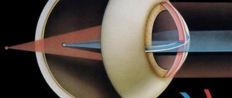
Normal visual acuity
is ensured by the operation of the optical apparatus of the eye. Using the optical media of the eye, a reverse reduced image of an object is projected onto the retina.
The optical or refractive apparatus of the eye includes:
cornea
anterior chamber of the eye
lens
vitreous body.
They work like collecting lenses.
The refractive power of the optical apparatus of the eye is called refraction. It is equal to 60 diopters (1 diopter is equal to the optical power of a lens with a focal length of 1 meter, i.e. a converging lens of 1 diopter focuses the rays at a point 1 meter behind itself).
Normally, refraction allows you to obtain a projection of the image of an object on the retina. The clarity of the image on the retina, in addition to the refractive apparatus of the eye, depends on the size of the eyeball.
Approximately 30% of modern people suffer from some kind of refractive error.
With refractive pathology, the refraction of light in the eye is impaired and, as a result, the image does NOT focus accurately on the retina. This means that a person with refractive errors cannot see surrounding objects clearly and clearly and needs certain methods of vision correction.
Various types of clinical refraction occur.
Emmetropia or commensurate refraction
is a state of vision when the focus of the optical system of the eye coincides with the retina. Disproportionate refraction is called ametropia.
Ametropia includes:
myopia
hypermetropia
astigmatism.
If the focus of the optical system of the eye is behind the retina, i.e. A clear image is formed not on the retina, but behind the retina; this state of refraction of the eye is called farsightedness or hyperopia.
If the focus of the optical system of the eye is in front of the retina, and a clear image is formed before the rays reach the retina, such eye refraction is called myopia or myopia.
With a normal eye refraction of 60 diopters, parallel rays will converge in the central fovea of the retina. Parallel rays enter the eye from infinity. But at closer distances they change their course. It is believed that parallel rays enter the eye from a distance of no more than 5 meters. If the object in question is located at a distance of less than 5 meters, then its image on the retina will be unclear. Then there is a need for the process of accommodation. Accommodation occurs due to increased refractive power of the lens.
The refractive power of the lens increases with increasing transverse size of the lens. The lens is attached to the ciliary muscle using a special circular ligament of cinnamon. When the ciliary muscle, which has the shape of a ring, contracts, the diameter of this ring decreases, the ligament of Zinn is weakened, the tension of the lens capsule is weakened and the lens acquires a more convex shape, increasing its refractive power. At the same time, the eye sees better at close distances. The closer the distance, the more the ciliary muscle should tense.
Simultaneously with accommodation, convergence occurs - bringing the visual axes of both eyes to one object. The nearest point of clear vision is determined by the amount of accommodation. The volume of accommodation depends on the amount by which the lens can increase its refractive power and is determined by the elasticity of the lens and the strength of the ciliary muscle.
With age, the elasticity of the lens decreases - age-related changes in vision occur - presbyopia.
In this case, the eye becomes unable to adapt to seeing close objects.
It is believed that by the age of 10 the lens can increase its refractive power by 14 diopters, and by the age of fifty it can increase its refractive power by only 2 diopters.
To correct vision for presbyopia, glasses with collective lenses are prescribed. Glasses are selected in the ophthalmologist's office.
- Ophthalmology in Israel
- Eye – Structure of the eye
- Eye – Functions of the eye
- Eye diseases
- Causes of poor vision
- Eyeball – Exophthalmos
- Eyeball – Injuries
- Eye burns
- Sclera – Diseases of the sclera
- Eye – Refraction of the eye
- Cornea
- Cornea – Congenital and dystrophic changes in the cornea
- Cornea – Inflammatory diseases of the cornea of exogenous origin
- Cornea – Inflammatory diseases of the cornea of endogenous origin
- Cornea – Herpetic keratitis
- Eye - choroid
- Choroid of the eye - Developmental anomalies
- Uveitis - Uveitis
- Uveitis of the eye – Anterior uveitis
- Uveitis of the eye – Posterior uveitis
- Uvea of the eye – Peripheral uveitis
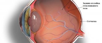
- Eye - Retina
- Retina – Retinal diseases
- Retina – Retinal detachment
- Retinal detachment - Treatment
- Myopia (Myopia)
- Farsightedness (Hypermetropia)
- Presbyopia (Age-related farsightedness)
- Astigmatism
- Spasm of accommodation
- Amblyopia (Lazy Eye)
- Asthenopia
- Hemeralopia
- Retinopathy
- Diabetic retinopathy
- Hypertensive retinopathy
- Atherosclerotic retinopathy
- Binocular vision
- Strabismus
- Strabismus – Paralytic strabismus
- Strabismus – Concomitant strabismus
- Optic Nerve – Developmental Anomalies
- Optic Nerve – Stagnant optic disc
- Optic nerve – Inflammatory diseases
- Optic Nerve – Optic Nerve Atrophy
- Optic nerve – Vascular lesions
- Optic nerve - Toxic lesions
- Optic Nerve – Optic Nerve Tumors
- Optic nerve – Tumors of the optic chiasm
- Eye - Lens
- Lens – Anomalies of lens development
- Lens – Lens luxation
- Lens – Cataract
- Cataract – Types of cataracts
- Cataract – Acquired cataract
- Cataract – Cataract Treatment
- Eye - Vitreous
- Vitreous – Vitreous diseases
- Eye – Conjunctiva
- Conjunctiva – Inflammatory diseases
- Conjunctivitis – Acute banal
- Conjunctivitis – Pneumococcal
- Conjunctivitis – Acute epidemic Koch-Wicks
- Conjunctivitis – Gonococcal
- Conjunctivitis – Diphtheria
- Conjunctivitis – Angular
- Conjunctivitis - Viral conjunctivitis
- Conjunctivitis – Chlamydial conjunctivitis
- Conjunctivitis – Autoimmune and allergic conjunctivitis
- Conjunctivitis – Chronic conjunctivitis
- Conjunctiva – Dystrophic diseases
- Lacrimal organs
- Lacrimal organs – Developmental anomalies
- Lacrimal glands – Inflammatory diseases
- Lacrimal drainage apparatus – Inflammatory diseases
- Eye - Eyelids
- Eyelids – Anomalies of the eyelids
- Eyelids – Blepharitis
- Eyelids – Stye
- Eyelids – Abscess, Cellulitis
- Eyelids – Chalazion
- Eyelids – Viral lesions
- Eyelids – Congenital benign neoplasms of the eyelids
- Eyelids – Acquired benign neoplasms of the eyelids
- Eyelids – Malignant neoplasms of the eyelids
- Glaucoma
- Glaucoma – Types of Glaucoma
- Glaucoma – Classification of glaucoma
- Glaucoma – Diagnosis of glaucoma
- Glaucoma – Glaucoma Treatment Methods
- Glaucoma – Drug treatment
- Glaucoma – Laser treatment method
- Glaucoma – Surgical treatment method
- Glaucoma - Recommendations
- Glaucoma - Prevention
- Keratoconus
- Sympathetic ophthalmia
- Light ophthalmia
- Endophthalmitis
- Panophthalmitis
- Colorblindness
- Nystagmus
- Computer vision syndrome
- Vision correction – Glasses
- Glasses – A little about glasses
- Vision correction – Contact lenses
- Contact lenses – A little about contact lenses
- Vision correction – Laser correction
- Laser vision correction – Laser operating principle
- Laser vision correction – LASIK
- Laser vision correction – Epi-Lasik
- PRK – Photorefractive keratectomy (PRK)
- Laser vision correction – Choice of surgery
- Conductive keratoplasty
- Department of Ophthalmology MC Ichilov (Souraski)
- Professor Anat Levinshtein – Ophthalmologist
- Professor Anat Kesler – Ophthalmologist
- Professor Yuval Yasur – Ophthalmologist