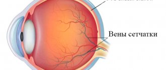
Eyes are one of the main sense organs. Thanks to them, all living beings have the opportunity to perceive the world around them. It is believed that vision provides us with about 90% of incoming information. As you know, in order to see objects normally, the combined work of both eyes is necessary. Thanks to binocular vision, we have the ability to perceive not only the size and shape of an image, but also its location in space. Unlike people, some living creatures (birds, reptiles, horses) see objects with each eye separately. In other words, they are characterized by monocular vision. In some cases, this is also observed in humans. Since such an ability is not characteristic of humans, this type of vision is considered pathological and requires treatment.
What does monocular vision mean?
The science that deals with vision problems is called ophthalmology. It covers not only eye diseases, but also studies the development and various types of vision in humans and other living beings. For example, you can learn about such a feature of birds as monocular vision. This type of vision allows some animals to distinguish objects with each eye separately. It is known that the vision of birds is several times better than that of humans. Due to the fact that their eyes are located on the sides, they see most of the space around them. Birds' field of vision is about 300 degrees. This gives them the opportunity to see images not only in front and to the side of them, but also behind them. Based on this, monocular vision refers to the ability to perceive objects with one eye. Normally, it occurs in all birds except owls, as well as in many animals.
What it is
The human visual system, which has developed in the process of evolution, is binocular (stereoscopic), which allows the brain to provide a three-dimensional image and create a qualitative sense of the location of objects in visible space. Binocular vision is characteristic not only of humans, but also of all mammals, as well as fish and birds. The ability to perceive the world with sufficient accuracy is ensured by the specificity of the paired visual organ - two eyes. Information entering the eyes in the form of electromagnetic radiation is initially processed separately by the brain.
And only then is it synthesized into a solid, detailed and three-dimensional visual image. High-quality visual perception requires the ability for the central nervous system to receive a “picture” from both the left and right eyes. This allows them to be compared and the “depth” of visible space to be measured. With correct vision, which must be ensured by the normal functioning of both eyes, the clearest perception is achieved. The overall field of view expands.
Read about training lateral vision in the material.
The image produced by looking at an object with only one eye can give a person an idea of the overall shape of the object, as well as its height and width. However, the functioning of such a truncated (or single) type of visual perception does not allow one to obtain a complete and high-quality picture of the relative position of objects in space.
This type of activity of the visual organs is called “monocular vision”. Actually, vision, in which static and moving objects can be “seen” with only one eye, is a pathology. This type of perception is characterized by a narrowing of the boundaries of the field, a significant drop in vision and the ability to perceive the features of the location of objects in space. Monocular vision allows for very little three-dimensional depth perception. The accuracy of estimating the depth of space in monocular vision in some cases deteriorates by approximately twenty times.
The characteristics of objects are recognized by a person only by indirect signs, such as differences in illumination or perspective. However, monocular vision is not an insurmountable obstacle to orientation in the world around us. The perception of volume and depth of spatial objects can be obtained by a person with such visual characteristics using familiar optical effects, which do not require the ability to binocular discrimination.
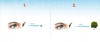
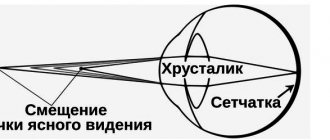
The habit of perception suggests that the size of objects does not change. This means that the human brain perceives images of small objects on the retina as a distant object, and vice versa.
The relative position of objects is also “understood” due to the fact that close objects can hide more distant ones. Brightness or color saturation also plays a role in the ability to navigate in space. The brightest of the objects (or the object whose color will be perceived as more saturated) “appears” to be closer. Distant objects are lost in the fog (the symptom of fog in the eyes refers to a decrease in visual acuity or sharpness that appears gradually). In monocular vision, objects that move at the same speed also differ: the one whose rate of change on the retina is slower is “seen” as further away.
There are two types of monocular vision:
- Information enters the brain from only one eye;
- The ability for a person to see alternately with the left and right eyes (monocular alternating vision).
For the normal functioning of the visual organs that provide binocular vision, a number of important conditions must be present:
- Coordinated work of all eye muscles;
- Correct placement of the two eyes in the same horizontal and frontal planes;
- Visual acuity sufficient to form a clear image on the retina of both eyes;
- Iseikonia (equal sizes of images on the retina of both eyes);
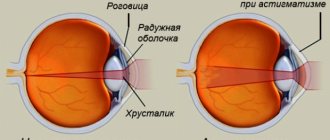
- A sufficient degree of transparency of the cornea, lens, and vitreous in both eyes;
- The absence of pathology in such important parts of the visual system as the retina, optic nerve and various parts of the brain responsible for visual perception (chiasma, optic tract, subcortical centers, cerebral cortex);
- Ability to fusion (bifoveal fusion).
Find out about the causes of visual impairment by following the link.
In cases where the functioning of the organs of vision is disrupted, a monocular type of perception occurs.
Difference between monocular and binocular vision
Thanks to ophthalmology, it is possible to answer the question of how monocular vision differs from binocular vision. Each of these types of vision has its own advantages and disadvantages. Monocular vision allows you to obtain information only about the shape and size of the image. Nevertheless, this type of vision is necessary for animals, since thanks to this they can view objects from two sides at the same time. As a result, their field of view increases. This is necessary for hunting and protection in the animal world.
The structure of the organ of vision in humans differs from that of birds and animals. The center of vision is located in the cerebral cortex. Thanks to the decussation of nerves, the information received from each eye is converted into a single image. That is, a person has binocular vision. In addition to the fact that this type of vision is distinguished by the ability to view an object with two eyes at once, it also has other features. Binocular vision is characterized by the perception of images in space. This means the ability to distinguish at what distance from the eyes an object is located, whether it is three-dimensional or flat.
Differences from binocular perception
Monocular visual perception does not make it possible to see the world holistically and three-dimensionally, like developed stereoscopic vision. The latter is also different:
- the ability to ensure full visibility of the observed picture, its relief;
- expanding the boundaries of visual perception;
- the appearance of binocular summation - a condition in which the functions of both visual organs are more developed than those of each of them separately;
- the ability to calculate the correct distance between visible objects.
The human visual organs can function correctly only if all conditions are met:
- coordinated activity of all eye muscles;
- correct placement of the eyes in the frontal and horizontal planes;
- normal visual acuity;
- identical sizes of images on the retina of the two visual organs.
The transparency of the biological lens, the corneal layer and the vitreous body, and the absence of defects in the parts of the brain responsible for the correct formation of images are also of great importance.
A person with binocular vision can work as an airplane pilot, professional driver, or surgeon. These positions are not available to those with monocular vision.
Pathologies in which monocular vision is observed
As is known, both monocular and binocular vision occur in humans. Nevertheless, the separation between vision, which is considered the norm for animals, is a pathology for humans. There are two types of monocular vision in humans. In the first case, it means the ability to see with only one eye all the time (right or left). This can occur with one-sided blindness. Another type of monocular vision refers to alternating vision with the right and left eyes. This type occurs with diplopia. The cause may be congenital eye defects or trauma.
In what pathologies does monocular human vision occur?
Humans have both binocular and monocular vision. However, the dissociation between the pictures created by the left and right eyes, which is a normal feature for an animal, is considered a deviation in humans. Human monocular vision is divided into two types:
- the ability to constantly see with only one eye - occurs in the case of one-sided blindness;
- vision alternately from one eye to the other - occurs in the case of diplopia, which can occur due to congenital abnormalities in the structure of the eye or due to injury.
Diagnosis of vision types
It is important to timely diagnose not only monocular vision itself, but also its cause. Often the source of the problem is eye injuries, vascular disorders, and congenital anomalies. Therefore, in addition to instrumental methods, it is necessary to ask the patient in detail about when the ability to see changed. The type of vision can be determined by performing a four-point color test. Thanks to this method, the presence of mono-, binocular or simultaneous vision is determined.
A different color filter (red and green) is placed in front of each person's eye. At some distance from the organs of vision there is a screen with 4 circles. Each of them has a color (white, red and 2 green). Depending on how many circles the patient sees, the ophthalmologist makes a conclusion about the type of vision. Normally, a person distinguishes all 4 figures. In this case, the white circle for him acquires a red or green color. With monocular vision, the subject sees only 2 or 3 figures on the screen. In some cases, the patient marks 5 circles. This is rare and is typical for simultaneous (both mono- and binocular) types of vision. This deviation from the norm does not require treatment.
Diagnostic methods
One of the diagnostic methods is the so-called “Hole in the Palm”. A piece of paper, previously rolled into a cylinder, is applied to one person’s eye. The palm is placed opposite the other eye at a distance of 15 cm from the face. With normal visual perception, there will be a small hole in it, through which the same picture will be visible as through a paper cylinder. If there is a monocular defect, a hole will not appear in the palm.
The other is the Kalf method. To conduct this experiment, the patient will need to take a horizontally located knitting needle in an outstretched palm and try to poke its end into a vertically placed pencil, which will be held by an assistant. With normal vision, this task will not be difficult. With monocular, a person will definitely miss.
Another way to determine monocular vision is to read with a pencil. A book with a pencil placed against its background is placed in front of the subject’s nose at a distance of 3 cm so that some lines become closed. The subject needs to read the sentences in the book without moving his head and fixing the piece of paper at one point. If there is a defect, you will not be able to see the text without peeking, since the pencil will partially cover it.
A four-point color test is also often used to detect monocularity. The visual fields of the eyes in this experiment are separated using color filters. The patient is asked to look through glasses with red and green lenses at four objects: two green, one red and one white. If a person has normal binocular vision, he will see green and red objects, and white objects will become red-green. If he has some kind of dominant eye, the colorless object will be either red or green.
There is also a diagnostic method called a prism test. It is used to determine monocular vision in young children. During this diagnosis, the ophthalmologist applies a prism with a power of 8-10 diopters to one organ of vision of the child being examined, while continuing to observe the other eye. After 20–30 seconds, the prism is removed, and the doctor begins to monitor the movements of the newly opened organ. During the specified time, the direction of the rays changes slightly. Because of this, the image on the retina of the visual organ is displaced. When the prism is removed from the field of vision, the image begins to double until the eyeball takes on its original appearance. With monocular vision, the gaze of the eye being examined will wander. Such diagnostics are carried out on both visual organs in order to identify the dominant eye of the patient being examined.
Why does strabismus develop with binocular vision?
It is known that during the newborn period a child does not have binocular vision. Its formation begins at the age of 1.5-2 months. At this time, divided vision is considered normal. At 3-4 months, the baby develops a reflex, according to which the images received by both eyes are perceived as a single one. However, the process of formation of binocular vision ends only at the age of 12. Based on this, a disease such as strabismus belongs to childhood pathologies. In this case, the child cannot focus his gaze on a specific object. Movements of the organs of vision may not occur simultaneously; one eye squints and squints in bright light. The cause of this pathology is incorrect or delayed formation of binocular vision. This is observed with myopia, astigmatism or farsightedness.
Binocular vision with strabismus: treatment of pathology
In order to develop binocular vision in a child, it is necessary to diagnose strabismus in time and begin treatment. First of all, you need to identify the cause of the pathology. This is required to choose a method for restoring binocular vision. Treatment of strabismus begins with a set of exercises necessary to strengthen the eye muscles. In order for a child to perform vision exercises correctly, parental or doctor supervision is needed. In some cases, wearing special glasses is indicated. If a child has developed amblyopia, then it is necessary to increase the load on one of the visual organs. To do this, one of the lenses of the glasses (on the side of the healthy eye) is sealed.
Methods for restoring binocular vision
Treatment methods for binocular vision can be divided into physical and surgical. In the first case, it means doing exercises for the eye muscles. They must be done regularly, several times a day. In some cases this method is sufficient. Most often, in addition to eye exercises, wearing glasses is required. They help get rid of the causes of strabismus (myopia, astigmatism, farsightedness). Another method of treatment for binocular treatment is surgery. It is used in cases where correction of strabismus by other means is impossible.