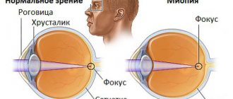
Not everyone knows what it is - moderate myopia, but this is ordinary myopia - a condition in which the image is focused not on the retina, but in front of it. The disease is manifested by a decrease in the quality of vision at a distance. For a long time, progressive myopia does not manifest itself in any way. Many people learn about it only during a consultation with an ophthalmologist. Second degree myopia begins from -3 to -6 diopters (diopters) - this is how experts determine the degree of myopia and further treatment.
What is moderate myopia?
Myopia is characterized by a shift in focus from the retina, which is responsible for visual perception. In this case, there is a noticeable approach of the further point of clear vision to the person. As a result, the image of distant objects is focused in front of the retina. A person complains of blurriness and double vision when looking into the distance, and his vision is noticeably deteriorating.
A shift in the main focus in moderate myopia most often occurs due to an increase in the anteroposterior size of the eyeball. This is facilitated by congenital weakness of the scleral membrane, prolonged visual stress, and spasm of accommodation. The length of the eye changes, but the refraction remains the same, which causes a certain discrepancy and shift in the main focus. An increase in the anteroposterior dimension by 1 mm causes a change in refraction of approximately 3D.
Less commonly, myopia is caused by changes in the thickness of the cornea or lens. This can be caused by cataracts, diabetes, pregnancy, and taking corticosteroid and sulfa drugs. A strong increase in refraction leads to excessive refraction of rays and focusing of the image in front of the retina. This myopia is called refractive myopia.
Classification and varieties
Regardless of the prerequisites for its occurrence, there are two main types of moderate myopia:
- Non-progressive. With this type of myopia, the patient's condition remains stable. At the same time, he can clearly see objects located nearby. The disease is well corrected with glasses and lenses;
- Progressive. The rate of decrease in visual acuity with this type of myopia is about a diopter per year. Rapid deterioration in quality requires mandatory correction and medication, and possibly surgery.
In turn, non-progressive myopia is conventionally divided into two more subtypes - slow progression and stationary. In the first case, the drop in sharpness is less than a diopter per year, in the second it is practically not observed.
Typically, the active development of such myopia occurs during school years, and at the age of 20-22 years the condition stabilizes.
Moderate myopia with astigmatism
Myopic astigmatism is characterized by a shift in the main focus in such a way that one part of the image falls on the retina, and the other is located in front of it (simple astigmatism). Most often, pathology occurs due to changes in the radius of curvature of the cornea. Complex myopic astigmatism is characterized by the projection of parts of the image at different distances from the retina.
The disease is manifested by decreased visual acuity, double vision, and image distortion. Surrounding objects may appear to a person to be curved, elongated, or asymmetrical. The patient complains of visual fatigue, headaches, and lacrimation. When examining certain objects, he may bring them very close to his face or tilt his head to the side.
Reasons for appearance
Myopic astigmatism in both eyes (causes and treatment affect the rate of disappearance of the symptoms of the disease) occurs due to a genetic predisposition. Also, the development of the disease may be associated with anatomical curvature of the lens or corneal dystrophy.
In some cases, astigmatism can be acquired and develops due to:
- mechanical eye injury;
- diseases characterized by impaired ocular circulation or increased eye pressure;
- complications after surgery or infectious diseases;
- negative environmental impact.
Causes of visual impairment
Myopia is a multifactorial, genetically determined disease. Most often, it develops in people with a hereditary predisposition under the influence of various provoking factors. It is often preceded by a spasm of accommodation or so-called “false myopia.”
Hereditary predisposition
According to statistics, the disease occurs in approximately half of people whose parents also had myopia. But in people with no family history, the risk of developing myopia is less than 10%. It should be noted that children do not inherit the disease itself from their parents, but only the tendency to it.
Visual load
Myopia most often occurs in schoolchildren, students, office workers, seamstresses, and book lovers. Its development is facilitated by excessive load on the visual organ, leading to asthenopic complaints. Soon the patient experiences a spasm of accommodation, and then true myopia.
Risk factors:
- prolonged sitting at the computer;
- reading in poor lighting;
- long-term work at close distances;
- failure to maintain distance between the eyes and a book, notebook or monitor;
- improper organization of the workplace;
- inconsistency between the height of the furniture and the height of the child.
Poor blood supply to the eyes
Myopia can be caused by deterioration of the blood supply to the membranes of the eyeball. This is observed with traumatic brain injuries, some infectious diseases and pathologies of the cardiovascular system. Decreased visual acuity also occurs when blood circulation in the retina deteriorates.
Poor nutrition
Myopia can develop due to a lack of certain vitamins and minerals. For the normal functioning of the visual organ, vitamins A, E, group B, chromium, zinc, copper, and manganese are necessary. With a deficiency of these substances, rapid progression of myopia is observed.
Incorrect vision correction
Overcorrection or undercorrection with diverging lenses can lead to an increase in the degree of myopia and a progressive decrease in visual acuity. Lack of correction is no less dangerous. But with false myopia, wearing glasses or contact lenses is strictly contraindicated.
Treatment
Treatment is indicated for people with any degree of myopia. Moderate myopia requires an integrated approach and regular visits to an ophthalmologist to monitor the dynamics of the disease.
Optical correction
Optical correction does not help restore vision, but it does make life easier for a person with myopia. With moderate myopia, wearing glasses or contact lenses should be constant.
Glasses
Spectacle correction is an affordable, safe and one of the most common methods of vision correction. A patient with moderate myopia is prescribed glasses with a negative optical power (with a “–” sign) from 3 to 6 diopters.
Spectacle correction has a number of features:
- financial inclusion;
- no risk of infection;
- durability;
- simplicity and ease of maintenance and operation.
One of the main disadvantages of glasses is that they are not able to cope with severe refractive errors, however, moderate myopia can be corrected in this way.
Lenses
The lens correction mechanism does not differ from spectacle correction: the patient is prescribed contact lenses with diverging eyepieces with an optical power of –3 to –6.
The advantage of lenses over glasses is their ability to cope with even severe refractive errors. In addition, it is impossible to forget or leave lenses somewhere; they do not affect your appearance or cause discomfort.
Disadvantages of contact correction:
- there is a risk of infection;
- the need for regular replacement;
- difficult to care for and wear.
Today, vision correction with lenses is the most common.
Eye exercises
Eye exercises should be performed regularly: it allows you to avoid eye strain and dry eye syndrome, does not require special equipment or skills, and does not take much time.
The most effective exercises for moderate myopia:
- Close your eyes for 3-5 seconds 10-20 times.
- Wide eye opening for 3-5 seconds 10-20 times.
- Alternately stopping the gaze on objects that are as far away and as close as possible; 5-10 times for 5-10 seconds.
- Circular movements of the eyeballs, 5-10 rotations in each direction.
- Move the eyeballs up, down, left and right 10 times in each direction.
Gymnastics must be performed daily, when working at a computer or otherwise straining the eyes - every hour.
Signs of the disease
The most characteristic symptom of myopia is blurred vision. Vagueness and double vision often appear. When looking into the distance, the patient has to squint, trying to focus his gaze, which causes rapid fatigue. At the same time, a person sees nearby objects clearly.
Symptoms of myopia
The development of a disease such as myopia may not be noticed immediately, since vision deteriorates gradually and many people attribute changes in the perception of objects to long work at the computer or fatigue.
Symptoms of moderate eye myopia:
- Blurred image of objects located in the distance and at a distance of up to 30 cm.
- The patient is still able to see objects located directly “under the nose” without correction means.
- Squinting of eyes. When you squint your eyelid, the sharpness of the image increases, since central vision increases by reducing the area of the pupil.
- In some cases, protrusion of the eye occurs due to an increase in the axis of the eyeball.
The effect of moderate myopia on pregnancy and childbirth
Progressive moderate myopia may be a contraindication to natural childbirth. This is because strong muscle contraction during pushing can lead to rupture, tearing, or detachment of the retina. As a rule, doctors recommend cesarean section for such women.
Doctors most often recommend that pregnant women with non-progressive moderate myopia give birth on their own. The final decision is made by the obstetrician-gynecologist together with the ophthalmologist. Natural childbirth is possible in the absence of pathological changes in the fundus and uncomplicated pregnancy.
If there is a risk of retinal detachment or previous eye surgery has been performed, a cesarean section is performed. Neglecting this rule can lead to blindness. Women with any degree of myopia should be monitored by an ophthalmologist throughout pregnancy.
Diagnostics
If symptoms of moderate myopia appear, you should be examined by an ophthalmologist. At the initial appointment, the doctor will measure the refraction value and check the condition of the patient’s cornea, lens, and retina. Diagnostic measures may include:
- ophthalmoscopy,
- fundus examination,
- biomicroscopy,
- refractometry.
If, according to the results of the examination, a myopia value of -3 to -6 diopters is detected in both eyes, the patient is diagnosed with “moderate myopia of both eyes.” Also, it can only be in one eye, and the degree of myopia in the two eyes can be different.
Restrictions
People with moderate myopia should not engage in weightlifting, boxing, gymnastics, wrestling and many other sports. However, it should be remembered that insufficient physical activity is no less harmful for people with myopia, since it can provoke the progression of the disease.
Myopic children should take an extremely responsible approach to choosing a profession. They should not choose a job that involves excessive visual stress (accountant, seamstress, programmer).
Symptoms
Moderate myopia is a noticeable visual impairment in which a person has difficulty seeing even not very distant objects, for example, those located at the other end of the room. This is the main symptom of myopia. The following phenomena may also be observed:
- floaters and flashes before the eyes,
- rapid eye fatigue,
- dryness and burning of the cornea,
- headaches,
- noticeable deterioration in the ability to see at dusk.
In children, you should pay attention to the fact that the child begins to squint and move closer to the TV when watching programs. This suggests that it is worth having your vision checked by an ophthalmologist to determine the presence of myopia and to determine its degree.
Moderate myopia in children
The disease most often occurs in childhood or adolescence. As a rule, myopia begins to appear at the age of 11-13 years. This is facilitated by prolonged reading, non-compliance with hygiene rules during classes, and improper organization of the workplace. Children who spend a lot of time at the computer often get sick.
The disease usually progresses until the age of 20-22, after which its progression slows down. In children, myopia can lead to the development of convergent strabismus and other unpleasant complications, so its treatment must be approached very responsibly.
Prevention
Some preventive measures for myopia include:
- regular examination by an ophthalmologist from the first months of life;
- proper organization of the workspace;
- maintaining personal hygiene;
- healthy diet with sufficient intake of vitamins and microelements;
- temporary restrictions on eye strain (working at a computer, reading, watching TV);
- timely consultation with a doctor if discomfort, pain or redness of the eyes occurs;
- protection from injury;
- moderate physical activity and regular walks in the fresh air.
Vision correction
An ophthalmologist, but not an optometrist (a person who has completed courses in selecting glasses), should select corrective agents and treat myopia. Therefore, if signs of myopia appear, you should go to the hospital, and not to the optician. The specialist will be able to make a differential diagnosis between true and false myopia, after which he will prescribe treatment and select glasses.
Myopia is corrected with diverging (minus) lenses. They help shift the main focus so that it falls on the retina. The patient is prescribed distance glasses, which he puts on if necessary. Myopia greater than -5 D requires constant wearing of glasses or contact lenses. For correct astigmatism, cylindrical lenses are selected.
General idea of the disease
Myopia (myopia) is one of the most common refractive errors, in which a person sees objects located at a short distance well, and poorly those that are far away.
Physiologically, this is due to a violation of accommodation - the ability to focus an image on the retina. With myopia, the image is focused in front of it.
People with myopia are advised to undergo vision correction, since difficulty seeing distant objects significantly impairs the quality of life. In the initial stages, it is recommended not to overstrain your eyes, take vitamins and regularly do eye exercises.
As vision deteriorates, glasses or contact lenses with a certain diopter value with a “–” sign are prescribed. The higher the digital value, the worse the vision, and in this regard, three degrees of myopia are distinguished:
- weak - up to –3 diopters;
- average - from –3.25 to –6 diopters;
- high - from –6 diopters.
If the degree is mild, glasses or contacts can only be worn under certain circumstances (for example, in a movie or museum). With moderate and high degrees, constant correction is required.
Surgical treatment
To prevent further progression of myopia, scleroplasty is often performed - strengthening the scleral membrane. This helps prevent further stretching of the eyeball, but does not correct the existing refractive error. To improve visual acuity, laser correction or keratoplasty can be performed. Refractive surgery is indicated only for stationary myopia.
Causes
They distinguish congenital, i.e. hereditary complex myopic astigmatism and acquired due to any factors. Causes of congenital astigmatism:
- uneven curvature of the cornea;
- irregular shape of the lens;
- uneven force of pressure of the eyelids and eye muscles on the eyeball.
These factors are inherited genetically, from generation to generation. If one or both parents suffer from vision problems, it is recommended that the child be seen by an eye doctor as early as possible.
Astigmatism acquired during life is often a consequence of eye injuries or infectious eye diseases.
Exercises
Specially selected exercises help improve blood circulation in the eyes, help increase muscle tone and eliminate spasm of the muscles that move the eyeball. This helps prevent further deformation of the eye and somewhat improves vision clarity.
Such exercises include squinting, circular movements of the eyes, alternating glances to the right-left, up-down, focusing the gaze on distant and near objects.
Some people also treat myopia with folk remedies. There are many recipes that supposedly help restore visual acuity. They often mention blueberries, walnuts, and carrots. Thus, blueberry juice diluted with water in a ratio of 1:2 is recommended to be dropped into the eyes every morning.
Experts consider folk remedies for improving vision in myopia as a way to prevent the development of the disease, which in itself, of course, is not a treatment.
Prevention of myopia
Myopia is a disease that can easily progress, especially in childhood and adolescence. It can reach a high degree within a few years, so efforts should be made to prevent myopia starting at a young age. Mainly for prevention it is required:
- organizing the correct regime of eye strain: avoid overstrain, take breaks from working at the computer every hour;
- sufficient lighting of the workplace;
- proper nutrition taking into account the daily need for vitamins;
- performing special exercises to relax the eyes.
By following these simple recommendations, you can prevent the appearance and development of moderate and high myopia.
Useful video about the causes and treatment of myopia:
By sharing our material on social networks, you will help other people learn more about this disease and how to treat it. Good health and good vision!
Why is moderate myopia dangerous?
Moderate myopia itself is not dangerous, but it causes a lot of discomfort to a person. He has to wear glasses or contact lenses almost all the time. A progressive form of myopia can develop into the so-called “myopic disease”. This disease can lead to retinal detachment and severe complications, including loss of vision.
With moderate myopia, some changes in the fundus (myopic cone) can be detected. Many patients experience an increase in intraocular pressure, which can subsequently lead to the development of glaucoma.
Treatment of moderate myopia includes optical correction, the use of medications, and surgical interventions. The progressive form of the disease, which causes serious complications, requires special attention. If left untreated, it can develop into malignant myopia, in which the change in refraction can reach 20-30 D.
Author: Alla Lomova, ophthalmologist, especially for Okulist.pro
Signs of myopia
The main symptom of myopia is decreased distance visual acuity and blurred images. Trying to restore clear vision, a person begins to squint, which causes:
- Fatigue and pain in the eyes resulting from prolonged tension in the muscles of the eyes and face.
- Headache.
When the disease occurs with existing astigmatism, which causes changes in the shape of the eyeball, stretching of the choroid and retina occurs, which often causes trophic disorders. Such disorders, for example, include the myopic cone, which occurs at the optic nerve head.
A myopic cone is a crescent-shaped formation that develops in the fundus of the eye, in close proximity to the border of the optic nerve. When it occurs, a disruption of the metabolic process in the eye muscles occurs, which causes a decrease in their motor activity. The progression of myopia provokes an increase in this formation and its transformation into a false posterior staphyloma.
As a result of this, the sclera near the optic nerve becomes thinner and the myopic cone begins to protrude to a limited extent. In this case, it is possible to develop hemorrhages in the retina and vitreous glass, and the appearance of a rough pigment focus in the macular zone.
Such changes often lead to retinal ruptures and detachment. They become the cause of a complicated form of cataract and vitreous opacification.
Types of myopia 2nd degree
Myopia of the 2nd degree can be progressive or non-progressive. The first type is especially dangerous; it appears at an early age. The second type poses less danger to humans.
Often, with 2nd degree myopia, astigmatism develops - a vision pathology that occurs due to deformation of the cornea of the eyes.
Myopia can also be further complicated by the following pathologies:
- Retinal dystrophy.
- Keratoconus.
In rare cases, malignant myopia occurs when the disease develops rapidly and becomes severe.
Non-surgical correction
In the treatment of moderate myopia, correction by optical means occupies a leading position. This happens because the deviation from the normal vision to this extent is still small and can be easily corrected using this method. It is also recommended for children and elderly people.
Advantages of optical correction:
- Speed – a good specialist will select the ideal lenses or glasses in 10 minutes, teach you how to use and store them.
- Painless - glasses and lenses, when properly selected, do not cause any pain or discomfort in the eyes.
- Price - this plus, of course, can be argued. The price for a pack of lenses is 20 times less than the price for laser surgery, but a new pair of lenses is needed every 2 weeks or a month. Laser surgery is done once for the rest of your life. Accordingly, everyone will make their own choice.
The disadvantages of optical correction can be divided between glasses and lenses. Children's and teenagers' complexes about wearing glasses are still alive, no matter how fashionable the glasses become. For this reason alone, many young people suffer and do not wear them.
The main reason why people are forced to stop using lenses is allergies and increased sensitivity of the eyes. They also cannot be used in the presence of infectious diseases of the organs of vision. Some people wearing contact lenses are put off by the moment of putting them on; they think it is painful and scary.
What happens in the visual system with myopia?
The human eye is a complex system that transmits images of the environment to the brain. It consists of:
- The outer shell is formed by the cornea and sclera. The cornea is located in the front and has a curved shape in the form of a hemisphere. It is transparent, so it easily transmits light rays. This is an important organ involved in the process of refraction; after light rays pass through it, they are collected at one point. The sclera is a white tissue covering the eyeball; it performs a protective function.
- The middle (choroid) membrane provides nutrition and blood supply to the visual system. It includes the iris. It is located behind the cornea and is a kind of diaphragm, in the central zone of which the pupil is located. The key task of the iris is to regulate the amount of light that enters the visual apparatus. If the lighting is too bright, the muscles of the iris contract, the pupil narrows, and the amount of light rays entering decreases. In poor lighting, the opposite process occurs - the pupil dilates, and the eye captures more light.
- The inner shell is the retina, which consists of a large number of photosensitive nerve cells. These cells capture photons (light particles) and convert them into nerve impulses that travel along special fibers to the brain and form a complete picture.
In addition to the organs described above, the visual system has other intraocular structures that are responsible for its proper functioning:
- The vitreous body is a formation of jelly-like consistency that fills the volume of the eyeball. Performs a fixing function and maintains its shape.
- The lens is a biconvex natural lens that is located behind the pupil. It is located in a special capsule and is secured by ligaments connecting it to the ciliary muscle.
- Ocular chamber - it is formed by small slit-like spaces that are located between the iris and cornea in the anterior part, and the iris and lens in the posterior part. Such spaces are filled with fluid that nourishes the structures of the visual apparatus.
- Other intraocular structures - eyelids, lacrimal glands, extraocular muscles and others.


