Causes of macular edema
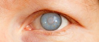
The diameter of the macula is approximately 5 mm, but it is this small part of the eyeball that consists of photoreceptors - light-sensitive cells responsible for central object vision in humans.
In this article
- Causes of macular edema
- Symptoms that occur with macular edema
- Diagnosis of Irvine-Gass syndrome
- Diabetic macular edema: laser treatment
- How does laser coagulation work?
- Rehabilitation period after laser treatment of macular diabetic edema
- Laser treatment of the retina: complications
If fluid accumulates in the macula area, macular edema occurs. It is also called Irvine-Gass syndrome, who first described this macular condition. Swelling is often a sign of one of the following ophthalmic diseases:
- diabetic retinopathy;
- thrombosis of the central vein of the retina;
- uveitis (various inflammations of the choroid);
- retinal detachment;
- glaucoma;
- neoplasms in the organs of vision (both benign and malignant).
Macular edema can also be a complication after eye surgery (for example, after cataract removal), the result of blunt trauma to the eyeball or poisoning with toxic substances.
There are also non-eye diseases accompanied by macular edema:
- infectious diseases (HIV, tuberculosis);
- kidney pathologies;
- brain diseases (meningitis) and head injuries;
- blood diseases;
- allergy;
- atherosclerosis;
- rheumatism.
There are many factors that provoke swelling of the central part of the retina. If such a symptom is detected, a comprehensive examination is prescribed. The patient will have to be checked by several specialists, not just an ophthalmologist.
Causes of retinal edema
The main reason is liquid ingress in the center of the inner shell. This happens due to various processes. There are different mechanisms for the development of symptoms. It was first described by the scientist S.R. Irwin. He considered edema as a consequence of surgery to remove the lens for cataracts. However, the mechanism of its origin is still not clearly understood. It is assumed that the development of macular edema is determined by the type of surgery performed on the eye.
Today other reasons are known. One of the most common is diabetes mellitus, which often causes diabetic retinopathy. Swelling occurs due to damage to the vessels through which fluid leaks into the macula.
Factors that provoke macular edema can be combined into two groups. The first includes ophthalmological diseases:
- glaucoma;
- uveitis;
- retinitis pigmentosa;
- retinal disinsertion;
- occlusion or thrombosis of the retinal veins;
- diabetic retinopathy;
- ametropia.
Reasons for development
The pathogenesis is based on the development of an autoimmune reaction. The immune system malfunctions, and leukocytes begin to attack healthy cells in the lungs, joints, and skin.
Löfgren syndrome most often develops in young women (20-35 years old). Children, as a rule, do not get sick from it. Löfgren's syndrome begins acutely, and then gradually the clinical manifestations may resolve on their own. A favorable course of the disease is often observed.
In addition to the viral and bacterial theory of the development of sarcoidosis, there is also an opinion that the disease develops in people who have long-term contact with volatile chemicals in factories. The hereditary factor should not be overlooked. That is why sarcoidosis is a polyetiological autoimmune disease.
Forms of edema (macular)
The cause of Irvine-Gass syndrome affects the form in which it develops. Thus, diabetic macular edema is a consequence of damage to the retinal vessels, which happens with diabetic retinopathy.
Retinal vein thrombosis causes cystoid macular edema. The fluid penetrates the perivascular region of the eye and remains in the center of the retina.
Inflammation of the retina and trauma to the eye lead to fusion of the connecting membranes connecting the vitreous body to the retina. It begins to exert a pulling effect on the retina, causing swelling.
Clinical manifestations
Löfgren's syndrome in sarcoidosis manifests itself polysymptomatically. Clinical manifestations begin acutely. A person develops erythema nodosum on the body, it tends to spread to the limbs, back, abdomen, and neck. The nodes have an oval shape, their diameter can reach up to 6 centimeters. It is worth noting that necrosis does not occur at the site of erythema formation.
The main manifestation is considered to be damage to the bronchial and tracheal lymph nodes. This poses a great danger to the patient:
- cough appears;
- occasionally body temperature increases;
- health worsens;
- night sweats occur;
- sleep quality deteriorates;
- weight loss occurs;
- the person is feeling anxious.
Later Löfgren's syndrome is characterized by joint damage. They become swollen and painful, and mobility worsens. Objectively, it is possible to notice an increase in the lymph nodes of the neck, as well as in the armpits.
Symptoms that occur with macular edema
Any form of the syndrome causes severe visual impairment. Patients complain about:
- blurry image in the central visual area;
- pinkish color of the image received by the eyes;
- photophobia;
- deterioration of visual acuity, which varies depending on the time of day;
- visual distortions of objects (straight lines of visible objects are deformed).
If the symptoms of edema are noticed in a timely manner, there will be no serious visual impairment, although it recovers very slowly (from two months to a year). Chronic edema (macular) can lead to irreversible damage to the retinal receptors, which are replaced by fibrous tissue, causing severe impairment of central vision without the possibility of further correction.
Symptoms
At the initial stage, macular edema develops asymptomatically. If left untreated, the area of swelling increases, resulting in the following symptoms:
- blurred image;
- distorted perception of straight lines;
- sensitivity of the retina to bright or daylight, which precedes the development of photophobia;
- altered color perception: the image is perceived by the eye in pink tones;
- Deterioration of vision, presumably in the morning.
Uncomplicated edema does not provoke visual impairment, but despite this, complete recovery of the retina is observed no earlier than three months after treatment.
Chronic macular swelling leads to damage to the visual receptors and proliferation of fibrous tissue, which precedes loss of central vision.
Classification
Sometimes macular edema develops in pregnant women against the background of late toxicosis.
Depending on the cause, retinal swelling occurs:
- Diabetic. The pathological process becomes a consequence of diabetic retinopathy that occurs against the background of diabetes mellitus. Disorders of carbohydrate metabolism are accompanied by damage to the blood vessels of all organs, including the eyes. As a result, the permeability of the vascular walls through which fluid penetrates into the tissue increases. The lesion can cover the entire retina or only its central part - the macula.
- Cystic. With cystoid edema of the retina, cavities form in the affected tissues in which fluid accumulates. The pressure in the membranes of the eye is disturbed, inflammatory processes develop, and infiltration accumulates in the area of the macula.
- Dystrophic. The cause of edema is the degenerative processes occurring in the retina. Most often, this type of pathology develops in older people due to age-related changes occurring in the body.
By area of damage there are:
- diffuse retinal edema – localized in the central region, spreading over a fairly large area;
- focal – does not affect the central zone, has a fairly limited area of damage.
Types of cystoid macular edema and prognosis
Macular edema, depending on the location and degree of development of the pathology, is classified into one of two types:
- Focal (swelling area no more than 2 mm, not central location);
- Diffuse (the area of tissue changes is more than 2 mm, the edema is located in the center of the macula).
The prognosis for macular edema is usually favorable. This pathology itself does not lead to vision loss. However, prolonged swelling can lead to disruption of tissue trophism and cause the development of secondary pathological processes and organic changes in visual structures.
Restoring normal hydrodynamics when macular edema occurs can last from 2 to 15 months. If the rate of improvement is low, careful monitoring of the condition of the retina and optic nerve head and timely adoption of adequate measures in case of complications occur is necessary.
Diagnosis of Irvine-Gass syndrome
Potentially, macular edema can lead to complete blindness, so it is important to start treating the pathology on time. Timely treatment can not only stop the progression of the disease, but also restore vision. To diagnose the syndrome, standard and specialized research methods are used:
- Ophthalmoscopy (examination of the fundus of the eye). This method will not detect local macular edema, so additional studies may be needed.
- The Amsler test (Grid or Amsler lattice) is a black square with many small squares inside on a white background and a black dot in the middle. The patient looks at the grid from a distance of 30 cm with one eye, after 5 seconds approaches the grid at 20 cm and again looks at its center point. After 5 seconds, the subject returns to the starting position. If central vision is not impaired, a person sees all lines and angles without distortion. When the retina is damaged, the lines of the square become bent.
- Optical coherence tomography (OCT) is an examination method that allows you to evaluate changes in the retina, its thickness, and volume.
- Fluorescein angiography (FA) is a test that helps identify areas of retinal ischemia and vascular abnormalities.
- Heidelberg retinal tomography is a laser scan of the retina that allows you to determine its thickness in the center.
To make a diagnosis, the doctor selects a technique individually for each patient.
Causes
When the retina swells, the visual perception of shapes and colors is impaired.
The main causes of retinal edema are vascular disorders and inflammatory pathologies of the visual analyzer. Most often, swelling of the retina is caused by:
- injury or surgery, after which the blood supply to the eye structures is disrupted;
- concussion, damage to areas of the central nervous system that are responsible for visual perception;
- angiopathy (usually diabetic);
- retinopathy arising from arterial hypertension;
- retinal venous thrombosis;
- inflammatory ophthalmological pathologies leading to degeneration of the structures of the visual analyzer;
- displacement or deformation of the vitreous body.
At risk are people suffering from chronic inflammatory eye diseases, diabetes mellitus, and arterial hypertension.
Treatment of retinal edema
Treatment of retinal edema must be directed to the cause that caused this complication. The causes of retinal edema and the mechanism of formation are as follows: the fluid circulating in the vascular system of the retina begins to accumulate in one place, forming stagnation. Based on diagnosis and medical history, the ophthalmologist may prescribe one of three techniques.
Drug treatment
In most cases, for minor swelling of the retina, the doctor prescribes medications that have an anti-inflammatory effect. Such drugs contain substances that help improve blood circulation in the vascular system. In the presence of inflammatory processes, it is recommended to use eye drops and ointments. The same drugs are prescribed for trophic disorders of the vascular system.
If there are no positive results when using medications, the doctor may prescribe special injections into the lens of the eyeball. These drugs have a powerful anti-edematous effect.
Surgical intervention
In most cases, to reduce swelling of the visual organs, it is sufficient to use medications that have a strong anti-inflammatory effect. However, when swelling is caused by pathology in the lens of the eyeball or retinopathy due to diabetes, immediate surgery is necessary.
Treatment tactics for retinal edema are developed individually, depending on the cause that caused it
If a deformation of the eyeball is detected, specialists prescribe a vitrectomy procedure, as a result of which the vitreous body is removed. As a result of this effect, the retina is relieved of increasing pressure, and blood circulation is normalized over time. Also, this procedure can be prescribed when the lens of the eyeball is displaced.
Laser coagulation
If focal swelling of the eyeball occurs, a laser coagulation procedure is prescribed. The essence of the method lies in soldering certain fragments of the circulatory system, through the walls of which moisture spreads. In addition, this technique restores blood circulation and improves nutrient exchange inside the eyeball. The method is considered effective if the disease is detected in the early stages. The likelihood of success and complete restoration of vision is very high.
This technique is also used to treat edema caused by diabetes mellitus. In such cases, special barriers are created that hold the retina and the vitreous together. The operation allows you to strengthen the retina and avoid its thinning and detachment.
Laser coagulation makes sense for pathologies in the vascular system of the eyeball.
Macular edema: treatment of the syndrome
Macular edema is treated in three ways: conservative (medication), surgical and laser. The choice of treatment method depends on the form the swelling has taken. The cystic type of syndrome is treated with anti-inflammatory drugs. Macular degeneration, accompanied by edema, is eliminated through surgery. For diabetic macular edema, the most effective laser treatment method is laser photocoagulation of the retina. Let's consider this method in more detail.
Useful video
In case of swelling in the macular area, the main thing is timely consultation with an ophthalmologist and diagnostics. There are no specific preventive measures for the pathology. The only way to prevent the development of swelling is the treatment of provoking diseases of the visual apparatus.
Surgery is not always necessary to relieve swelling. If indicated, the elimination of edema occurs with the help of drug treatment. It is possible to completely restore vision after a period of 2 months to 1 year.
Author's rating
Author of the article
Alexandrova O.M.
Articles written
2031
about the author
Was the article helpful?
Rate the material on a five-point scale!
If you have any questions or want to share your opinion or experience, write a comment below.
Treatment
Treatment of macular edema may involve surgery. There are also conservative techniques and new methods using laser equipment. The choice of treatment method depends on the cause of the pathology and the neglect of the patient’s condition.
During conservative therapy, the patient is prescribed medications in injections, tablets and drops. These are corticosteroids and non-steroidal anti-inflammatory drugs (NSAIDs). Treatment with NSAIDs is more desirable, since unexpected side effects do not occur with this course of therapy. Such unpleasant effects may occur while taking corticosteroids. This includes an increase in IOP, a decrease in local immunity, and the appearance of ulcers in some areas of the cornea. NVPS is often used before eye surgery to improve the effectiveness of cataract surgery. In the postoperative period, both NSAIDs and corticosteroids are used as anti-inflammatory therapy. These drugs also act as prevention of postoperative macular edema.
If positive changes are not observed during conservative treatment, then certain substances intended for intravitreal administration can be introduced into the vitreous cavity. In case of pronounced changes in the vitreous body, vitrectomy is performed - removal of the vitreous body.
Diabetic macular edema can only be cured using a single method - laser photocoagulation of the retina. It is very important to start laser therapy as early as possible. This treatment method is also very effective for focal macular edema. True, there is an opinion that in this case, even with successful completion of laser treatment of diffuse edema, there is a very high probability of rapid deterioration in visual function. With laser therapy of the retina, all defective vessels that allow fluid to pass through their walls are coagulated. The center of the macula is not touched.
The most favorable outcome of treatment for macular edema is the gradual (over several months) disappearance of the problem and the same gradual restoration of visual functions. The question of how successful the treatment will be depends entirely on the timeliness of contacting an ophthalmologist. Even if the ophthalmologist recently examined you, and minor symptoms have just appeared, you need to go to the doctor urgently.
No matter what anyone says or promises, macular edema of the retina cannot be treated with folk remedies. Such therapy simply will not give any positive results. You can use traditional methods in combination with pharmacological and surgical ones. For example: adhere to proper nutrition to stabilize blood sugar levels if there is a risk of diabetic macular edema.
The appearance in the macula area, or the so-called macula, located in the central region of the eye, is a fairly common occurrence in ophthalmology. The macula is the area of the retina that is responsible for central vision. Often among specialists you can hear the term “Irwin-Gass syndrome”, which characterizes macular edema. The pathology owes this name to ophthalmologist S. R. Irwin, who first characterized the disease that appeared after cataract removal.
This syndrome is not an autonomous disease; it is diagnosed as a concomitant of diseases such as diabetic retinopathy, retinal vein thrombosis, and uveitis. Pathology can also appear as a result of surgical interventions and eye injuries.
In rare cases, this complication can lead to vision loss. Basically, with timely and correct treatment, swelling can be eliminated.
Cystic macular edema
Description
Cystic macular edema (COM)
is the accumulation of fluid in the central region of the retina, a common cause of decreased central vision, often reducing vision after long-term inflammatory diseases or after surgery on the eye.
Anamnestic signs of the disease
Cystic macular edema
- a consequence of an underlying ocular or systemic disease or a combination of both. Most often it is detected in the following conditions:
- In the postoperative period after cataract extraction
- For diabetic retinopathy
- With the exudative form of senile maculopathy
- In case of occlusion of the central retinal vein or its branches
- After filling procedures, etc.+, creating deformations of the sclera
- For chronic uveitis, including the flat part of the ciliary body
- For vascular diseases and collagenoses
- For aphakic glaucoma using adrenaline drugs
- For tumors of the choroid (melanoma, hemangioma)
- For local retinal detachment in the proliferating form of diabetes
- For retinitis pigmentosa
- In the presence of preretinal membranes
- With chronic use of certain medications
- For toxic eye lesions
- With perimacular telangiectasia of retinal vessels.
, two mechanisms for the occurrence of edema
have been consistently proposed :
inflammatory and hypoxic
. However, none of these mechanisms has so far been unconditionally accepted for the following reasons.
Inflammatory theory
is able to explain cases of edema associated with surgical trauma, scleral deformations and chronic uveitis, and the hypoxic mechanism of occlusion, thanks to the known trophic connection of the choriocapillaris layer with the outer layers of the retina, satisfactorily explains the conditions associated with primary vascular pathology. It is possible that in each specific case of cystic retinal edema these two mechanisms are intertwined, which is to some extent confirmed by the uniformity of the observed clinical picture of the lesion, regardless of the reasons that caused it.
Retinitis pigmentosa and perimacular telangiectasias also cause disruption or shunting of retinal blood flow.
Choroidal tumors create multiple occlusions of retinal capillaries with secondary hypertrophy of vascular endothelial cells. These phenomena are followed by edema or the formation of cysts, due to the expansion of extracellular spaces by serous exudates, limited to the limits of the inner plexiform and inner nuclear layers. However, most often cystoid edema is a consequence of cataract surgery, especially the intracapsular method
.
It is believed that surgical trauma to the anterior eye either stimulates the synthesis and release of prostaglandins or prevents the deactivation of prostaglandins and their removal outside the eye. According to various researchers, after intracapsular cataract removal, COM occurs in 40%-60% of cases. Extracapsular cataract extraction
results in COM in approximately 10% of cases.
Indirect evidence of the correctness of the assumption about the role of prostaglandins is provided by clinical experience in recent years. When prostaglandins like latanoprost are used to treat glaucoma, an increased incidence of cystoid macular edema and anterior uveitis has been found.
Recent studies have shown that macular edema, anterior uveitis, or both occur simultaneously in 15% of patients using such drugs.
of the intraocular lens
can also contribute to the development of COM . Implantation of lenses with pupillary fixation is a greater risk factor compared to implantation of an anterior chamber lens. The lowest percentage of development of COM is observed with implantation of intraocular lenses into the posterior chamber and capsular bag.
True, in most cases, macular edema goes away after 3-6 months without additional therapeutic measures, however, if the anterior hyaloid membrane of the vitreous body is destroyed or the fibers of the vitreous body are pinched in the corneoscleral wound, the condition can turn out to be chronic.
Very often, the true extent of the lesion becomes clear only after an angiographic examination, since the detached retina and subretinal fluid, which remain transparent, do not reveal their boundaries.
Diabetic retinopathy
ranks second in frequency among the primary pathologies leading to the development of COM. This complication is often observed in adult diabetics over 30 years of age, whose disease duration exceeds 10 years. Accompanying the course of diabetes with arterial hypertension or atherosclerosis makes COM an even more likely complication of diabetic retinopathy.
The question of the role of light lesions
in the occurrence of cystic edema has been raised by many medical specialists. Light from the coaxial illuminator of an operating microscope falling on the retina of the posterior part of the aphakic eye creates a scotoma observed during the first week after surgery. Light conducted inside the eye to the retina using fiber-optic illuminators during posterior vitrectomy surgery can also, due to its energy characteristics, cause damage to the retina. And although it is still unclear how important the role of retinal irradiation during surgery is in the appearance of postoperative cystoid edema, it is more reasonable take measures to reduce the amount of light entering the eye. Technical means for this have long been introduced by manufacturers into lighting devices.
Treatment and prevention
The use of anti-inflammatory drugs has been shown to reduce the incidence, severity and duration of macular edema, regardless of whether it was caused by surgical trauma or systemic disease.
Currently, a list of antiprostaglandins
- non-steroidal and steroidal anti-inflammatory drugs, with the help of which good results are achieved.
The most repeatable treatment results were when using one of the following drugs
:
- Fenoprofen (Nalfon)
60 mg every 4-6 hours - Ibuprofen (Brufen)
400 mg every 4-6 hours - Prednisone
20 mg (1 gab, 4 times) per day for 1 week, tapering to 40 mg/week for 2 weeks, then maintaining the dose at 20 mg/week for 2-3 weeks.
There is a report of the cure of chronic coma with hyperbaric oxygenation - inhalation of oxygen at a pressure of 2 atmospheres for 1 hour once a day* A significant decrease in the severity of retinal edema and an increase in visual acuity from 0.2 to 0.8-1.0 are noted.
The beneficial effect of this treatment is explained by the narrowing of the capillaries supplying the macular area under the influence of oxygen. At the same time, the cell density of the endothelial layer increases and the formation of a collagen layer is stimulated, isolating leak sites. Local treatment
Reducing the degree of cystoid edema of the macular area can be achieved by using ocular forms
anti-inflammatory drugs such as:
- Water or oil eye drops of 1% indomegacin - 1 drop 3-4 times a day. This remedy can be prescribed prophylactically 1 day before cataract extraction surgery and 3-4 times a day after surgery for 4-6 weeks.
- Eye drops 0.5% trimethamine ketolorac - 1 drop 4 times a day.
- Eye drops 0.1% diclofenac - 1 drop up to 4 times a day.
- Eye drops 1% prednisolone acetate - 1 drop 4 times a day,
- Subtenon's or retrobulbar injection of 40 mg triamsinolone.
When using eye drops, it is important to instruct the patient to close their eyelids immediately after instillation and not to open their eyes for 10-15 seconds after instillation of the drops.
It is very useful to create slight pressure on the inner canthus to prevent the outflow of the medicine through the lacrimal ducts into the nasopharynx. Most patients recover with improved visual acuity within 1-2 weeks from the start of intensive corticosteroid therapy; Some studies have shown that a single retrobulbar injection of a corticosteroid was as effective as three two-week courses of sub-Tenon injections of the same drug. Visual acuity during recovery in the two compared groups turned out to be very close. Of course, as with all cases of corticosteroid use, periodic monitoring of intraocular pressure remains necessary.
Evidence of usefulness as an adjunct to corgicosteroid therapy
carbonic anhydrase inhibitors, for example, diacarb (diamox). This drug promotes the outflow of fluid through the retinal pigment epithelium into the choroid. In the course of a specially conducted angiographic study in chronic uveitis, a statistically significant decrease in retinal edema under the influence of diacarb was noted, although it was not always accompanied by an increase in visual acuity.
It was also found that in patients with retinitis pigmentosa, oral administration of diacarb reduces retinal swelling and increases visual acuity. The discovery of this fact raised a natural question - whether topical use of carbonic anhydrase inhibitor eye drops, such as dorzolamide hydrochloride, is appropriate to improve the effectiveness of the treatment of retinitis pigmentosa. Angiographic studies have found that local application of dorzolamide improves the condition of the retina, but an increase in visual acuity was not achieved. Oral administration of diacarb was effective in both improving the condition of the retina and increasing visual acuity.
Adverse reactions are often observed when taking anti-inflammatory drugs orally. Most often, adverse reactions manifest themselves in the form of gastrointestinal disorders (dyspepsia, anorexia, nausea and vomiting, constipation, flatulence), headache, drowsiness, dizziness, tremor, skin rash, tinnitus, and instability of body posture.
To reduce the likelihood and severity of side effects, these drugs should be taken 30 minutes before meals. In addition, the side effects that occur with oral administration of diacarb can be significantly reduced by simultaneously prescribing oral baking soda - sodium bicarbonate
(1 g three times a day).
Corticosteroids
in oral form should not be prescribed to patients suffering from peptic ulcers or osteoporosis. Before starting a course of subconjunctival, retrobulbar or sub-Tenon injections of corticosteroids, local use of the drug should ensure that there are no adverse reactions to steroids (primarily acute ocular hypertension). It should be remembered that after injection the drug has an effect on the eye for a month and it is impossible to stop its effect.
Laser treatment
The issue of using laser treatment for cystoid edema of the macular area must be considered, first of all, from the perspective of its possible influence on the immediate cause of its occurrence. Thus, in case of diabetic macudopathy, occlusion of a branch or the most central retinal vein, the main efforts should be made to find the place or places of fluid leakage from the vessels. The duration of edema and the degree of loss of visual acuity are secondary arguments in favor of laser treatment, although some authors believe that a drop in visual acuity to 0.2 or lower is already the basis for the use of lasers in coagulating mode. It should still be remembered that laser treatment can only bring unconditional benefit when identifying and specifically blocking areas of blood vessels that allow leakage. In addition, the possibility of using laser devices in the mode of subthreshold radiation, which stimulates the phagocytosis of dying segments of the sensory epithelium by pigment epithelial cells, cannot be discounted.
There are often studies devoted to the issue of choosing the type of laser. Here is one example. Diode infrared laser compared
(emission wavelength 810 nm) and
argon green laser
(emission wavelength 514 nm).
Both settings were compared in the treatment of diabetic retinopathy with disseminated macular edema. The treatment procedure was the creation of a coagulation "mesh" on the retina
. The authors found no differences in the effect of the two laser systems - neither in final visual acuity, nor in the final condition of the retina, nor in the number of additional treatment sessions performed during the one-year follow-up period. The only factor significantly influencing the outcome of treatment was the duration of existence of a systemic disease - diabetes, regardless of the type of laser used for treatment.
The conclusions of such studies can hardly be considered final. First of all, it is not clear why the “mesh” was chosen as the procedure - a procedure whose adequacy in relation to different stages of diabetic retinopathy is very doubtful, and based on the above findings, the “length of experience” of the disease in the patients of the group was different. The advantages and disadvantages of lasers affect the solution of precisely defined tasks, and not in greater or lesser ease of use or thermal effect, and, as already indicated above, it is unlikely that any laser with a constant wavelength of radiation can be universal for all cases and departments retina.
Therefore, a sufficient basis for performing laser photocoagulation for cystic retinal edema can be considered the possibility of meeting the following conditions:
- Detection and documentation of the presence of a point or points of fluid leakage from the retinal vessels.
- Localization and “linking” of leakage points to landmarks visible on the retina during ophthalmoscopy or ophthalmoscopy using a contact lens,
- The technical ability to block leakage points by this surgeon and the available laser device without damaging functionally significant areas of the macular area.
Treatment with panretinal photocoagulation
of the retina for cystoid macular edema due to occlusion of the central retinal vein must be resorted to in one of two discovered circumstances:
- If the formation of new vessels is noted on the optic nerve head or in other parts of the retina.
- If the angiogram shows signs of blocking the perfusion of the peripheral retina.
If COM after occlusion of the central retinal vein proceeds without the formation of new vessels, and the patient’s visual acuity more than two months from the onset of the disease turns out to be 0.2 or lower, the retina is coagulated in the form of a lattice with a constant “step” of coagulates equal to 5-6 diameters of an individual coagulum.
In the treatment of cystoid macular edema
sometimes it is necessary to decide on the advisability of
surgical treatment
.
This question is especially relevant in cases where COM is detected in the postoperative period after abdominal eye surgery. Then the main goal of surgical treatment is to restore normal anatomical relationships in the anterior segment of the eye, removing all detected pathological adhesions of the vitreous body. In case of aphakia with strangulation of the vitreous body in a wound without the presence of an intraocular lens, it is recommended to perform anterior vitrectomy
through the limbal approach or the pars plana of the ciliary body, which in 75% of cases brings an effect expressed in improving the condition of the eye and visual functions. However, since surgery itself can lead to serious complications, it should be resorted to only after making sure that other treatment methods are ineffective.
Utility of pars plana vitrectomy
in cases resistant to treatment of pseudophakic cystoid edema, was confirmed by different authors by analyzing the outcomes of operations on eyes with adhesions of the vitreous body and anterior chamber structures. The result of the operations was a significant increase in visual acuity (on average by 0.4-0.5) after treatment.
Vitrectomy through the pars plana of the ciliary body was ineffective in that part of the cases of chronic cystoid edema that developed after uncomplicated extracapsular extraction and implantation of an intraocular lens into the capsular bag. In addition, vitrectomy surgery should only be performed in cases of inflammation of the pars plana or chronic uveitis, and after less invasive approaches to treatment are considered untenable in their results.
Cryotherapy of the pars plana may be attempted before a decision is made to proceed with vitrectomy.
, as an alternative treatment for failure of systemic and local treatment of chronic uveitis. Caution in the transition to surgical treatment is dictated by the fact that possible complications of any operation can be very serious.
However, when considering the issue of surgical treatment of cystoid macular edema, one should take into account and weigh not only the likelihood of possible postoperative complications. The technical support of ophthalmic surgery has been improved in recent years, and not to a small extent. Thanks to the use of computer technology and other modern technology products, many surgical interventions have acquired a new level of safety. This gives reason to believe that where previously we had the right to expect serious postoperative complications, fatal changes in the anterior segment of the eye and dislocation of the vitreous body will now be absent, which allows us to resort to invasive techniques in treatment with greater confidence in the well-being of the eye and vision.
—
Article from the book: Macula. Treatment methods, main lesions, laser treatment, low vision (clinical essay) | Skitsyuk S.V., Pristash I.V.
Diabetic macular edema: laser treatment
Laser coagulation of the retina is a minimally invasive procedure that is performed on an outpatient basis under local anesthesia. The essence of the operation is as follows: the retina is cauterized with a laser and soldered to the choroid. After this, the vessels stop growing and bleeding. In people with diabetes, after this procedure, visual acuity ceases to decrease.
Laser coagulation is used to treat retinal tears and detachments caused by a variety of reasons. This procedure is prescribed even to pregnant women who have a retinal detachment. After photocoagulation, women are allowed to give birth naturally.
However, there are a number of contraindications to such laser treatment:
- retinal hemorrhages (blood effusions on the retina and areas of the eye next to it);
- clouding of the cornea, vitreous body, lens (impaired transparency of the optical media will prevent the doctor from monitoring the fundus of the eye during the procedure);
- proliferation of iris vessels;
- hemorrhage into the vitreous body due to damage to the retinal vessels (hemophthalmos);
- visual acuity is less than 0.1 (this contraindication is relative and the doctor may decide in favor of the procedure).
Any form of cataract, cataract, corneal dystrophy, destruction of the vitreous body - all this becomes a limitation for the purpose of the procedure.
Diabetic macular edema
- Pathogenesis of diabetic macular edema
- Clinical picture
- Treatment
Macular edema (MO) is one of the main causes of vision loss in patients with diabetes mellitus (DM). Many studies have shown that the prevalence of MO in type 1 and type 2 diabetes strictly correlates with the duration of the disease. The WESDR1 study found that the prevalence of MO increases with the severity of retinopathy and duration of diabetes. Thus, in patients with non-proliferative diabetic retinopathy, MO was detected in 2-6% of cases, with preproliferative retinopathy - in 20-63%, and with proliferative retinopathy - in 70-74% of cases.
In patients with type 1 diabetes lasting less than five years, MO was not found, but in patients with an experience of 20 years or more, it was diagnosed in 29% of cases. In people suffering from type 2 diabetes, the prevalence of MO ranged from 3% with a duration of the disease of less than 5 years to 28% with a duration of diabetes of more than 20 years. In patients with type 2 diabetes on insulin, MO was detected more often (15%) than among patients with type 2 diabetes who received tableted glucose-lowering drugs (4%). Over a 10-year observation period, according to WESDR, the development of MO was found in 20.1% of patients with type 1 diabetes, 25.4% of patients with type 2 diabetes on insulin, and 13.9% of patients with type 2 diabetes not receiving insulin.
Pathogenesis of diabetic macular edema
In the development of diabetic MO, the main role is played by a breakthrough of the internal hematoretinal barrier, which is secondary to anatomical damage at the level of capillary endothelial cells (disruption of intercellular contacts or endothelial damage) caused by local retinal hypoxia, osmotic stress and increased expression of vascular endothelial growth factor - VEGF2 and proinflammatory cytokines.
It is the internal hematoretinal barrier, which regulates the metabolic exchange between the blood and the retina, that maintains it in a dehydrated, and therefore transparent, state. The concentration of proteins that provide tight junctions between vascular endothelial cells is reduced in the early stages of experimental diabetes, which may explain increased vascular permeability.
Fluorometric studies of the vitreous in patients with diabetes show that a breakthrough of the internal blood-retinal barrier prevails over changes in the external blood-retinal barrier when macular edema occurs. Subsequently, with the long-term existence of MO, the external hematoretinal barrier also suffers.
These anatomical lesions may be accompanied by a functional factor (impaired autoregulation of blood flow): a local increase in blood flow in the capillaries of the macular retina, caused by local hypoxia, leads to an increase in perfusion pressure in the capillaries and promotes diffusion from them. Increased intravascular hydrostatic pressure tends to force fluid through the vessel wall (Starling's law). Autoregulatory dilatation of arterioles causes a decrease in intravascular pressure in arterioles and an increase in venules (Poiseuille's law). An increase in hydrostatic pressure causes an increase in the diameter of arterioles and venules (Lapplace's law), and also leads to an increase in their length and tortuosity.
Serial observations in patients with diabetes have shown that the diameter of retinal vessels and their length (tortuosity) increase even before the appearance of clinically significant changes, and also decrease after laser coagulation of the retina for MO and proliferative retinopathy. Often the source of leakage is microaneurysms that are located adjacent to areas of non-perfusion. Histologically, they are localized protrusions of the capillary wall with focal proliferation of endothelial cells and loss of pericytes. Factors contributing to the formation of microaneurysms likely include loss of pericytes, hemodynamic disturbances (increased capillary pressure), and local production of vasoproliferative factors. Most likely, it is damage to the components of the capillary wall, combined with a violation of autoregulation, that leads to a breakthrough of the internal blood-retinal barrier and to retinal edema.
In general, hemodynamic disturbances in the retina are similar to those in the kidneys in the early stages of diabetes mellitus—increased blood flow and glomerular permeability, resulting in albuminuria. Fluid that passes through the capillary wall should normally be reabsorbed by the pigment epithelium and adjacent retinal capillaries. When diffusion exceeds the potential of the pigment epithelium and capillaries to reabsorb fluid, clinical signs of MO occur. Significant accumulation of fluid in the intercellular spaces of the retina leads to the formation of cystic MO.
“Solid” exudates arise as a result of diffusion through the walls of microaneurysms and dilated capillary segments of plasma components (for example, lipoproteins) and their deposition in the retina. In addition, MO may be associated with a sharp disruption of capillary perfusion or changes in the vitreomacular interface. Thus, in the pathogenesis of diabetic MO there are three (often concomitant) primary lesions.
Clinical picture
A standard ophthalmological examination includes: visometry, biomicroscopy, tonometry, ophthalmoscopy using a high diopter aspheric lens or Goldmann contact lens. In addition to the standard examination, a number of additional studies are carried out (FA, OCT).
Currently, there is no generally accepted classification of diabetic MO, however, most researchers, depending on which of the following lesions is predominant, identify its main clinical forms:
- capillary hyperpermeability associated with an isolated breakthrough of the internal blood-retinal barrier and impaired autoregulation (focal edema), or in combination with a violation of the external blood-retinal barrier (diffuse edema);
- disruption of vitreoretinal relationships or the density of the internal limiting membrane of the retina (traction edema);
- microocclusion of capillaries (ischemic edema).
In diabetes-related studies, macular edema is characterized as retinal thickening or the presence of hard exudates within 1 disc diameter of the center of the macula.
The Early Treatment of Diabetic Retinopathy Study (ETDRS), a multicenter clinical trial, coined the term “clinically significant macular edema.” Macular edema is clinically significant if one of the following conditions is present:
- retinal edema within 500 mm of the center of the macula;
- hard exudates within 500 mm of the center of the macula if accompanied by retinal thickening (which may extend beyond 500 µm);
- retinal edema within 1 disc diameter or more, that is, any area of edema must fall within 1 disc diameter from the center of the macula.
Cystic diabetic macular edema results from the accumulation of fluid in the outer plexiform and inner nuclear layers of the retina centrally near the fovea, forming fluid-filled cyst-like structures.
The introduction of optical coherence tomography has changed the view of DME and its classification.
Visualization of the macular area and the border between the vitreous and the retina allowed the classification of macular edema as follows:
- Spongy: Sponge-like retinal swelling is present in 88% of cases with DME. This form is mainly limited to the outer layers of the retina, the retina becoming hyporeflective at these levels.
- Cystic macular edema: Cavity formation begins in the outer plexiform layer. Cystic macular edema, isolated or in combination with diffuse lesions, occurs in 47% of cases of DME. On OCT, cystoid macular edema is represented by decreased intraretinal reflectivity. “Fresh” cystoid macular edema is characterized by the presence of pseudocysts in the outer layers; the inner layers remain relatively intact. With the long-term existence of cystic edema, the walls of the cysts dissolve, and larger drain cavities are formed.
- Serous retinal detachment: accounts for 15% of all forms and is visible as a hyporeflective area in the subfoveal region of the retina. This form is inevitably related to one of the first two forms described.
- Tractional: Vitreomacular traction can lead to foveal detachment. This can be easily diagnosed on OCT, which shows how the posterior vitreous causes traction on the macula, resulting in a tractional retinal detachment.
- Induration of the posterior hyaloid membrane: the diagnosis can be easily made on OCT even in the subclinical form. In advanced cases, it can be diagnosed clinically as a dense, shiny retinal membrane on biomicroscopy.
In conclusion, OCT provides us with in vivo histopathology of the retinal layers, which provides greater insight into disease pathogenesis and monitoring of treatment effectiveness.
Fluorescein angiography (FA) can detect the earliest signs of DME. This method is sensitive to the qualitative detection of fluid leakage. FA allows us to evaluate macular edema by identifying fluorescein leakage and ischemic areas.
DME is also classified into focal and diffuse depending on the picture on FA. In focal DME, local hyperfluorescence (leakage of intravascular fluid into the interstitial space due to vascular permeability) is detected on FA due to local leakage from the microaneurysm. Typically, these microaneurysms are surrounded by deposits of hard exudates. Multifocal macular edema is sometimes confused with diffuse macular edema. This form is detected on FA as several foci of leakage due to the presence of multiple microaneurysms. In diffuse DME, there are areas of diffuse leakage on FA due to leakage from intraretinal capillary beds and/or intraretinal microvascular abnormalities (IRMAs) and/or from arterioles and venules without areas of leakage from microaneurysms.
FAH divides DME into four categories:
- focal/multifocal edema—well-defined local or multifocal areas of leakage from the microaneurysm are identified;
- diffuse edema is defined as the presence of diffuse leakage from the retinal capillary bed or any intraretinal microvascular abnormality (IRMA);
- cystoid macular edema - areas of diffuse dye leakage combine into cystic spaces of the macula in the late phase of angiography;
- ischemic maculopathy is an area of ischemia that can be considered as an area of increased foveal avascularity. The presence of macular ischemia is an important finding when deciding on the type of treatment to help those patients who suffer from loss of visual acuity of unknown origin.
Treatment
As has been proven in most multicenter studies, the main treatments for diabetic retinopathy and MO are:
- the most stable compensation of diabetes mellitus (DCCT 3 - compensation of diabetes mellitus with intensive insulin therapy reduces the risk of progression of retinopathy by 63%, development of proliferative retinopathy - by 47%, development of MR - by 26%, the need for laser coagulation of the retina - by 51%);
- normalization of blood pressure (WESDR - the presence of arterial hypertension at the onset of the disease increases the risk of developing proliferative diabetic retinopathy by 91% and MO by 210%; UKPDS4 - strict control of blood pressure can reduce the risk of progression of retinopathy by 34% and decreased vision by 47%, necessity in laser coagulation of the retina by 35%);
- correction of lipid metabolism disorders (FIELD5 - treatment with fenofibrate reduces the risk of progression of retinopathy by 79%, the need for laser coagulation of the retina - by 31%).
Laser coagulation of the retina currently remains the mainstay of treatment for diabetic MR. Despite the fact that the experience of many specialists confirmed the high effectiveness of laser coagulation in the treatment of diabetic motility, only after large-scale studies carried out by the Early Treatment of Diabetic Retinopathy Study Group - ETDRS, the method received worldwide recognition, since its effectiveness was reliably proven. According to the ETDRS study, retinal laser coagulation reduces the risk of severe vision loss by 50%, and an increase in visual acuity of more than one line can be achieved in 16% of patients.
Thus, laser coagulation of the retina currently remains the only method of treating diabetic MM, the effectiveness of which has been proven in large multicenter studies. For local edema, focal laser coagulation is usually used; for diffuse leakage, various modifications of “grid” type laser coagulation are performed. The effectiveness of treatment is especially high when laser coagulation is performed at an early stage of diabetic maculopathy with high visual functions and minimal deposits of “hard” exudates.
Today, it remains not entirely clear why laser coagulation of the retina can lead to resolution of diabetic MO. According to some researchers, focal coagulates promote obliteration of microaneurysms with pathological leakage, which is accompanied by minimal impact on the underlying retinal pigment epithelium. Obliteration of microaneurysms leads to a decrease in MR. Other researchers believe that laser treatment destroys part of the photoreceptors, which consume most of the oxygen needed for normal functioning of the retina. Scarring after exposure causes thinning of the retina, which facilitates the diffusion of oxygen from the choroid. Thus, laser exposure reduces the retina’s need for oxygen and increases its supply from the choroid.
The caliber of capillaries in the macular zone decreases after laser exposure, which possibly leads to a decrease in MO. There is also a theory that laser exposure causes the destruction of the affected retinal pigment epithelium, which is then replaced by “healthy” ones that are able to absorb more fluid from the retina, which leads to a decrease in swelling. Laser exposure of the outer retina has also been shown to induce endothelial proliferation in the capillaries and veins in the inner layers of the retina. Through the “activated” vessels there is less sweating, which helps to reduce MO. The effect is maximum in the affected areas, but also manifests itself in areas quite distant from the place of laser exposure. Despite the fact that the mechanism of action of laser coagulation on macular edema is not completely clear, focal exposure or “grid” coagulation remains the main methods for preventing a decrease in central vision in patients with diabetes, being the “gold standard” of treatment over the past 30 years. However, despite the proven effectiveness of laser coagulation, some patients continue to lose vision. This may be due to both complications of laser treatment (development of creeping atrophy and subretinal fibrosis) and resistance to the effects. In addition, there are a number of limitations in the use of laser coagulation: the presence of high MO, fibrosis of the internal limiting membrane of the retina or vitreomacular traction syndrome.
It was the understanding that laser coagulation is not an ideal method of treatment that led to an active search for treatment options for MM that would have fewer negative consequences, which was greatly facilitated by the development of pharmacology and surgical technologies. The beginning of the 21st century was marked by the active introduction into clinical practice of the technique of intravitreal injections of various drugs. One of the first drugs that were introduced into the vitreous body to treat MO were crystalline corticosteroids. The mechanism of their action is associated with the influence on the cascade of inflammatory reactions, which play a significant role in the pathogenesis of MO. In diabetes, there is an increase in the level of pro-inflammatory cytokines, an increase in the adhesion of leukocytes to the capillary endothelium, which leads to impaired blood flow and increased hypoxia. Corticosteroids reduce the production of inflammatory mediators and, to some extent, VEGF, cause apoptosis of leukocytes, increase the production of tight junction proteins, which leads to a decrease in vascular permeability. Thus, they protect the blood-retinal barrier through various mechanisms.
For many years, ophthalmologists around the world have been using triamcinolone acetonide (Kenalog 40, Bristol-Myers, Italy) to treat MO. Numerous studies have shown a significant decrease in MO and an increase in visual functions after intravitreal administration of Kenalog (IVVC), even with pronounced cystic changes in the retina. According to various authors, after administration of the drug, the thickness of the retina is reduced by an average of two times, and visual acuity increases by 2 lines. The main side effects of IVVC include increased intraocular pressure and progression of cataracts, which, according to various studies, occur in 18–35% and 25–40%, respectively. Many authors note the effectiveness of IVVC both in the treatment of primary MR and edema refractory to previous treatment, proposing the use of this method both as the main method and as an addition to laser coagulation of the retina.
The duration of action of triamcinolone acetonide is approximately 3–6 months, and a significant proportion of patients require repeated injections, increasing the risk of side effects. Attempts to improve the delivery of corticosteroids to the posterior segment of the eye and reduce the need for repeated injections have led to the development of slow-release intravitreal implants. To date, an intravitreal implant, Ozurdex (Allergan, USA), has been developed and is being used. By releasing the required amount of dexamethasone over a certain period of time, the implant gradually undergoes biodegradation and hydrolyzes, dissolving in the vitreous body.
A qualitatively new stage in the treatment of MO was the use of drugs that inhibit the production of VEGF, which plays a key role not only in the processes of angiogenesis, but also in vascular permeability. Already in the early stages of diabetes in the retina, not only the expression of VEGF increases, but also the receptor sensitivity to it, which subsequently leads to the appearance of vascular abnormalities characteristic of non-proliferative retinopathy. In combination with oxidative stress, this leads to the formation of large areas of ischemia, which further stimulates VEGF expression, promoting the appearance of MO and neovascularization. Effects on VEGF can have different points of application: binding the molecule, blocking the receptor, reducing expression.
Another cytokine involved in the pathogenesis of MO is TNFα 13. Theoretically, blocking it can reduce the inflammatory process and thus promote regression of MO. The drug infliximab (Remicad, Centocor Ortho Biotech, USA), which is an antibody to TNFα, blocks it, eliminating all the effects of the cytokine.
, indications for surgical treatment have expanded significantly . If previously posterior vitrectomy was performed only in cases where MR was associated with the presence of vitreoretinal traction, now MR that is refractory to laser and drug treatment can serve as an indication for surgery. In the first case, the effectiveness of the method is associated mainly with the direct elimination of vitreoretinal traction, in the second with the removal from the vitreal cavity of inflammatory mediators, vasoactive factors for which the vitreous body is a reservoir, as well as with increased oxygenation of the retina. In the presence of vitreomacular traction, vitrectomy is performed to remove the posterior hyaloid membrane of the vitreous body, but if there is fibrosis of the internal limiting membrane of the retina, then its removal is also necessary.
A non-surgical method for the treatment of MO associated with pathology of the vitreomacular interface has been proposed and is being actively studied - pharmacological vitreolysis. The purpose of this method is to eliminate partial detachment of the posterior hyaloid membrane, since it is one of the common causes of the development of MO. A drug is injected intravitreally, which causes complete detachment of the posterior hyaloid membrane, liquefies the vitreous, acting selectively without damaging the retinal tissue. Vitreolytic drugs are currently being actively developed and studied. One of these drugs is Microplasmin (ThromboGenics, Belgium), a recombinant drug that is a shortened form of human plasmin, a protein with fibrinolytic activity. In the studies conducted, the drug showed high efficiency, leading to complete detachment of the posterior hyaloid membrane, without causing any mechanical or toxic effects on the retina.
Despite the large number of promising treatment methods described, MO remains one of the main causes of vision loss in patients with diabetes. Currently, laser coagulation of the retina remains the most effective in the treatment of MO. Its combination with intravitreal administration of crystalline corticosteroids and angiogenesis inhibitors can improve functional results and reduce the need for repeated laser intervention.
Diagnostics
What diagnostic methods are used to determine retinal disease? Visual examination and even examination of the fundus do not always give a clear picture of the health of the eye shell. Therefore, if pathology is suspected, the following is prescribed:
- coherence tomography;
- fluorescein angiography;
- laser tomography.
Coherence tomography is considered the most accurate diagnosis of fundus problems. The method allows you to determine the layer thickness, structure and volume of the retina. Tomography is completely harmless to health, so it is used to monitor the results of treatment.
The subject of antigiography research is the vessels of the orbit. In this case, the patient is injected into a vein with a sparkling contrast substance and high-frequency imaging is performed. The image clearly shows a vascular pattern, providing information about the presence of changes and areas of edema.
Laser diagnostics provides comprehensive information about the condition of the fundus organs, including the retina.
Causes of retinal dystrophy
● Dystrophy of the retina (eyes) occurs when the vascular system of the eye is disrupted, during which the process of scarring of the central part of the retina begins.
Most often, this disease affects older people due to pathological changes in the body due to malnutrition, alcohol abuse and smoking. changes in the immune status, scarring of the layers of the retina begins.
The main symptom of retinal dystrophy is the absence of central vision: the patient sees objects located in the area of peripheral vision, and a black spot hangs in the center.
But even side objects are not clearly visible, although it is possible to distinguish day from night.
● Moderate and high degrees of myopia can lead to retinal dystrophy. It should be remembered that retinal detachment and retinal dystrophy are completely different things.
In old age (over 60 years), it is not retinal detachment that occurs, but retinal dystrophy, which is manifested by the replacement of the retina with fibrous tissue with the formation of scars.
Quite often, an edematous form of retinal dystrophy is recorded. In this case, vision decreases in direct proportion to the increase in edema, in the presence of which hemorrhages are possible.
Treatment of retinal dystrophy with folk remedies
● In case of edematous retinal dystrophy, the first step is to make adjustments to the daily diet. Salty foods, as a rule, increase the accumulation of fluid in tissues, including the retina, with the subsequent formation of edema and hemorrhages.
Therefore, if you suspect that you have an incipient form of the disease, avoid very salty foods, pickles and marinades. Before eating herring, soak it in milk for a couple of hours.
Drinking beer is out of the question! 1.5-2 hours before bedtime, do not drink any liquid, not even tea. Because the fluid that does not have time to leave before you go to bed increases swelling of the arms and legs and even the retina.
● To nourish the retina, it is necessary to consume foods rich in vitamins. For retinal dystrophy, the use of diuretics is indicated. The following collection has a good diuretic effect:
- Chop well and mix three parts of knotweed herb. two parts of bearberry leaf. one part each of tricolor violet and St. John's wort; pour one tablespoon of the mixture into 200 ml. boiling water and leave for half an hour, strain and take 1/3 cup of infusion half an hour before meals three times a day.
● When treating an illness, you should not abuse the use of diuretics. They can be used for no more than 7-10 days in a row. And remember that while taking diuretics, you need to use products containing potassium, which is essential for normal heart function and is excreted in the urine.
Sharp upward increases in blood pressure significantly increase retinal edema, so stability of these indicators should be achieved, which should not exceed 135/85 and not lower than 120/70.
Traditional medicine recipes for the treatment of retinal dystrophy
Pour half a liter of boiling water over three tablespoons of the mixture and leave in a thermos for 2 hours. Drink the entire infusion throughout the day in equal portions. The duration of treatment is 45 days.
● Pour 250 grams of peeled garlic into a liter of vodka and leave for 14 days at a temperature of 30 degrees. in a well-sealed jar (without air!), shaking several times a day.
Then strain and drink 10-15 drops before meals three times a day for two months in a row, after a 10-day break, repeat the treatment.
Be healthy, and may the Lord God help you in this.
Macular edema treatment with folk remedies.
How does laser coagulation work?
Laser treatment of macular edema is completely painless and bloodless. The procedure lasts only 20-30 minutes. The patient does not need to take vacation or sick leave. A couple of hours after the procedure he will be able to leave the clinic. Before starting laser treatment, pain-relieving drops and a mydriatic drug are instilled into the patient's eyes to dilate the pupil. After 10-15 minutes, the doctor begins coagulation. During it, the patient sits motionless and looks in front of him at one point. Moving your eyes is prohibited. The doctor aims the laser at the retina. Under its influence, the temperature of the eye tissues increases, which leads to their coagulation (coagulation). The laser creates a tight connection between the retina and the choroid, eliminating the cavity caused by detachment or rupture.
To completely penetrate the internal environment of the eye, a Goldmann mirror lens is installed on it. It helps to accurately localize the laser impact area. During the procedure, the person being operated on can see flashes of light emitted by the laser, but he does not feel pain or other discomfort. The doctor observes the progress of coagulation through a stereomicroscope, which provides volumetric perception.
Treatment of macular edema with folk remedies
Treatment with folk remedies can give results if the swelling is not too severe. The following recipes exist:
- To eliminate cystic edema, take calendula internally and externally. Pour 50 g of dried flowers into 180 ml of boiling water and let it brew for 3 hours, then strain. Take 50 ml orally three times a day, at the same time instill the decoction into the eyes, 2 drops 2 times a day. Continue treatment for at least 5 weeks
; - Pour 40 g of dry celandine into a glass of cold water and bring to a boil, simmer over low heat for 10 minutes. Strain through several layers of gauze, drop 3-4 drops into the eyes three times a day. The course of treatment is 1 month
; - Brew fresh nettle in the proportion of 1 tbsp. l. raw materials per glass of boiling water. Leave overnight, strain, dissolve 1 tsp in the broth. baking soda. Use for cold gauze compresses, apply them to the eyelids for 15 minutes
; - mix 2 tbsp. l. chopped onion peels and 2 tbsp. l. hawthorn berries, pour 1 liter of boiling water, cook for 10 minutes. Take the decoction daily, 150 ml once a day, for 3 weeks;
Medicinal herbs are known for their anti-inflammatory properties. In high concentrations, they are able to soothe irritated areas, which is why celandine, nettle, calendula and other plants are widely used in folk recipes. Before carrying out any manipulations, you need to thoroughly wash your hands, clean your face and eyelids of decorative cosmetics. Traditional medicine suggests eating as much celery, spinach, fresh herbs and cabbage of any variety as possible.
If symptoms persist, you should seek help from a qualified ophthalmologist.
Treatment methods
Determining the optimal treatment regimen is the responsibility of the attending physician. Depending on the degree of damage to the macula, edema is treated with medication or surgery.
With the help of medications
Detection of edema at an early stage of development is an indication for drug therapy. Swelling is treated using the following groups of drugs:
- Anti-inflammatory drugs are used in forms such as tablets, eye drops, and ointments. For diabetic edema, the patient is prescribed the drugs Avastin and Diclogen, which are administered by injection;
- hormonal drugs of the corticosteroid group (Ozurdex) are prescribed by an ophthalmologist if swelling is accompanied by complications. Since medications containing hormones have side symptoms, the patient must adhere to the dosage during their use.
If drug treatment does not provide a beneficial effect, the patient is indicated for surgical intervention.
Surgery
Surgical treatment is performed if the cause of the pathology is central vein thrombosis. During surgery, the doctor removes the vitreous humor. To prevent the development of pain, the patient is anesthetized with an anesthetic before surgery.
In order to eliminate cystic and diabetic edema, the doctor performs a vitrectomy. During this minimally invasive operation, the surgeon excises the vitreous body and removes pathological membranes that contribute to increased pressure on the retina. If swelling has formed as a result of age-related changes, surgery is based on resection of newly formed blood vessels.
Laser technique
Laser treatment of edema is based on soldering the affected vessels and reducing their permeability, as a result of which the infiltrate is not able to penetrate the structure of the macula.
The advantage of this procedure is that there is no direct effect on the retina.
Folk remedies
Provided there is no allergic reaction to medicinal herbs, the patient is allowed to use traditional methods of treatment.
- To relieve diabetic edema, it is recommended to use a decoction of celandine . The recipe is as follows: pour two tablespoons of dried herbs with purified water, and then simmer the liquid over low heat until it boils. Next, the container with the decoction is wrapped in a towel and infused for half an hour: at the final stage, the infusion is decanted and used as eye drops. It is recommended to instill three drops into the affected eye once a day.
- If swelling is accompanied by redness of the eyeball, it is recommended to use onion decoction . To prepare the drug, you need to mix pre-chopped pine needles, rose hips and onion peels in a ratio of 5:2:2. The resulting mixture must be poured with water and boiled for at least 15 minutes over low heat. After the decoction is ready, it is taken orally: it is recommended to drink at least one liter of medicinal liquid per day.
- Another effective remedy against swelling is caraway decoction . The recipe for preparing the infusion is as follows: one tablespoon of crushed seeds is steamed with 500 ml of boiling water and boiled for 10 minutes. The finished decoction is infused for half an hour and used in the form of eye drops. To eliminate swelling, you need to instill two drops of medicine into the affected eye daily.
Rehabilitation period after laser treatment of macular diabetic edema
Usually, after surgery, the patient has to limit himself for some time when playing sports, working at the computer, and cannot eat certain foods. After laser coagulation, you need to put drops prescribed by an ophthalmologist into your eyes for the first two to three days. You should also protect your eyes from ultraviolet rays of the sun and bright light. It is better to go outside in the daytime with sunglasses. It is also better to avoid visual stress in the first few days. In the next two to three weeks, you will have to limit physical activity. Physical education is not contraindicated. However, lifting weights is very dangerous. This may lead to relapse of the disease.
Since diabetic macular edema is caused by diabetes mellitus, the patient needs to constantly monitor his diet and blood glucose levels.
After the laser procedure, the doctor will schedule the next examination. You can't miss it. Laser coagulation is a safe treatment method, but it is not without its drawbacks. Some complications may occur.
Prevention
- What products do doctors recommend for age-related macular degeneration? — It has been proven that people who consume large quantities of vegetables and fruits and berries are less susceptible to the disease (blueberries are the most beneficial). Greens, carrots, spinach, cabbage, tomatoes, apples - they should always be present in the daily diet. Fat intake should be optimized. Eat fish and seafood at least twice a week.
- See an ophthalmologist regularly. After 60 years of age, it is recommended to visit a doctor at least once a year.
- Maintain visual hygiene: you cannot read, write, or do other things in rooms with insufficient lighting;
- Avoid eye strain;
- Protect your eyes during hazardous work (for example, welding) from ultraviolet rays. Sunglasses must have a high degree of protection.
- Take vitamins and minerals.
- Do not smoke or stay in smoky areas.
When a person is helpless, his quality of life decreases. To be active and independent, you need to take care of your health, especially your eyes. An active lifestyle, feasible physical exercise, and giving up bad habits will help delay vision loss for many years.
Video:
Related articles:
- Eye keratitis: symptoms and treatment, photo
- Demodicosis of the eye (eyelid) in humans: symptoms and treatment, photo, massage
- Episcleritis. Symptoms and treatment
- Anisocoria: causes, treatment, ICD-10 code
Prognosis and complications of the disease
Löfgren's syndrome occurs individually. How long it lasts and what the forecasts are, it is possible to answer only according to certain criteria. It is known that the older the person, the worse the prognosis. If you do not consult a doctor in time, the syndrome will drag on and complications will arise:
- bronchial obstruction;
- respiratory failure;
- development of “pulmonary heart”;
- emphysema;
- attachment of bacterial flora.
Respiratory failure develops after bronchial obstruction. The patient's condition gradually worsens as the lungs begin to be replaced by connective tissue; the process may stop spontaneously after 1-2 years.
Gradually, the heart begins to enlarge due to lack of oxygen in the blood. However, you should not ignore seeking medical help, because Löfgren's syndrome is now treatable.
Laser treatment of the retina: complications
Any operation involves certain risks. Possible consequences of laser coagulation may include:
- development of an inflammatory process in the conjunctiva, resulting from the patient’s failure to comply with hygiene during the recovery period (inflammation is treated with drops for 4-5 days);
- corneal edema, which is accompanied by a temporary decrease in visual function;
- disruption of the outflow of intraocular fluid caused by edema of the ciliary body.
These complications appear almost immediately after the procedure. Later, the patient may experience radiation cataracts, worsening twilight vision, and defects in the visual field. In these cases, you will need to undergo a new course of treatment. To avoid complications, you must follow all the recommendations of the ophthalmologist.
Diabetic macular edema is a very serious disease, but it is not a death sentence for the patient. If a patient with diabetes leads a healthy lifestyle and is regularly examined, he will be able to prevent or detect eye and other pathologies in time.
Causes
Macular edema can develop due to the following reasons:
- inflammatory eye diseases: – damage to the choroid, – inflammatory process in the iris and ciliary body;
- glaucoma – persistent increase in intraocular pressure;
- central retinal dystrophy;
- benign or malignant tumors of the eye;
Factors contributing to the occurrence of edema are infectious diseases, cardiovascular pathologies, and concussions.
A particular risk factor is diabetes mellitus.