Perimetry is an instrumental examination method used in ophthalmology to determine the boundaries of the visual field. The field of view is the part of the surrounding space that the human eye can see when the gaze is completely motionless.
The two most commonly used types of perimetry are kinetic and threshold static automated. The purpose of testing is to identify patterns of vision loss that indicate specific problems, including retinal and optic nerve pathologies, glaucoma, brain tumors, and stroke. The goal is to control the progression of visual field loss.
Static perimetry
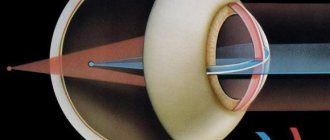
Static perimetry allows you to determine the field of view by projecting it onto a round surface. Diagnostics is based on observation of a stationary object, which is recorded in different parts of the field of visual perception.
This technique makes it possible to identify many diseases at the initial stage of development. Perimetry is carried out using automatic computer perimeters. The device helps determine the indicators of the central field of vision and peripheral.
The following perimeters are used for diagnostics:
- Rodenstock;
- Humphrey;
- Octopus;
- Perikom.
These devices are used in ophthalmology offices of district clinics. The devices are large and expensive. They are used for inpatient examination of patients.
The patient is placed in a motionless state and fixed. By changing the intensity of the vertical border of the optic colliculus, it determines the light sensitivity of the retina.
With suprathreshold static perimetry, photosensitivity thresholds are set in advance at different points of the visual field. This examination reveals obvious pathologies of the retina.
What you need to know about perimetry
The visual field is the space that a person recognizes when his gaze is fixed and his head is still. If you look at a certain object, in addition to its clear image, a person sees other objects located around. This is called peripheral vision, and it is not as clear as central vision.
Perimetry is an ophthalmological study that allows you to examine the boundaries of the visual fields through a projection onto a spherical surface. There are kinetic and static perimetry. Kinetic research involves using a moving object, while static research involves varying the illumination of an object in one position.
The study helps to analyze changes in the visual field and determine the localization of the pathological process (retina, optic nerve, visual pathways, visual centers in the brain). Most often, a narrowing of the visual field and loss of some areas (scotoma) are detected.
Indications for perimetry:
- retinal pathologies (tears and detachments, dystrophy, hemorrhages, burns, tumors);
- diagnosis of macular pathologies, including toxic lesions;
- detection of retinitis pigmentosa;
- diseases of the optic nerve (neuritis, trauma);
- diagnosis of pathologies of the visual pathway and cortical centers in the presence of neoplasms, injuries, stroke, severe malnutrition;
- a brain tumor;
- hypertonic disease;
- traumatic brain injuries;
- signs of cerebrovascular accident;
- confirmation of glaucoma, tracking the dynamics of the process;
- checking patient complaints (aggravation factors);
- preventive examination.
Perimetry is contraindicated if the subject is under the influence of alcohol or drugs, or has mental illness. The procedure does not cause any complications.
What can distort perimetry results:
- drooping eyebrows;
- deep planting of eyeballs;
- drooping eyelid;
- height of the bridge of the nose;
- exposure of the irritant to large vessels near the optic nerve head;
- low visual acuity;
- poor quality correction;
- rim glasses.
False visual field defects can also appear due to the structural features of the face and pupil width. To eliminate false defects, repeat testing is carried out in the same program. In order for dynamic observation to be reliable, the same perimetry conditions must be observed (size of objects, lighting, time and colors).
Computer perimetry
This technique evaluates the visual field without the participation of an ophthalmologist. An automated algorithm is used to carry out the procedure.
Method:
- The windows in the room are closed, it should be darkened.
- The patient is asked to sit in front of the device and fixate the eye on the light point.
- Colored spots appear and disappear sequentially on the device's monitor. When one of them is fixed, you need to press the button.
- The visual organs are not checked at the same time. To study visual fields, the eyes are checked one at a time. Thus, a complete picture of the boundaries emerges. The button is pressed by the patient.
- The program independently changes the speed at which dots appear, their size and brightness. They appear in random order from different directions.
- The procedure ends with decryption using a computer program installed in the device. It displays all received data. Analyzing the data obtained, the doctor draws up a conclusion.
Computer perimetry is the best opportunity to identify most eye diseases at an early stage and take appropriate measures. Regular perimetry helps determine changes for better or worse. The study allows you to adjust the course of therapy and make a decision on further treatment.
The duration of the procedure is no more than 30 minutes. The patient receives the result in printed form.
How to perform perimetry
Depending on which device is used to perform the procedure, the technique for examining visual fields differs.
Förster perimeter
Perimeter survey
First, a study is carried out on white :
- The patient is asked to sit next to the device with his back to the light source. The chin is placed on the device's stand. One eye is covered with a blindfold, and the other looks at a white mark placed in the central part of the perimeter. It is at this point that a person will have to look throughout the entire procedure.
- After several minutes allocated for habituation, the patient is informed that he is fixing his gaze on a stationary mark, and after he notices a moving point on the periphery, he needs to tell the specialist about it.
- The doctor begins to move the mark along the meridians in the direction from the periphery to the central part, and the person being examined lets him know when he sees the object.
- The doctor turns the device alternately by 45˚ and 135˚.
- The same actions are carried out with the other eye as with the first.
Upon completion of the examination, the specialist creates a schematic representation of a person’s visual fields.
Then perimetry is carried out using color marks .
- The subject should not know which color will be used for the procedure. Therefore, during the examination, a person needs not only to mark the mark, but also to correctly determine its color.
- After this, the boundaries are indicated on the schematic representation of the visual fields. If the patient makes a mistake with the color, the tag moves further until the specialist gets the correct answer.
The most commonly used colors are red, yellow, green and blue. The procedure is performed with 8 meridians and an interval of 45˚ or 12 meridians and 30˚.
Computer-assisted research
- The patient sits near the device. A shutter is installed over one eye, and the chin is placed on a stand.
- Objects move chaotically on the monitor, and the patient, having seen the object, must press the mouse button.
Computer perimetry of the eye takes longer - about 5-10 minutes. The essence of the procedure is that the brightness and size of a static object are constantly changing. The study determines the sensitivity of the retina to color in any of its zones.
The data is considered more accurate than the study conducted by Förster's perimeter. The results obtained are saved on the computer, and if necessary, you can view and evaluate them again.
What may prevent you from receiving correct data:
- Ptosis of the upper eyelid;
- Overhang of eyebrows into the visual area;
- Deep-set eyes;
- The presence of a high bridge of the nose.
If a person has such symptoms, it is recommended to undergo an examination using a computer device and perimeter. This will allow you to get more accurate results.
Decoding the results
The results are interpreted by an ophthalmologist. Normal field of view:
- limited from above to 55 degrees;
- from inside and below - up to 60 degrees;
- outside - up to 90 degrees.
These results are considered standard, but a person’s normal field of vision varies up to 5–10 degrees. These boundaries are the limit of visibility of the achromatic stimulus.
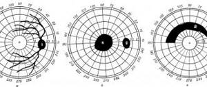
If there are disturbances in visual perception, then areas of loss appear - scotomas. They are relative and absolute.
The field of view is not a continuous zone. Even in a healthy person, some areas fall out where the points are not fixed. The norm is the absence of scotomas, that is, the patient’s visual organ perceives the entire visible area. If they are, this does not mean the beginning of optic nerve atrophy. Such points are indicated by congenital defects that remain invisible throughout life and do not affect the acuity of visual perception.
Some factors listed in the “Preparation for perimetry” section can simulate field distortion; they should be taken into account when interpreting the results obtained.
Types of perimetry
Perimetry is carried out using several different methods. The simplest is the Donders test, which allows you to evaluate the boundaries of the visual field. The patient is placed a meter away from the doctor and asked to focus on the examiner's nose. The patient first closes one eye, and the doctor shows a visible object and traces it in one of the meridians. A healthy person notices an object with both eyes simultaneously. The action is repeated in 4-8 meridians to roughly determine the boundaries of the field of view. A prerequisite for the Donders test is the preservation of boundaries.
To study the central field, the Amsler test is used - an even simpler examination method. The test makes it possible to evaluate a zone up to 10° from the center of the visual field. When diagnosing, a grid of horizontal and vertical lines is used, with a dot in the center. The patient must fix his gaze on a point from a distance of 40 cm. Signs of pathology according to the Amsler test: curvature of lines and the appearance of spots. The method is indispensable for the primary diagnosis of macular pathologies.
The central field of vision can be examined using the campimetry method. The patient must close one eye and fixate his gaze on a black board located a meter away. The board (1x1 m) has a white dot in the center. White objects of different diameters (1-10 mm) are moved along the studied meridians until they disappear. Scotomas are first noted on the board, and the results are transferred to a form.
In theory, the results of different methods should match, but in practice, moving objects are viewed better than stationary ones. This is especially noticeable in areas with defects, which is called the Riddoch phenomenon.
Methods for determining visual fields
The field of view is determined monocularly - one at a time for each eye, while the second is covered with a bandage. The principle for all methods is common: the subject must fix his gaze motionlessly at a certain point, and at the same time note the appearance of an object in the field of view (or its disappearance).
Control method
Does not require special conditions or equipment. The patient's field of vision is compared with the doctor's field of view: the doctor sits opposite the patient at a distance of one meter, and the doctor and the subject cover the opposite eye, the doctor moves an object, for example, a pen, in the space around him from the periphery to the center, the patient must determine the appearance of a handle in the field of view. The doctor’s task is to compare the patient’s sensations with his own. The method does not have accuracy and subtlety, but it allows you to identify gross violations and loss of large segments of the visual field.
Kinetic perimetry
With kinetic perimetry, the patient must see a moving object moving from the periphery to the center (or vice versa). The points of appearance of a moving object in the field of view, when connected, form a line - an isopter. This type of perimetry is isopter perimetry or quantitative perimetry.
Static perimetry
Static perimetry is carried out on the arc projection perimeter, which are of two types:
- with white or colored objects moved manually along a perimeter arc;
- projection arc perimeter (the test object is a light spot from the projector).
With static perimetry, the object is motionless, but its brightness changes. The patient should note the differences in illumination. Such perimetry is called static or profile, as well as differential light threshold.
Test objects in the projection perimeters have varying brightness, size, contrast, and exposure time.
Automated computer perimetry
Modern perimeters or visual field analyzers provide the ability to more accurately determine visual fields in certain areas, save study results, and conduct examinations over time. The latest generation perimeters can perform both static and kinetic perimetry; these two methods complement each other, but if early diagnosis is needed, priority is given to the static method.
Computer campimetry
Computer campimetry can be achromatic and color. The method is more often used to diagnose and evaluate the results of treatment of diseases of the retina and optic nerve. Features of modern campimetry:
- The computer screen acts as a flat screen;
- The received information is subjected to computer processing and storage in accordance with the established program.
Examination price
Computer perimetry can be carried out as a separate study or as part of a comprehensive vision diagnostics.
The cost of computer perimetry in our clinic starts from 1,500 rubles.
The determining factors for obtaining a high-quality keratometry result are the quality and correctness of the choice of equipment and the practical training of the doctor. The Moscow Eye Clinic employs specialists with a high level of professional training who masterfully use the modern equipment we have for vision diagnostics.
You can find out the cost of a particular procedure or make an appointment at the Moscow Eye Clinic by calling 8 (800) 301-62-07 (daily from 9:00 to 21:00, free for mobile phones and regions of the Russian Federation) or by filling out the form online entries.
Kinetic perimetry
The following types of kinetic perimetry are distinguished: Perimetry using a white test object, color, topographic, objective, ophthalmoscopic perimetry.
Perimetry using a white test object is most common in wedge practice in the USSR and abroad. The study is carried out in turn for each eye (the second eye is covered with a light bandage). The subject should sit comfortably near the perimeter, placing his chin on a special stand of the device so that the eye being examined is opposite the fixation point located in the middle of the perimeter arc. Looking at the fixation point, the subject must note the moment when he notices the appearance of a moving test object in the field of view. This position of the test object on the arc corresponds to the point of the retina where its sensitivity is threshold in relation to the test object; it is marked on the diagram of the visual field. The movement of the test object must be continued until the point of fixation to ensure that the visual field is preserved throughout the entire meridian. By rotating the perimeter arc, a study is carried out along the meridians at 15°, 30° or 45°. When examining individuals with sufficiently high visual acuity, a test object with a diameter of . 3 mm. To identify minor defects and slight narrowing of the visual field, P. is carried out using a test object with a diameter of 1 mm.
Color perimetry is carried out similarly to P. using a white test object, but in contrast to it, test objects of blue, red and green colors are used, diameter. 5 or 10 mm; At the same time, the moment of correct differentiation by the subject of the color of the presented object is noted. To exclude congenital anomalies of color perception, before performing color P., it is necessary to examine patients using the polychromatic tables of E. B. Rabkin (see Color vision).
Topographic perimetry (isopperimetry) is carried out using several test objects of various sizes and brightness. As a result of the study, several isopters are obtained, respectively - lines connecting points on the diagram of the visual field, which correspond to points of the retina with the same light sensitivity. This type of P. allows a detailed examination of the visual field and is used for accurate diagnosis of diseases of the visual analyzer. To study spatial summation in the field of view, two objects of different sizes are used, which are aligned in such a way with light filters that the amount of light reflected by them becomes the same. Normally, the isopters obtained when studying using these two objects coincide, but in pathology they diverge.
Objective perimetry is based on determining the boundaries of the visual field using pupillography (see Pupilography), which records the pupillary reactions of the subject, or encephalography (see) by assessing the alpha rhythms of the EEG.
Ophthalmoscopic perimetry is carried out using an ophthalmoscope (see Ophthalmoscopy), registers a rough projection of light onto the retina of the subject and is used to determine the degree of preservation of the visual field and the advisability of surgical treatment for clouding of the optical media of the eye (for example, a cataract, cataract, etc.).
Progress of the procedure
The patient himself is the main sensitive sensor that helps in performing computer perimetry. It is placed near a special eyepiece and fixes the gaze at a luminous central point. He puts his hand on the joystick. Light signals appear in the field around this target point in a chaotic manner and at different time intervals. The patient, seeing this flash, must immediately press the button. After this, the computer processes the data and compares the received signal with the location of the light mark.
Each eye is examined separately, that is, the procedure is repeated twice. The duration of the study does not exceed 20 minutes. During diagnosis, there are no external influences on the eye, so there is no risk of complications or side effects. The patient must be very careful and follow all instructions.
The result of computer processing can be displayed on the screen or printed in the form of a detailed map of the field of view. This information is very useful and valuable for an ophthalmologist.
The perimetry of the eye is
These are studies of peripheral vision. The decoding gives an idea of the consistency of the visual function and whether it is within normal limits. So, in a healthy state, the eye should cover a certain surrounding space.
Often, perimetry violations go unnoticed by humans. He perceives objects in a narrowed range and does not notice it. Therein lies a serious danger. After all, pathology can develop in the future.
Visual field assessment is required. A computer method is used for this. The study is carried out on special equipment, which is a hemisphere. The patient needs to place his head there. Inside it there is a field on which luminous points appear. They flare up in different places in this space.
Accordingly, when the patient sees such a point, he presses a special button. In this case, it is not allowed to shift your gaze; it must be directed strictly to the middle of the field. Based on the results of the study, a color scheme is drawn up. This is a transcript of the study, where those parts of the visual fields that are no longer accessible to the patient are visible. This means a deviation from the norm.
| This kinetic examination principle allows for a very accurate assessment of the state of the visual fields. |
Application area
Computer perimetry is primarily intended to identify eye diseases, but since signals along the optic nerve are transmitted directly to the brain, this method allows you to diagnose some other pathologies:
- Brain disorders associated with stroke;
- Traumatic brain injuries;
- Neoplasms in the brain.
If a patient feigns vision problems and claims that he sees poorly, for example, in order to avoid being drafted into the army, then computer perimetry will immediately prove the inconsistency of such a statement. Some patients tend to exaggerate their symptoms, but it is almost impossible to deceive computer diagnostics.
Normal perimetry readings
The field of view can be represented as a three-dimensional visual hill. Its base is the boundaries of the field, and the height of the hill determines the degree of light sensitivity of individual areas of the retina. Normally, the height decreases from the center to the periphery. To simplify the analysis, perimetry results are displayed as a map on a plane. The areas of the fundus of the eye on such a map are presented in such a way that disturbances in the lower parts of the retina are reflected by changes in the upper ones.
The center of the visual field (fixation point) is the photoreceptors of the fovea. Since the optic disc does not contain light-sensitive cells, it is represented on the map as a “blind” spot. It is also called physiological scotoma or Mariotte's spot. The blind spot is located in the outer part of the field in the horizontal meridian (10-20° from the center of the field). Normally, angioscotomas can also be detected, that is, projections of retinal vessels that are associated with Mariotte's spot and are shaped like tree branches.
Norms of peripheral boundaries:
- top – 50°;
- lower – 60°;
- internal – 60°;
- external – less than 90°.