Pupils of different sizes - what is it?
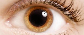
Anisocoria is a condition in which the pupils of the right and left eyes differ in size or diameter. The pupil is the round black area in the center of the iris. Depending on the lighting, it can have dimensions from 1mm to 6mm in diameter.
In the presence of general or ocular pathology, anisocoria is always combined with the following manifestations:
- restriction of eye movement, or the eye on which the pupil is larger
- drooping upper eyelid (ptosis)
- Pain in the eyes
- increased temperature or fever
- headache
- decreased vision
- double vision
Diagnostic methods
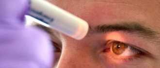
Anyone can be diagnosed with miosis. To do this, just look in the mirror and pay attention to the state of your pupils. If it does not go away for a long time, you need to undergo a full examination. Obviously, it is necessary to exclude alcohol and drugs.
If such facts occur, then it is better to wait until the effect of the substances wears off and not take them anymore. If this is not the case, then serious research will be required. It is necessary to measure intraocular pressure, check the blood and the condition of blood vessels. In addition, do an MRI, which will assess intracranial pressure.
Causes of anisocoria
There are two types of anisocoria:
- physiological. Normally, every fifth person has a slight difference in the size of the pupils.
- pathological Eye diseases that can lead to anisocria: glaucoma, inflammatory diseases of the eye (iritis, uveitis), eye tumors
- pathological in general human diseases: viral infection, syphilis, brain tumors, cranial nerve palsy, Horner's syndrome, migraine, cerebral aneurysm.
14.1.2. Pupil. Norm and pathology of pupillary reactions
In children of the first year of life, the pupil is narrow (2 mm), reacts poorly to light, and dilates poorly.
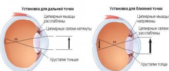
In the sighted eye, the size of the pupil constantly changes from 2 to 8 mm under the influence of changes in illumination. In room conditions with moderate lighting, the pupil diameter is about 3 mm, and in young people the pupils are wider, and with age they become narrower. Under the influence of the tone of the two muscles of the iris, the size of the pupil changes: the sphincter contracts the pupil (miosis), and the dilator ensures its dilation (mydriasis). Constant movements of the pupil - excursions - dose the flow of light into the eye.
The change in the diameter of the pupillary opening occurs reflexively:
- in response to irritation of the retina by light;
- when setting to clearly see an object at different distances (accommodation);
- with convergence (convergence) and divergence (divergence) of the visual axes;
- as a reaction to other irritations.
Reflex dilation of the pupil can occur in response to a sharp sound signal, irritation of the vestibular apparatus during rotation, or with unpleasant sensations in the nasopharynx.
Observations are described confirming the dilation of the pupil under great physical stress, even with a strong handshake, when pressing on individual areas in the neck, as well as in response to a painful stimulus in any part of the body. Maximum mydriasis (up to 7-9 mm) can be observed during painful shock, as well as during mental stress (fear, anger, orgasm). The reaction of pupil dilation or constriction can be developed as a conditioned reflex to the words dark or light. The reflex from the trigeminal nerve (trigeminopupillary reflex) explains the rapidly alternating dilation and contraction of the pupil when touching the conjunctiva, cornea, eyelid skin and periorbital region.
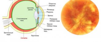
The reflex arc of the pupillary reaction to bright light is represented by four links. It starts from the photoreceptors of the retina (I), which received light stimulation. The signal is transmitted along the optic nerve and optic tract to the anterior colliculus of the brain (II). The efferent part of the arc of the pupillary reflex ends here. From here, the impulse to constrict the pupil will go through the ciliary node (III), located in the ciliary body of the eye, to the nerve endings of the sphincter of the pupil (IV). After 0.7-0.8 s the pupil will contract. The entire reflex path takes about 1 s. The impulse to dilate the pupil comes from the spinal center through the superior cervical sympathetic ganglion to the pupillary dilator (see Fig. 3.4).
Drug dilation of the pupil occurs under the influence of drugs belonging to the mydriatic group (adrenaline, phenylephrine, atropine, etc.). The most persistent dilation of the pupil is a 1% atropine sulfate solution. After a single instillation in a healthy eye, mydriasis can persist for up to 1 week. Short-acting mydriatics (tropicamide, midriacil) dilate the pupil for 1-2 hours. Constriction of the pupil occurs when miotics are instilled (pilocarpine, carbachol, acetylcholine, etc.). The severity of the reaction to miotics and mydriatics varies from person to person and depends on the ratio of the tone of the sympathetic and parasympathetic nervous systems, as well as the state of the muscular apparatus of the iris.
Changes in the reactions of the pupil and its shape can be caused by an eye disease (iridocyclitis, trauma, glaucoma), and also occurs with various lesions of the peripheral, intermediate and central parts of the innervation of the muscles of the iris, with injuries, tumors, vascular diseases of the brain, upper cervical ganglion, nerve trunks .
After a contusion of the eyeball, post-traumatic mydriasis may occur as a consequence of sphincter paralysis or dilator spasm. Pathological mydriasis develops in various diseases of the thoracic and abdominal organs (cardiopulmonary pathology, cholecystitis, appendicitis, etc.) due to irritation of the peripheral sympathetic pupillomotor pathway.
Paralysis and paresis of the peripheral parts of the sympathetic nervous system cause miosis in combination with narrowing of the palpebral fissure and enophthalmos (Horner's triad).
For hysteria, epilepsy, thyrotoxicosis, and sometimes in healthy people. The width of the pupils changes independently of the influence of any visible factors at uncertain intervals and inconsistently in the two eyes. In this case, other eye pathology may be absent.
Changes in pupillary reactions are one of the symptoms of many general somatic syndromes.
If the reaction of the pupils to light, accommodation and convergence is absent, then this is paralytic immobility of the pupil due to pathology of the parasympathetic nerves.
Methods for studying pupillary reactions are described in Chapter 6.
How to treat anisocoria
Physiological anisocoria does not affect vision or eye health. Therefore, it does not require treatment.
With pathological anisocoria, the cause of the appearance of different pupils is first identified. Then they carry out treatment.
For example, for a brain infection, treatment is carried out in a specialized hospital. A course of antibiotics and antiviral drugs is prescribed.
Head tumors and aneurysms of the head vessels require surgical treatment.
For glaucoma, treatment is aimed at normalizing eye pressure and preventing the development of glaucoma attacks.
In case of inflammatory eye diseases, a course of treatment with antibiotics is carried out.
For eye tumors, surgical treatment is indicated.
Constricted pupils in humans
When the pupils are narrowed, this indicates certain processes in the body:
- The natural reason is bright light, because of this the size of the pupil changes and it narrows. This occurs in blinding sun or directional light.
When the irritant is removed, the apple of the eye will expand again and return to its normal state. This is a reaction that occurs in all healthy people.
- This effect may be associated with taking medications or other medications. They sometimes cause temporary narrowing of this organ of the eye.
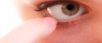
A constricted pupil is often examined during ophthalmic procedures.
- In case of injury, the effect of expansion of the pupil occurs. But there are cases when the opposite process occurs.
If the organ is narrowed and does not return to normal for a long time, this indicates a strong decrease in intracranial pressure. And this condition is expressed in the narrowing of the organ in question,
- When a small dot remains instead of a pupil, this indicates the possible use of psychotropic drugs, anesthetics, alcohol or narcotic drugs. For example, opium, marijuana.
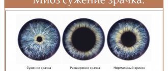
| There is also a medical term for severe constriction of the pupil. This is called miosis. It is characteristic of both eyes, but can also occur in one of them. |
Moreover, the difference is so pronounced that even the interlocutor will notice it. Thus, the effect of miosis is determined by a variety of reasons. Return to contents
Symptom Definition
a normal pupil narrows in daylight and dilates in poor lighting.
As a rule, this pathology is detected at an early age, and signs of anicozoria are usually visible to the naked eye.
If the excess pupil diameter in one eye is significantly pronounced, then this fact can be noticed immediately. Often with this pathology, nausea and vomiting occur.
Sometimes distortions of objects, things that a person is looking at are possible. The contours of objects may blur and take on distorted, irregular shapes. In addition, the phenomenon of bifurcation of objects often appears.
The eyes begin to react too intensely to light, especially sudden and bright ones.
Phenomena of a febrile nature, chills and temperature can also accompany anicozoria.
Headache, eye pain, and vomiting are possible. In children with this disease, torticollis is often observed.
You should consult a doctor immediately if the following symptoms are added to the existing discrepancy in pupil size:
- fever;
- photophobia;
- headache;
- nausea;
- swelling and drooping of the eyelid (upper);
- rapid decline in vision.
Typically, anisocoria manifests itself at an early age (like other similar diseases, for example, heterochromia), and the symptoms manifest themselves quite clearly. Firstly, by the appearance of the pupils, a departure from the normal state becomes clear. Attentive parents may notice other symptoms, for example, the disease in some cases is accompanied by nausea and vomiting.
An older child and an adult, in addition to noticeable asymmetry, may experience disturbances in eye function, which are expressed in blurred images, darkening of the eyes, double vision and discomfort. If you notice these negative signs, you should immediately consult a doctor, as they can be serious disorders and diseases of the eye organs.
The causes of foggy eyes are described here.
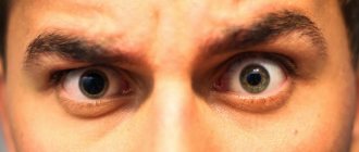
Example of anisocoria
Treatment
Therapy depends on the cause of the symptom:
- No treatment is required for congenital or physiological anisocoria.
- For inflammatory eye pathologies, treatment includes local and systemic antibacterial drugs.
- For tumor formations, surgical treatment is indicated.
- For meningitis and encephalitis, treatment is complex.
Thus, anisocoria can occur either with disturbances in the structure of the iris, or with inflammation of the structures of the eyeball (including the pupillary sphincter or dilator), or be associated with diseases of the nervous system: its autonomic part, peripheral nerve fibers, central nervous system or iris receptors.
In any case, anisocoria is always a reason for medical consultation, and its treatment depends on the identified cause of the pathology.