Causes of microphthalmos
The main causes of microphthalmia are associated with intrauterine developmental disorders of the fetus.
The main provocateurs of the disease are infectious pathologies of the reproductive organs in women. In addition, hereditary pathologies are an increased risk factor. The causes of the isolated nature of the pathology include intrauterine infections:
- toxoplasmosis;
- rubella;
- herpes.
The cause of microphthalmia can be isolated or genetic pathologies.
The main etiological provocateurs are:
- TORCH infections.
The four most dangerous intrauterine diseases (toxoplasmosis, rubella, herpes). Infectious agents of these pathologies penetrate the fetal bloodstream, thereby provoking the development of many defects; - Teratogenic effects.
Microphthalmos is caused by the influence of ionized rays, smoking, alcohol consumption, and specific medications on the fetus; - Amniotic fusions.
These are constrictions in the form of dense threads that are localized in the amniotic sac. Such threads compress the fruit. Such an effect entails a reduction in the eyeball or a complete absence of development of the fetal organ at various stages of pregnancy; - Genetic pathologies.
Pathologies associated with genetic mutations (Temple Al-Ghazali, Delleman, Peters, Rieger syndromes). These diseases lead to the development of many abnormalities, including microphthalmia.
Nevertheless, the main cause of microphthalmia is teratogenic factors. The high-risk group includes women whose work involves interaction with toxic substances. Also, the consumption of narcotic substances significantly contributes to the development of underdevelopment of the visual organs of the fetus.
The stage of disruption of the embryogenesis process depends on the time of influence of pathogenic factors on the fetus.
Anatomical causes of congenital farsightedness
In addition to physiological factors, hypermetropia can be caused by a number of anomalies. For example, various disorders in the structures of the eye can cause farsightedness in children:
- Lenticonus is a change in the shape of the surface of the lens when it becomes cone-shaped and protrudes in different directions. With anterior lenticonus, the lens is extended into the anterior chamber of the eye, with posterior lenticonus, towards the vitreous body, and with internal lenticonus, the cone protrudes into the lens itself. With lenticonus, high levels of farsightedness may occur. Preventive measures (to avoid the occurrence of amblyopia) consist of special exercises and hardware treatment. If the lenticonus is significant, the lens can be removed.
- Microphthalmos is a shortened eyeball that does not develop to an “adult” state. Medical statistics say that in 75% of cases, the development of an abnormality of the organ of vision is provoked by the pathological course of pregnancy, and can also occur during difficult childbirth. Only in other situations is microphthalmos a consequence of a genetic syndrome. This pathology is often the cause of congenital farsightedness in children.
- Aphakia is the complete absence of the lens in a newborn baby. It is often caused by disturbances during intrauterine development. With aphakia, the refractive power of the eye is significantly reduced, visual acuity is approximately 0.1, in which case correction with strong plus lenses or implantation of an intraocular lens is required, but this method is only available from the age of 18.
- Hypermetropic astigmatism is the presence of farsightedness on the two main meridians of the eyes. In children, it manifests itself in the form of asthenopia - rapid fatigue during work, and can also lead to convergent strabismus and amblyopia.
- The hereditary form of farsightedness is caused by the presence of sufficient degrees of this disease in both parents. In this situation, you should be especially attentive to the child’s vision condition and visit an ophthalmologist regularly. Often in cases of congenital hereditary farsightedness, it begins to progress, and such development threatens with negative consequences.
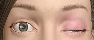
Symptoms of microphthalmos
The most common symptom of microphthalmos is a unilateral reduction of the eyeball.
Basically, similar symptoms are observed in infants immediately after removal from the mother’s womb, since the etiology of the disease is congenital. In addition to deformation of the eyeball, microphthalmos is characterized by the following painful effects:
- decreased visual acuity;
- frequent tearing;
- facial asymmetry;
- discomfort;
- pain in the eye, with cysts formed between the membranes of the visual-sensory organ.
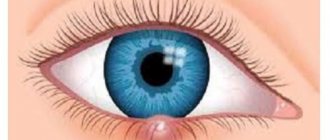
In the presence of microphthalmos, a defect may develop in which part of the eye shell is missing (coloboma).
This pathology provokes damage in various areas of the eye organ, including:
- iris;
- retina;
- vision receptors that transmit visual information to the brain.
The presence of coloboma occurs in both one and both eyes. Pathology of direct images contributes to impaired visual perception.
{banner_horizontalnyy2}
Classification of microphthalmos
The disease is classified into 2 categories: unilateral and bilateral microphthalmos. The most common form is the first option. The second occurs in 10% of the total number of sick children.
In addition, the types of pathology are divided into idiopathic and hereditary disorders. The first option means that doctors were unable to identify the etiological factors of the disease. Hereditary microphthalmos manifests itself in newborn infants when the development of pathology occurs in an autosomal dominant or autosomal recessive manner.
There are two degrees of severity of the pathology: ordinary and combined microphthalmia. Pathology of a simple type occurs without anomalies and third-party diseases. The complex form means the presence of other concomitant disorders affecting the anterior region of the eye. Consequently, the latter option is characterized by a more complex form.
Approximately up to 50% of patients have concomitant ophthalmological diseases. In most patients, along with microphthalmia, cryptophthalmia develops (complete, incomplete, abortive) proliferation of an orbital cyst.
Another form of microphthalmos is nanophthalmos. This form of pathology is characterized by a reduction in the eyeball with thickening of the scleral capsule, the manifestation of severe farsightedness and an increased risk of developing glaucoma.
Diagnosis of microphthalmos
Microphthalmos has a pronounced visual character. Consequently, the presence of the disease is noticed very easily.
The final diagnosis is established by an ophthalmologist or pediatrician during a postpartum external structural examination of the child’s eye. To record the disease, the pediatrician opens the baby’s eyelids and examines the outer tissue of the eyeball. If a unilateral type of lesion is detected, the other eye is extensively examined for the presence of additional defects and anomalies that may lead to deterioration of visual functions.
It is not always possible for a doctor to determine the cause of the development of pathology. Therefore, often an additional influencing factor is determined through diagnostic methods using hardware equipment - ultrasound, MRI.
Considering the fact that microphthalmia is often caused by genetic pathologies, a DNA analysis can be performed, in which a blood sample and buccal epithelium are examined.
To diagnose the disorder, the following medical and ophthalmological methods are used:
- Biomicroscopy.
To diagnose the disease, the condition of the palpebral and orbital conjunctiva is assessed, the anterior part of the eyeball is examined, and the level of opacification of the optical media is determined; - Visometry.
Allows you to evaluate visual acuity using special tables (for example, Sivtsev - Golovin); - Refractometry.
The procedure is aimed at an objective assessment of the optical functions of the eye and identification of refractory anomalies using special refractometer devices.
After confirmation of the diagnosis, consultative participation of a geneticist is indicated. Modern medicine has the capabilities of prenatal examination of the visual apparatus. Microphthalmos is diagnosed differentially with underdevelopment of the eyelids and eyeball (cryptophthalmos) and complete or partial absence of the eyeball (nanophthalmos).
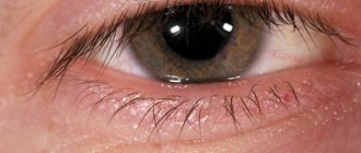
Enophthalmos of the eyeball
Enophthalmos of the eyeball often occurs due to injury. Mechanical injury to the eye and orbit can lead to destruction of soft tissue or the temporal lobe. The volume of the orbit is 30 ml. The eyeball occupies only 6.5 ml of this volume. Because of this, severe trauma can easily cause the eye to become displaced. As a rule, this is accompanied by ptosis (drooping) of the upper eyelid and miosis, when the muscles of the lens atrophy and the pupil narrows regardless of external factors and remains in this state. When the eye sinks into the orbit, we are talking about enophthalmos. When the eyeball protrudes (bulging eyes), exophthalmos is diagnosed.
Enophthalmos can be congenital or acquired. The first form of this pathology is caused by factors such as:
- Abnormal structure of the skull bones.
- An increase in the size of the anatomical axis of the eyeball - the line that connects the axes of the eye.
- Lipodystrophy is a pathology characterized by degeneration of adipose tissue.
- Violation of trophism - the process of cellular nutrition.
The causes of acquired enophthalmos can be of an ophthalmological nature, and it also happens when there is no connection with diseases of the organs of vision.
The first group of factors includes:
- Microphthalmos is a reduction in the size of the eye. Usually develops in a unilateral form. The causes of this pathology can also be congenital and acquired. Microphthalmos can be detected during an initial eye examination.
- Orbital bone fracture.
- Atrophy of the orbit caused by mechanical trauma.
- Subatrophy of the eye, manifested in the slow drying of its tissues.
Sometimes enophthalmos is caused by reasons that are not related to the organs of vision. The eyeballs can shift due to severe exhaustion or dehydration. This is usually caused by diseases such as cholera, peritonitis, and anorexia. Oncological diseases, including brain tumors, are often accompanied by enophthalmos. Damage to the cervical spine can also lead to displacement of the eye in the orbit.
Treatment of microphthalmos
The doctor thinks over the treatment tactics for microphthalmia, based on the degree of underdevelopment of the eye tissues and the duration of the pathology. The therapy paradigm is based on surgical correction of the structures of the eyeball.
Surgical correction
Surgical correction methods include:
- Scleroplasty.
The indication for this operation is due to slight hypoplasia of the eye organ, namely a decrease in the eye lumen to 3 mm. During surgery, strips of special scleroplastic tissue are implanted into the posterior wall of the sclera; - Cosmetic ocular prosthetics.
It is advisable when the size of the eyeball is reduced to 5 mm. During the operation, a thin-walled prosthesis is inserted to maintain normal lability of the eyeball. For patients with increased sensitivity of the cornea, de-epithelialization of the cornea is used. During the operative procedure, the cornea is lined with a rounded allograft; - Implantation technique.
The essence of the operation is the placement of a skin-fat graft in the retro-orbital space with delayed prosthetics. Surgery is indicated when the size of the anteroposterior vector of the eyeball does not exceed 10 mm.
Important!
Surgery is permitted for patients at least 7 years of age. Surgical intervention is necessary for diagnosed glaucoma, cataracts, and retinal detachment. In addition, the operation is performed to improve the appearance of the visual organ; prosthetics are used for this purpose.
Conservative therapy
Conservative methods of therapy are used for preventive purposes aimed at relieving residual complications with the installation of antiseptic solutions, such as:
- Miramistin;
- solutions for treating contact lenses;
- Chlorhexidine.
{banner_horizontalnyy3}
Surgery as a method of treating enophthalmos
Surgery is prescribed immediately for injuries if a fracture of the orbital bones has occurred. During the procedure, the surgeon must remove all bone fragments. This method of treatment is also used to eliminate enophthalmos with a strong displacement of the eye into the orbit. The essence of the operation is as follows: the doctor inserts an implant under the lower wall of the orbit that will support the eye in the desired position. Such implants are made from silicone, polymers and various metals, including titanium. If treatment is started in a timely manner, the pathology can be eliminated quickly enough and visual functions can be preserved.
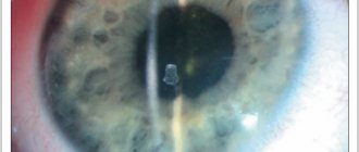
Complications of microphthalmos
The consequences of microphthalmos are complicated by dangerous pathologies that lead to significant visual impairment. The prolonged course of the disease provokes the formation of cysts, inflammation of the horny and conjunctival membranes (keratitis, conjunctivitis). The outcome in severe cases is complete blindness.
Additionally, microphthalmos in children leads to eye damage in the form of cataracts (clouding of the lens). Other complications of microphthalmos are narrowing of the palpebral fissure (eye opening). Pathology increases the risk factor for the development of glaucoma. Doctors have proven that congenital eye pathologies can cause malignant neoplasms in the intraorbital segment.
Prevention of congenital farsightedness
There are many reasons for farsightedness in children. They can be divided into three groups:
- Natural physiological ones that go away over time;
- Associated with disorders during intrauterine development of the fetus;
- Genetic.
Unfortunately, modern medicine is not yet able to prevent gene mutations. Thus, a child with Franceschetti syndrome can be born from two completely healthy parents. Pregnant women should be attentive to their health, follow all doctor’s recommendations and not use drugs, alcohol, or nicotine that are harmful to the baby’s health.
Congenital hyperopia is corrected with glasses or contact correction, as well as with hardware treatment in children under 18 years of age. Upon reaching this age, microsurgical operations on the eyes are already permitted, which will help cope with severe impairments and restore high quality vision for many years.