Description
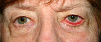
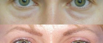
Drooping of the upper eyelid (ptosis).
Abnormal position of the upper eyelid, leading to partial or complete closure of the palpebral fissure. Ptosis is manifested by a low position of the upper eyelid, irritation and increased fatigue of the eye, the need to tilt the head back for better vision, the development of diplopia and strabismus. Diagnosis of drooping upper eyelid includes measuring the height of the eyelid, checking the symmetry and completeness of eyelid movements in both eyes. Treatment of ptosis of the upper eyelid is carried out surgically using resection or creation of a levator duplicator, etc.
Upper eyelid ptosis
The normal condition is a slight overlap of the iris of the eye by the upper eyelid. When the overlap exceeds two millimeters, the initial stage of ptosis of the upper eyelid is diagnosed.
Unilateral or bilateral congenital ptosis of the upper eyelid is more often diagnosed in childhood. With a slight prolapse, the disease is sometimes left for further monitoring and observation. If there is significant prolapse, mandatory medical intervention is required.
If treatment is not timely, there is a risk of complications such as a significant decrease in visual acuity and strabismus.
The most common symptoms of the development of ptosis of the upper eyelid include:
- Drooping eyelid in one or both eyes.
- Irritated eyes. Unpleasant sensations intensify with each closing of the eyelids.
- It becomes difficult to keep your eyes open, which leads to rapid eye fatigue. Prolonged course of this condition leads to frequent headaches.
- To improve visibility, a person is forced to tilt his head back.
Drooping eyelids cause double vision. Due to disruption of the blinking process, the cornea dries out, which leads to dry eye syndrome or infectious lesions.
The main causes of noncongenital drooping eyelids are age-related disorders or acquired injuries.
Additional facts
Normally, the iris is covered by the edge of the upper eyelid by about 1.5. Ptosis (blepharoptosis) is said to occur if the eyelid drops below the upper edge of the iris by 2 or more millimeters or is below the eyelid of the other eye when comparing them. Drooping of the upper eyelid can be either congenital or a condition that develops during life, so blepharoptosis is quite common among children and adults. Drooping of the upper eyelid is not only a cosmetic defect, but also interferes with the normal development and functioning of the visual analyzer, causing mechanical difficulty in vision. Plastic surgery and ophthalmology deal with the correction of drooping upper eyelids.
H02.4 Eyelid ptosis
Detection of pathology
The cosmetic defect is visible to the naked eye. When determining ptosis, the ophthalmologist calculates the distance between the center of the pupil and the edge of the upper eyelid. This indicator is called MRD. The eyelid crossing the middle of the pupil indicates zero MRD. If the eyelid is above or below the middle of the pupil, then it is equal to the interval from +1 to +5 or from -1 to -5, respectively.
To determine the degree of visual deviation, the doctor conducts the following studies:
- Determination of visual acuity.
- Determination of the boundaries of the visual field.
- Examination of the fundus using an ophthalmoscope.
- Examination of the cornea.
- Determination of the degree of production of the lacrimal glands.
- Assessing the condition of the tear film using biomicroscopy.
During the examination, the patient should relax as much as possible. If he is in a tense state, mistakes cannot be ruled out when drawing up a picture of the disease.
Classification
Based on the time of development, congenital and acquired blepharoptosis are distinguished. Taking into account the degree of severity, drooping of the upper eyelid can be partial (the edge of the eyelid covers the upper third of the pupil), incomplete (the edge of the eyelid is lowered to half the pupil) and complete (the upper eyelid covers the entire pupil). Ptosis can be unilateral (69%) or bilateral (31%). Depending on the etiology of drooping of the upper eyelid, the following types of ptosis are distinguished: aponeurotic, neurogenic, myogenic, mechanical ptosis and pseudoptosis (false).
Classification of blepharoptosis
The table will help to trace the dependence of the classification on the reasons that led to the development of ptosis:
| Types of ptosis | Causes of pathology |
| Aponeurotic | The result of age-related changes, including skin aging. This disease is called involutional. Its other name is senile or senile ptosis. |
| Injuries affecting the functionality of the upper eyelid muscle or the consequences of surgery. | |
| Using steroids for a long time. | |
| Neurogenic | Receiving injuries that affect the functioning of the nervous system. |
| Bacterial or viral infections. | |
| Neurological diseases (stroke, multiple sclerosis, etc.). | |
| Diabetic neuropathy. Aneurysm inside the skull. Migraine due to muscle paralysis. | |
| Damage to the sympathetic cervical nerve caused by Horner's syndrome. | |
| Myogenic or myasthenic | Increased muscle fatigue. |
| Mechanical | The appearance of tears and scars in the area of the upper eyelid or its internal/external commissure. |
| Insertion of a foreign object into the eye. | |
| False or pseudoptosis | The presence of excess skin folds on the upper eyelid. |
| Decreased elasticity of the eyeball. | |
| Displacement of the eyeball. | |
| Oncogenic | The appearance of neoplasms in the orbital area. |
| Anophthalmic | Lack of support for the upper eyelid due to the absence of the eyeball. |
According to how severe ptosis is, it is divided into the following types:
- Partial - a third of the pupil is covered.
- Incomplete – 2/3 of the pupil is covered by the eyelid.
- Full – the pupil is not completely visible.
Accordingly, the 1st, 2nd and 3rd degrees of severity of ptosis.
Causes
The lifting of the eyelid is carried out due to the functioning of a special muscle that lifts the upper eyelid (levator), which is innervated by the oculomotor nerve. Therefore, the main causes of drooping upper eyelid may be associated either with an abnormality of the levator palpebral muscle or with pathology of the oculomotor nerve. Congenital drooping of the upper eyelid may be based on underdevelopment or complete absence of the levator muscle; in rare cases, aplasia of the nuclei or pathways of the oculomotor nerve. Congenital blepharoptosis is often familial in nature, but can also be caused by the pathological course of pregnancy and childbirth. Congenital drooping of the upper eyelid in most cases is combined with another pathology of the organ of vision: anisometropia, strabismus, amblyopia. Aponeurotic blepharoptosis most often develops against the background of involutional changes associated with the natural aging process of the body. Sometimes the cause of drooping upper eyelid is injury to the levator aponeurosis or its damage during ophthalmological operations. Neurogenic ptosis of the upper eyelid is a consequence of diseases of the nervous system: stroke, multiple sclerosis, paresis of the oculomotor nerve, meningitis, tumors and abscesses of the brain, etc. Drooping of the upper eyelid of a neurogenic nature is observed in Horner's syndrome, characterized by paralysis of the cervical sympathetic nerve, retraction of the eyeball (enophthalmos) and constriction of the pupil (miosis). The causes of myogenic blepharoptosis can be myasthenia gravis, muscular dystrophy, congenital myopathy, and blepharophimosis. Mechanical drooping of the upper eyelid can be caused by retrobulbar hematoma, eyelid tumors, orbital damage, eyelid deformation as a result of ruptures, injury from foreign bodies of the eye, scarring. Pseudoptosis (false, apparent drooping of the upper eyelid) occurs with excess skin on the upper eyelid (blepharochalasis), strabismus, and hypotony of the eyeball.
Diagnostics
The primary diagnosis of drooping upper eyelid is carried out during a visual examination. During a physical examination, the height of the eyelid, the width of the palpebral fissure, the symmetry of the location of the eyelids of both eyes, the mobility of the eyeballs and eyebrows, the strength of the levator muscle, the position of the head, and other functional indicators are assessed. Of the specialized ophthalmological tests, visual acuity testing, biomicroscopy, and perimetry are of greatest diagnostic interest. If necessary, the strabismus angle, exophthalmometry, determination of the volume of accommodation, convergence study, and binocular vision study are performed. In case of mechanical ptosis, in order to exclude damage to the bone structures in the levator area, a survey X-ray of the orbit is indicated. If the neurogenic nature of drooping upper eyelid is suspected, a CT scan (MRI) of the brain is performed, and a neurologist and neurosurgeon are consulted.
Treatment
First of all, treatment of ptosis of the upper eyelid is aimed at eliminating functional pathology and only then at correcting the cosmetic defect. In the case of a neurogenic nature of the drooping of the upper eyelid, the underlying pathology is treated; In addition, local physiotherapy is prescribed - galvanization, UHF, paraffin therapy. In case of congenital drooping of the upper eyelid, as well as the lack of effectiveness of conservative therapy for acquired ptosis for 6-9 months. , resort to methods of surgical ophthalmology. The timing of correction of congenital blepharoptosis is established differentially: partial drooping of the upper eyelid is operated on at 13-16 years of age; Complete ptosis, due to the likelihood of developing amblyopia, should be eliminated in preschool childhood. Surgeries for drooping upper eyelid (ptosis correction) are aimed either at shortening the muscle that lifts the upper eyelid (congenital ptosis) or at shortening the levator aponeurosis (acquired ptosis). With congenital ptosis, the levator is isolated, plication (shortening) of the muscle is performed by excision or creation of a duplication. In cases of severe blepharoptosis, the levator palpebral muscle is sutured to the frontalis muscle. Standard surgery for acquired blepharoptosis involves removing a thin strip of skin from the upper eyelid, resection of the aponeurosis and fixing its lower edge to the cartilage of the upper eyelid. In plastic surgery, correction of upper eyelid drooping can be combined with upper blepharoplasty.
DISEASES OF THE VITROUS AND EYEBALL (H43-H45)
H43 Vitreous diseases
H43.0 Vitreous prolapse (prolapse) Excludes: vitreous syndrome after cataract surgery ( H59.0 ) H43.1 Vitreous hemorrhage H43.2 Crystalline deposits in the vitreous H43.3 Other vitreous opacities H43.8 Other diseases of the vitreous Vitreous: • degeneration • detachment Excludes: proliferative vitreoretinopathy with retinal detachment ( H33.4 ) H43.9 Disease of the vitreous, unspecified
H44 Diseases of the eyeball
Includes: disorders affecting multiple structures of the eye H44.0 Purulent endophthalmitis. Panophthalmitis. Vitreous abscess H44.1 Other endophthalmitis. Parasitic endophthalmitis NOS. Sympathetic uveitis H44.2 Degenerative myopia H44.3 Other degenerative diseases of the eyeball. Chalkosis. Siderosis of the eye H44.4 Hypotony of the eye H44.5 Degenerative conditions of the eyeball. Absolute glaucoma Atrophy of the eyeball. Wrinkling of the eyeball H44.6 Unremoved (long-standing in the eye) magnetic foreign body Preserved old magnetic foreign body in: • anterior chamber • ciliary body • • lens • posterior wall of the eyeball • vitreous body H44.7 Unremoved (long-standing in the eye) magnetic foreign body eye) non-magnetic foreign body Unremoved (non-magnetic) (old) foreign body in: • anterior chamber • ciliary body • iris • lens • posterior wall of the eye • vitreous body H44.8 Other diseases of the eyeball. Hemophthalmos. Luxation of the eyeball H44.9 Disease of the eyeball, unspecified
H45* Lesions of the vitreous body and eyeball in diseases classified elsewhere
H45.0 * Hemorrhage into the vitreous body in diseases classified in other headings H45.1 * Endophthalmitis in diseases classified in other headings Endophthalmitis in: • cysticercosis ( B69.1 +) • onchocerciasis ( B73 +) • toxocariasis ( B83 . +) H45.8 * Other lesions of the vitreous body and eyeball in diseases classified elsewhere
Forecast
The aesthetic and functional results of blepharoptosis correction with correctly chosen surgical tactics usually last a lifetime. For drooping upper eyelids caused by ophthalmoplegia, treatment can achieve only a partial effect. Surgical treatment of myogenic ptosis caused by myasthenia gravis is ineffective. Left untreated for drooping upper eyelids over time can lead to the development of amblyopia and vision impairment.
Source
RCHR (Republican Center for Health Development of the Ministry of Health of the Republic of Kazakhstan)
Version: Clinical protocols of the Ministry of Health of the Republic of Kazakhstan - 2015
ICD categories: Congenital ptosis (Q10.0), Eyelid ptosis (H02.4)
Sections of medicine: Ophthalmology
Degrees and symptoms
As the name implies, the main symptom is varying degrees of drooping of the upper eyelid. This may result from the fact that the patient is forced to constantly make efforts to lift it. Such actions lead to increased fatigue and emotional lability of patients. They often throw their heads back, especially children.
Drooping of the eyelid interferes with blinking, which leads to dryness and irritation of the mucous membrane of the eye. The tendency to infection of the organ of vision in such cases also increases.
Patients often develop asymmetry of the eyebrows, and children often develop strabismus and the “lazy eye” symptom. Visual acuity decreases, visual fields narrow. Diplopia often develops with ptosis.
There is Marcus-Gunn syndrome , which is also characterized by drooping of the upper eyelid. Its peculiarity is that ptosis disappears as soon as the patient opens his mouth wide or clenches his jaw tightly.
Concomitant symptoms depend on the disease that caused the ptosis. For example, with myasthenia gravis , patients complain of excessive muscle weakness and increased fatigue. With stroke and multiple sclerosis, paresis and paralysis and difficulty walking are noted.
Ptosis can be complete (the eye is completely closed) or partial. The degrees of partial ptosis are as follows:
- Weak is characterized by drooping, in which the eye is open by 5-7 mm.
- With an average degree, the distance is 3-4 mm.
- Severe ptosis is the distance from the upper to the lower eyelid 1-2 mm long.
As already mentioned, ptosis can be acquired or congenital (diagnosed in an infant). It can also be one- or two-sided.
general information
Short description
Recommended by the Expert Council of the RSE at the PVC "Republican Center for Health Development" of the Ministry of Health and Social Development dated September 30, 2015 Protocol No. 10
Ptosis (blepharoptosis) is an abnormally low position of the upper eyelid, which can be congenital or acquired [1,2,6 ,7,9].
Protocol name – Ptosis (blepharoptosis) Protocol code: code(s) : Q10.0 Congenital ptosis H 02.4 Ptosis of the eyelid Abbreviations used in the protocol:
ALT–alanine transferase AST–aspartate transferase HD–biochemical analysis VB–congenital blepharoptosis HIV–human immunodeficiency virus PB–post-traumatic blepharoptosis CBC–complete blood count OAM–general urine test PGDG–transverse horizontal diameter of the eye PZR–posterior size of the eyeball PTI–prothrombin index Ultrasound–ultrasound examination ECG–electrocardiogram ERG–electroretinography EPI–electrophysiological study Date of protocol development/revision: 2015. Patient category: adults, children. Users of the protocol: therapists , pediatricians , general practitioners, neurologists, ophthalmologists, ophthalmic surgeons.
Mobile application "MedElement"
— Professional medical reference books. Standards of treatment
— Communication with patients: questions, feedback, making an appointment
Download the application for ANDROID / for iOS
Mobile application "MedElement"
— Professional medical reference books
— Communication with patients: questions, feedback, making an appointment
Download the application for ANDROID / for iOS
Classification
Clinical classification.[2,7,9,12,14] By origin: Congenital: · simple ptosis caused by impaired differentiation of peripheral; muscles, which is combined with damage to the superior rectus muscle of the eyeball; · ptosis with blepharophimosis caused by impaired muscle differentiation; · ptosis caused by external ophthalmoplegia; · ptosis combined with epicanthus; · ptosis combined with myasthenia gravis; · ptosis caused by paralysis of the sympathetic nerves; · synkinetic ptosis, which is based on the internuclear connections of the levator and masticatory muscles. Acquired: · due to traumatic lesions; due to diseases of the central nervous system. According to clinical manifestations: · complete; · partial. By localization: · one-sided; · double-sided. According to the degree of manifestation; · mild ptosis of the eyelid, drooping by 2 mm, does not cover the pupil; · moderate ptosis - the eyelid covers the 12th pupil; · severe ptosis - the eyelid is drooped by 4 mm, the pupil is closed, there is obscurative amblyopia. Special forms: · Horner's syndrome: ptosis of the upper eyelid, miosis, enophthalmos; · Marcus-Hun phenomenon: ptosis of the upper eyelid; movement of the lower jaw (when the mouth opens, ptosis disappears, and when it closes, it appears); · pseudo Graefe phenomenon: - when looking straight, as well as when the eyeball is abducted towards the lesion, mild ptosis persists; · Graefe syndrome: ptosis of the upper eyelid, atrophy of the external muscles of the eye; · atrophy of the muscles of the tongue, soft palate, larynx, and masticatory muscles.
What is ptosis and its ICD 10 code
Blepharoptosis is manifested by an abnormally low position of the upper or lower eyelid. A pathological condition is a cosmetic defect with an increased risk of complications. A child with an anomaly suffers from strabismus, myopia, and diplopia.
Overhanging skin folds reduce visual function.
Usually the disease develops on the left or right side. There are cases of the formation of bilateral ptosis.
The pathology is based on changes in the m.levator ani, the muscle responsible for raising the upper eyelid. The anomaly appears when the structure and functioning of the levator or oculomotor nerve is disrupted.
According to statistics, the anomaly occurs equally often in men and women.
Diagnostics
List of basic and additional diagnostic measures. Basic (mandatory) diagnostic examinations carried out on an outpatient basis [3,4,15]: · visometry (without correction and with correction)*; · biomicroscopy; · measurement of palpebral fissure width* · measurement of levator function (levator excursion)*. Additional diagnostic examinations performed on an outpatient basis: · ultrasound examination of the eyeball; · ophthalmoscopy. Diagnostic criteria for making a diagnosis [3,4,5,6]: Complaints and anamnesis: · drooping of the upper eyelid, cosmetic defect - due to uneven width of the palpebral fissure of both eyes · changes in the position of the eyelids - (retraction and ptosis) In case of damage to the oculomotor system of the eyes . · decreased vision due to partial or complete blocking of the visual axis by the eyelid. Physical examination: · external examination; · measuring the width of the palpebral fissure; · measurement of levator function (levator excursion). Laboratory tests: · general blood test: normal; General urine test: normal. Instrumental studies: · visometry: decreased visual acuity; · biomicroscopy: examination of the anterior segment of the eye; · Ultrasound examination of the eyelids and orbit allows us to exclude tumor formations and the presence of foreign bodies. Indications for consultation with specialists: · therapist – to assess the general condition of the body before surgery; · pediatrician – to assess the general condition of the body before surgery; · neurologist – to exclude systemic and neurological diseases; · oncologist – if the presence of malignant tumors is suspected; otorhinolaryngologist - to exclude inflammation in the paranasal sinuses.
Treatment
Treatment goals:
restoration of the position of the century and its excursion. Treatment tactics: treatment of ptosis is carried out promptly, regardless of its origin: Non-drug treatment: · General regimen 3, · table 15 Drug treatment: Antibacterial for the prevention of inflammation of the anterior segment of the eye: · sodium sulfacetamyl - eye drops 30%, 2 drops 2-3 once a day for 7-10 days. Anti-inflammatory drug for the prevention of inflammation of the anterior segment of the eye: · dexamethasone - eye drops 0.1%, 2 drops 2-3 times a day for 5 days. · neomycin + dexamethasone – eye drops, 2 drops 2-3 times a day for 7 days. Anesthetics: · oxybucaine - eye drops 0.4%/5ml; 2 drops three times before surgery; · lidocaine – injection solution 2%, 0.5 ml once during surgery. · diclofenac sodium – eye drops 0.1%, 2 drops 2-3 times a day for 5 days Other types of treatment: none. Surgical intervention - the goal of the operation is to shorten the levator to obtain the correct position of the upper eyelid and restore the cosmetic effect. The choice of surgery depends on the degree of preservation of the levator function. correction of ptosis (blepharoptosis) by suturing the frontal muscle (ICD 9 - 08.31). Indication: mild, moderate ptosis. Contraindications: · somatic condition of the patient; · uncompensated diabetes mellitus; · oncological diseases. correction of ptosis (blepharoptosis) by resection or relocation of the levator muscle or aponeurosis (ICD 9 - 08.33); Indication: · severe ptosis. Contraindications: · somatic condition of the patient; · uncompensated diabetes mellitus; · oncological diseases. correction of ptosis (blepharoptosis) by other means, all degrees of ptosis (ICD 9 - 08.36). Indication: · for all types of ptosis. Contraindications: · somatic condition of the patient; · uncompensated diabetes mellitus; · oncological diseases. Surgical intervention provided on an outpatient basis: · shortening of the levator muscle by suturing the frontal muscle. surgical intervention provided in a hospital setting: · shortening of the levator by resection or relocation of the levator muscle or aponeurosis; · correction of ptosis (blepharoptosis) in other ways. Further management: · follow-up examination by an ophthalmologist 2 times a year (monitoring the position of the upper eyelid) Indicators of treatment effectiveness : · correct position of the upper eyelid, cosmetic effect; · increased visual acuity.
Drugs (active ingredients) used in treatment
| Dexamethasone |
| Diclofenac |
| Lidocaine |
| Neomycin |
| Oxybuprocaine |
| Sulfacetamide |
Exercises for eyelid ptosis
Acquired ptosis is best treated at home. If you start gymnastics at the first signs of drooping eyelids, you can significantly improve your appearance and avoid the need for surgical intervention.
Four simple exercises that will help with eyelid ptosis:
1.
Make several circular movements with your eyes, then close your eyes tightly. Repeat at least three times.
2.
Open your eyes as wide as possible and maintain the position for 15 seconds. After this, close your eyes tightly for 10 seconds. Repeat 5 times.
3.
Press the brow ridges with your index fingers and try to bring your eyebrows together. Do the exercise until your muscles become tired.
4.
Massage your eyebrows with your index fingers. You should start with light touches, gradually increasing the pressure. Finish the massage with the appearance of unpleasant sensations.
Therapeutic exercises have a positive effect on blood circulation and stimulate the facial muscles. Exercises can be done at any convenient time, preferably at least three times a week.
Information
Sources and literature
- Minutes of meetings of the Expert Council of the RCHR of the Ministry of Health of the Republic of Kazakhstan, 2015 List of used literature: 1) Avetisov S.E., Kashchenko T.P., Shamshinova A.M.
 Visual functions and their correction in children. M.: Medicine. - 2005. - P. 85. 2) Kashchenko T.P. Problems of oculomotor and binocular pathology // Vestn. ophthalmol. 2006. - No. 1. - P. 32-3 3) Kashchenko T.P., Fedchenko O.T., Antonova E.G. Experience in the treatment of concomitant convergent strabismus in adults with a large deviation angle // Ophthalmosurgery. 2011., - No. 1. - P. 70-75. 4) Kashchenko T.P., Shapovalov S.JL, Pospelov V.I. Problems of oculomotor and binocular pathology // 8th Congress of Ophthalmologists of Russia: Abstracts. report -M., 2005. pp. 740-741. 5) Kovalevsky E.I. Eye diseases in common diseases in children. - M.: Medicine, 2003. P. 16-18. 6) Kopaeva V.G. Eye diseases. M.: Medicine, 2002. - P. 49, 401. 7) Rozanova O.I., Shchuko A.G., Ilyin V.P., Malyshev V.V. Convergent concomitant strabismus in adults. - Irkutsk, 2005. P. 83-84. Rozanova O.I. Shchuko A.G., Ilyin V.P., Malyshev V.V. Resistance of the pathological system of visual perception in adult patients with concomitant strabismus // Bull. VSSC SB RAMS. 2004. - No. 2. - P. 37-44. 9) Tayguzin R.Sh., Kanyukov V.N. Optimization of methods of surgical treatment of strabismus // Scientific-practical. conf. with international participation “Fedorov Readings”, 7th: Abstracts. M., 2009.-S. 191. 10) Taylor D., Hoyt K. Pediatric ophthalmology (translation from English). St. Petersburg: Binom, 2002. - P. 27. 11) Chernysheva S.G., Veklich Ya.O. Causes of development and results of surgical treatment of secondary divergent strabismus // Scientific-practical. conf. with international participation “Fedorov Readings”, 5th: Abstracts. M., 2007. - pp. 376-377. 12) Khvelidze T.Z., Features of concomitant strabismus occurring at different age periods: Abstract of thesis. dis. .cand. honey. nauk., -M., 2001.-P. 3, 16. 13) Archer SM, Musch DC, Wren R:A., GuireK.E. et at Social and "-emotional impact of strabismus surgery on quality of life in children // J. American Association of Pediatric Ophthalmology and Strabismus. 2005; 9: 148-151. 14) Beauchamp GR, Felius J., Stager DR, Beauchamp CL The utility of strabismus in adults // Trans. Am. Ophthalmol. Soc. December 2005; 103: 164-172. 15) Estermann SS, Mojon D. Opinions of German-speaking experts about strabismus surgery // Klein. Monatsbl.Augenheilkd. 2009. -Vol. 226.-No. 6. -475-484.
 Visual functions and their correction in children. M.: Medicine. - 2005. - P. 85. 2) Kashchenko T.P. Problems of oculomotor and binocular pathology // Vestn. ophthalmol. 2006. - No. 1. - P. 32-3 3) Kashchenko T.P., Fedchenko O.T., Antonova E.G. Experience in the treatment of concomitant convergent strabismus in adults with a large deviation angle // Ophthalmosurgery. 2011., - No. 1. - P. 70-75. 4) Kashchenko T.P., Shapovalov S.JL, Pospelov V.I. Problems of oculomotor and binocular pathology // 8th Congress of Ophthalmologists of Russia: Abstracts. report -M., 2005. pp. 740-741. 5) Kovalevsky E.I. Eye diseases in common diseases in children. - M.: Medicine, 2003. P. 16-18. 6) Kopaeva V.G. Eye diseases. M.: Medicine, 2002. - P. 49, 401. 7) Rozanova O.I., Shchuko A.G., Ilyin V.P., Malyshev V.V. Convergent concomitant strabismus in adults. - Irkutsk, 2005. P. 83-84. Rozanova O.I. Shchuko A.G., Ilyin V.P., Malyshev V.V. Resistance of the pathological system of visual perception in adult patients with concomitant strabismus // Bull. VSSC SB RAMS. 2004. - No. 2. - P. 37-44. 9) Tayguzin R.Sh., Kanyukov V.N. Optimization of methods of surgical treatment of strabismus // Scientific-practical. conf. with international participation “Fedorov Readings”, 7th: Abstracts. M., 2009.-S. 191. 10) Taylor D., Hoyt K. Pediatric ophthalmology (translation from English). St. Petersburg: Binom, 2002. - P. 27. 11) Chernysheva S.G., Veklich Ya.O. Causes of development and results of surgical treatment of secondary divergent strabismus // Scientific-practical. conf. with international participation “Fedorov Readings”, 5th: Abstracts. M., 2007. - pp. 376-377. 12) Khvelidze T.Z., Features of concomitant strabismus occurring at different age periods: Abstract of thesis. dis. .cand. honey. nauk., -M., 2001.-P. 3, 16. 13) Archer SM, Musch DC, Wren R:A., GuireK.E. et at Social and "-emotional impact of strabismus surgery on quality of life in children // J. American Association of Pediatric Ophthalmology and Strabismus. 2005; 9: 148-151. 14) Beauchamp GR, Felius J., Stager DR, Beauchamp CL The utility of strabismus in adults // Trans. Am. Ophthalmol. Soc. December 2005; 103: 164-172. 15) Estermann SS, Mojon D. Opinions of German-speaking experts about strabismus surgery // Klein. Monatsbl.Augenheilkd. 2009. -Vol. 226.-No. 6. -475-484.
 Information
List of protocol developers:
1) Uldanov Oleg Galimovich – Candidate of Medical Sciences, Associate Professor of the Department of Ophthalmology of the Kazakh National Medical University. S.D. Asfendiyarov, (Almaty). 2) Dzhumataev Erik Asylkhanovich – Candidate of Medical Sciences, doctor of the highest category, ophthalmologist at the Consultative and Rehabilitation Department of the Kazakh Research Institute of Eye Diseases (Almaty). 3) Niyazov Ilzat Azimzhanovich – doctor of the highest category, ophthalmologist at the prosthetics office of the Kazakh Research Institute of Eye Diseases. (Almaty). 4) Mazhitov Talgat Mansurovich – Doctor of Medical Sciences, Professor of Astana Medical University JSC, clinical pharmacologist of the highest category, general practitioner of the highest category. Conflict of interest: none. Reviewer: Irina Anatolyevna Dolmatova – Doctor of Medical Sciences, head of the ophthalmology course at the Kazakh-Russian Medical University Conditions for reviewing the protocol: Review of the protocol 3 years after its publication and from the date of its entry into force or in the presence of new methods with a level of evidence.
Attention!
If you are not a medical professional:
- By self-medicating, you can cause irreparable harm to your health.
- The information posted on the MedElement website and in the mobile applications “MedElement”, “Lekar Pro”, “Dariger Pro”, “Diseases: Therapist’s Guide” cannot and should not replace a face-to-face consultation with a doctor. Be sure to contact a medical facility if you have any illnesses or symptoms that concern you.
- The choice of medications and their dosage must be discussed with a specialist. Only a doctor can prescribe the right medicine and its dosage, taking into account the disease and condition of the patient’s body.
- The MedElement website and mobile applications “MedElement”, “Lekar Pro”, “Dariger Pro”, “Diseases: Therapist’s Directory” are exclusively information and reference resources. The information posted on this site should not be used to unauthorizedly change doctor's orders.
- The editors of MedElement are not responsible for any personal injury or property damage resulting from the use of this site.
Source