Causes of appearance and pathogenesis
Even at the stage of nuclear formation, cells appear in the epithelium of the lacrimal gland ducts that differ from healthy ones by abnormal indicators. It is from such cells that mixed tumors are formed. About 4-10% of them can eventually degenerate into cancerous tumors - adenocarcinomas. This type of cancer is more common than another primary malignant tumor - sarcoma.
Symptoms of lacrimal gland tumors
A benign tumor is localized in the upper outer quadrant of the orbit. Typically, the formation appears on only one side of the face, and is more often diagnosed among older patients. The tumor increases in size at a slow pace. Its shape is usually irregular and the consistency is dense. The tumor is not fused to the bone wall of the orbit.
Sometimes the pressure of the formation on the nerve trunks leads to pain in the head and eyes.
If the tumor is significant, the eyeball may move from its normal position downwards and inwards, which negatively affects the mobility of the eye. Optic nerve atrophy is a rare complication, but as the tumor grows, it can develop eye dysfunction, congestive disc syndrome, etc.
Another clinical picture accompanies the appearance of malignant tumors of the lacrimal gland or malignancy of a primarily benign formation.
The rapid increase in size of the tumor and its invasion of the gland capsule contributes to the appearance of the following symptoms:
- protrusion of the eyeball;
- immobility of the eye due to tumor infiltration of the muscle fibers of the ligamentous apparatus;
- swelling of the conjunctiva;
- severe pain;
- increased intraocular pressure;
- congestive disc syndrome;
- partial loss of vision;
- disintegration of the orbital walls.
In addition to metastatic damage to the skull and nasal sinuses, tumors of the lacrimal gland are dangerous due to the appearance of distant cancerous foci.
Diagnosis of the disease
It is impossible to make a final diagnosis solely after examination by an ophthalmologist, since this clinical picture requires more detailed diagnosis. Its basis is histological examination, which is shown in all clinical pictures.
However, a complex of instrumental studies is also required, represented by the following procedures:
- X-rays allow you to detect areas of bone destruction and visualize the focus of pathology, its size and shape;
- computed tomography carefully examines the pathogenic neoplasm, determines the unevenness of its boundaries and the contours of the bone wall of the orbit;
- Ultrasound determines the density and consistency of the tumor, as well as the presence of its shadow;
- a radioscintigram confirms the presence of a malignant tumor in the lacrimal gland;
- remote thermography;
- a biopsy allows a histological diagnosis to be made.
Additionally, it is recommended to examine the nervous system, since complications can extend specifically to it. When all the results are ready, the doctor gathers a consultation and makes a final diagnosis, after which he selects an adequate treatment regimen.
Diagnosis and treatment of lacrimal gland tumors
Data for making a diagnosis are obtained from an external examination and anamnesis, information obtained using radiography, ultrasound, and radioisotope scanning.
If the tumor is benign, a depression with smooth contours is found in the area of the orbital wall.
When the tumor is malignant, these contours have blurred contours, and thinning of the bone is visible.
Treatment is surgical. Tumors are excised with the lacrimal gland. If a cancerous formation is detected, orbital exenteration is performed, after which the patient undergoes a course of radiation therapy. The prognosis for recovery in the presence of a benign tumor is favorable, but in the case of a malignant tumor it is poor.
Treatment
For eye cancer, complex treatment is performed. Its task is not only to destroy the tumor, but also to prevent its recurrence.
Surgery
Surgical methods are especially widely used in the fight against tumors. During treatment, every effort is made to save the eye. If the tumor is already widely localized or has reached advanced stages of development, then the eye may not be saved. In this case, they resort to implantation, that is, they use an eyeball prosthesis.
Surgical treatment methods include microsurgery , laser and radio wave techniques. These options assume that healthy tissue is left untouched and the intervention affects only the infected area.
Radiation therapy
Another method of treating cancer is radiation therapy. It can be internal (contact) and external .
- In the first case, under local anesthesia, a radioactive plate is installed inside the eye, which is removed after a few days. This method is called brachytherapy.
- In external beam radiation therapy, radioactive rays are applied to the affected area.
This treatment is most often used for melanoma. Radiation therapy can have consequences such as glaucoma and cataracts.
Symptoms of lacrimal sac tumors
The clinical picture of tumors of any type is similar in the early stages. The first symptoms are usually profuse and frequent lacrimation, slight swelling in the area near the lacrimal sac. When palpated, you can detect a dense, mobile, sometimes elastic formation.
In the initial period, the skin is easily separated from the tumor, later fuses with it, and becomes hyperemic (with the development of a cancerous tumor).
Strong pressure on the lacrimal sac leads to the leakage of drops of serous-purulent fluid. In general, the symptom complex of a disease with a benign course has much in common with that of chronic dacryocystitis.
Malignant tumors, even at small sizes, can grow outward through the skin and spread to the nasal sinuses. When pressure is applied to the lacrimal sac, the malignant neoplasm causes bleeding (blood flows out of the lacrimal canals).
Kinds
Like other types of cancer, eye lesions can be malignant or benign. These formations include a lot of varieties, each of which has its own characteristics:
- Tumor lacrimal gland It takes a very long time to develop. The eyelids are very swollen, the eyes are watery, there is a sensation of a foreign object. At a late stage, the eyeball loses mobility, droops and moves;
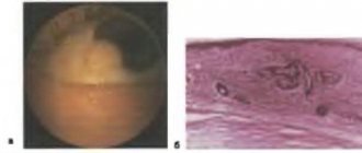
Tumor of the lacrimal gland: photo
- Cancer retina called retinoblastoma. At an early stage, a glow in the pupil can be seen in photographs of the patient. With such a lesion, strabismus progresses and severe pain appears. Retinoblastoma is most often diagnosed in children aged 1-1.5 years. Detection of this type of cancer at an early stage almost always (90%) means successful treatment and complete restoration of vision;
Eye cancer: photos in childrenRetinoblastoma: photo
- eyelid mass may be adenocarcinoma, basal cell carcinoma, or squamous cell carcinoma. The first is characterized by a growth in the thickness of the eyelid, with the second, metastases spread to the nearest lymph nodes, and the third is manifested by ulcers or the formation of nodes. Basal cell carcinoma occurs more often in people over 40 years of age. In this case, a lump appears at the bottom of the eye. This form of cancer often appears after a sunburn;
- Damage to the cornea of the eye is called melanoma. In this case, vision gradually begins to decrease, the shape of the pupil changes less frequently, and a dark spot (maybe in the plural) appears in the field of vision or on the iris;
- conjunctival lesions : papillomatous and pterygoid. In the first case, nodules of different sizes appear, and in the second, a whitish film with clearly outlined vessels;
Photo of conjunctival melanoma
- Meibomian gland cancer is called carcinoma. Most often the tumor appears in the upper part of the eye and is yellow in color. The use of drugs leads to an increase in education. Carcinoma has a high risk of recurrence (it develops extremely quickly).
Among eye tumors, the fastest growing is sarcoma . It quickly destroys the optic nerve and the mobility of the eyeball. This causes severe pain.
Treatment and prognosis
The disease is treated surgically. On the operating table, the lacrimal sac is opened and a biopsy is taken for emergency histological analysis. Benign tumors are completely excised and subsequent dacryocystorhinostomy is performed.
If a malignant process is detected, the lacrimal sac is also removed, after which several courses of radiation therapy are carried out.
Benign neoplasms of the lacrimal sac have a favorable prognosis for recovery. The prognosis for cancerous tumors is questionable due to their possible spread to nearby tissues and the appearance of distant metastases.
Causes
Diseases of the lacrimal gland can be congenital or acquired, resulting from any inflammatory diseases, tumors or injuries:
- Acute inflammation of the lacrimal gland (acute dacryoadenitis) most often develops against the background of mumps (mumps) in children, and less often - another infectious disease (influenza, pneumonia, scarlet fever, typhoid fever, etc.).
- Chronic dacryoadenitis occurs with certain blood diseases, syphilis, and tuberculosis.
- Sjögren's syndrome (“sicca syndrome”), characterized by damage to the lacrimal and salivary glands, is autoimmune in nature and can be observed in systemic connective tissue diseases.
- Congenital diseases include hypoplasia (underdevelopment), aplasia (absence) and hypertrophy (increase in size) of the lacrimal gland.
Diseases of the lacrimal ducts can be congenital (developmental abnormalities) or acquired, associated with nerve damage, infectious diseases, tumors, inflammatory diseases of the eyes (conjunctivitis, etc.) and nose; autoimmune disease (Sjögren's syndrome). Foreign bodies (for example, eyelashes) can also cause disruption of the outflow of tears.
Canaliculitis (inflammation of the lacrimal duct) most often has a fungal nature, but sometimes develops as a result of the introduction of a foreign body. It can also complicate the course of chronic conjunctivitis.
Dacryocystitis (inflammation of the lacrimal sac) usually occurs when there are disturbances in the outflow of tears, which are caused by narrowing and fusion of the nasolacrimal duct (congenital or acquired, for example, as a result of inflammation). In this case, the tear fluid stagnates in the lacrimal sac, and conditions are created for the development of infection in the lacrimal sac. Since the disruption of tear outflow is permanent, dacryocystitis often becomes chronic.
Dacryocystitis (inflammation of the lacrimal sac) can be provoked by: injuries, diseases of the nose and paranasal sinuses, decreased immunity, diabetes mellitus, occupational hazards, sharp fluctuations in ambient temperature, etc. An important cause of the development of dacryocystitis are pathological processes in the nasal cavity and paranasal sinuses. Sometimes the cause of obstruction of the nasolacrimal duct is damage due to trauma, often surgical (during puncture of the maxillary sinus, maxillary sinus).