Most people experience pathological changes in the lens over the years. They are also called age-related. Although it is difficult to avoid disorders, this does not mean that they do not need to be treated. Many people don't pay attention to their eyes. In this age of technological advancement, when phones and computers make up a large part of life, the eyes suffer the most. One of the most unpleasant vision pathologies is phacosclerosis - age-related hardening of the lens nucleus.
Causes
The lens is a biconvex lens of a transparent color, it is elastic, and begins to turn yellow over time. This lens is capable of changing structure and shape depending on the percentage of room illumination.
The main cause of the development of phacosclerosis is considered to be age-related changes in the visual analyzer. More than 70% of patients have this pathology.
Scientists have concluded that phacosclerosis can be caused by several other factors, including:
- hereditary predisposition;
- senile cataract;
- nuclear opacification;
- glaucoma;
- iridocyclitis;
- diabetes;
- myopia of the last stage;
- corneal ulcer;
- angiomatosis or retinal hamartoma.
Content:
- 1 Cataracts
- 2 The main drugs used in the treatment of cataracts
Description
The lens
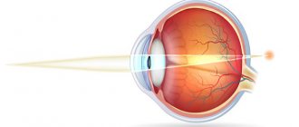
is part of the optical system of the eye and is involved in the act of accommodation. It has the appearance of a biconvex lens, the refractive power of the lens averages about 20 diopters, in a state of accommodation it increases significantly and can reach 30-33 diopters. The lens is located inside the eyeball between the iris and the vitreous body in the frontal plane. Together with the iris, it makes up the iridolens diaphragm, which divides the eyeball into anterior and posterior sections.
Distinguish between the anterior and posterior surfaces of the lens
; the line of transition of the front surface to the back is called the equator. The center of the anterior surface of the lens is the anterior pole, the center of the posterior surface is the posterior pole. The line connecting both poles is called the axis of the lens.
Radius of curvature of the anterior surface of the lens
at rest, accommodation is 10 mm, posterior - 6 mm. The length of the lens axis is usually 3.6 mm. A narrow gap separating the posterior surface of the lens from the vitreous body forms the retrolenticular (retrolenticular) space. The lens is held in the eye by the ligament of cinnamon, which consists of thin fibers. They attach to it at the equator. The other ends of the ligament of cinnamon are attached to the processes of the ciliary body.
The lens is covered by a membrane called the lens capsule
. This is a transparent elastic membrane. The part of the capsule covering the anterior surface of the lens is called the anterior capsule; covering the posterior surface - the posterior capsule. The thickness of the anterior capsule is 11-15 microns, the posterior one - 4-5 microns. Under the anterior capsule is a single-layer cuboidal epithelium, which extends to the equator of the lens, where its cells acquire an elongated shape.
The equatorial zone of the anterior capsule of the lens is a growth zone (germentative zone), since throughout a person’s life, young lens fibers are formed from its epithelial cells.
Lens fibers located in the same plane are connected to each other by an adhesive and form radial plates. The soldered ends of the fibers of adjacent plates form seams on the anterior and posterior surfaces of the lens. Connecting with each other, these seams form the so-called lens star
.
The outer layers of the lens substance adjacent to the lens capsule form its cortex (subcapsular layers), and the deeper layers form the nuclear zone of the lens
.
An anatomical feature of the lens is the absence of nerve fibers
n, blood and lymphatic vessels. The lens consists of a protein substrate and water. Water accounts for about 65%, proteins - about 35%.
In a normal lens there are
nucleoprotein, mucoprotein, compounds of potassium, sodium, calcium, phosphorus, sulfur, chlorine, magnesium, traces of copper, iron, manganese, boron and zinc. The tripeptide glutathione and ascorbic acid actively participate in redox processes. The lens also contains lipids, many vitamins (A, B1, B2, PP) and other necessary substances for proper metabolism.
Metabolism in the lens occurs slowly through diffusion and osmosis.
. In this case, the lens capsule plays the role of a semi-permeable biological membrane. All substances necessary for the normal functioning of the lens come from the intraocular fluid that washes the lens.
Throughout a person’s life, the size, shape, consistency and transparency of the lens change. In a newborn it is almost spherical in shape, soft in consistency, transparent and colorless. In an adult, the lens has the shape of a biconvex lens.
with a flatter front surface. It acquires a yellowish tint while maintaining transparency. The intensity of the yellowish tint increases with age.
At the age of 40-45 years, the nucleus of the lens becomes dense, the lens loses its elasticity. By this time, accommodation weakens and presbyopia occurs.
Around the age of 60, the ability to accommodate is almost completely lost.
. This is due to severe sclerosis of the lens nucleus - phacosclerosis. During this period, due to age-related changes - deterioration and slowdown of metabolism and tissue respiration - cloudiness of varying sizes and severity may appear in different layers. Initial opacities can be detected with maximum pupil dilation with short-acting mydriatics and slit-lamp examination.
Research methods.
Clinical methods for examining the lens consist of examining it under lateral illumination using a 20 diopter lens and examining it in transmitted light. Significant information is obtained from biomicroscopy of the eye, which makes it possible to establish the position and shape of the lens, identify the initial, smallest opacities and determine their localization. Valuable data can be obtained by echobiometry of the lens with ultrasound.
Symptoms
The compaction of the lens fibers is difficult for the average person to notice. The clinical picture is similar to sicca syndrome, conjunctivitis and some other conditions at an early stage of development. To confirm the disease, not only the medical history is important, but also the results of diagnostic tests.
With their help, a final diagnosis is made. The following signs indicate the development of phacosclerosis:
- degradation of nerve fibers due to the death of nerve endings and subsequent muscle atrophy;
- increased eye fatigue, especially after watching TV and reading;
- dry mucous membranes or lacrimation;
- pressing pain;
- increased sensitivity to light;
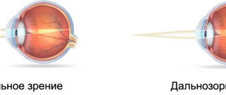
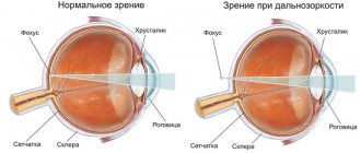
- hypermetropia;
- small details are difficult to see, the picture shows blurry spots.
Due to poor vision, pain is felt in the temples and radiates to the frontal part. As the disease progresses, the patient loses the ability to distinguish colors; when reading, letters float before the eyes.
Phakosclerosis itself does not cause deterioration in visual acuity. This symptom is characteristic of other ophthalmological problems that caused its development.
Diagnostics
To confirm the disease, a full examination of the ocular structures is carried out; it is important to study the condition of the biconvex lens.
Research methods:
- Ultrasound echobiometry involves the use of ultrasonic waves. The procedure lasts up to 30 minutes. The study is carried out with a special sensor, the patient's eyes must be open. Echobiometry is not performed in case of arterial thrombosis, inflammatory processes, foreign objects in the eye and alcohol intoxication. Echobiometry determines the depth of the anterior chamber of the eye, the length of the eyeball and the thickness of the biconvex lens.
- Biomicroscopy is a non-contact way to study the visual analyzer. A beam of light is directed into a person's eye using a special device, its angle and intensity are adjusted. The person is in a motionless state. Sometimes drops are used to dilate the pupil; they allow a more detailed examination of the fundus of the eye.
- View using a side lens. A 20 diopter lens is used. Thanks to this test, the doctor is able to determine the shape of the lens and the stage of deformation.
The results obtained are sent to an ophthalmologist; after studying them, the doctor will make a final diagnosis and prescribe appropriate treatment.
Hardening of the lens with age
Another feature of the eye is the hardening of the lens. According to research, in a fourteen-year-old teenager, the core is just beginning to harden and changes 450 times over time. In some cases, 1000 times hardening occurs. To determine the degree of hardening, a mechanical analyzer is used, which allows one to study different points and parts of the eye. This is how the stiffness of the eyeball is measured.
In parallel with the nucleus, the cortical parts of the eye also harden. They change 20 times during life. Until the age of 30, the cortical parts are normally harder than the nucleus of the eye, but by the age of 35 the parameters are equal.
After 35 years, hardening of the core predominates. The reasons for these phenomena have not yet been studied, but there are several opinions. Some scientists argue that the hardness of the lens increases due to the compaction of its constituent substances. However, such a process is always accompanied by dehydration of the eye, but this does not happen with phacosclerosis. With age, the amount of fluid in the lens does not change.
Treatment
Medicines are not used to treat the disease if the cause is age-related changes. Correction is carried out using glasses, which are selected individually depending on the optical power of the eyes.
Medicines intended to treat cataracts or glaucoma are also not prescribed. Medicines do not have any effect on the course of phacosclerosis; they do not eliminate it. Even drops intended to improve metabolism in the eyes will not help restore tissue respiration, since the lens has already hardened and this process is irreversible.
Vitamin drops will not help improve the condition of the disease, as well as drops for the treatment of other diseases.
Since the causes of the disease remain unknown, scientists have not found methods to combat the pathology.
Medicines are used if the cause of a disease that can lead to deterioration of visual perception is identified. Such pathological conditions include cataracts, glaucoma, and iridocyclitis. In this case, medications are prescribed to eliminate the cause and prevent the progression of the pathology.
The lens remains compacted; drops, ointments and medications for internal use will not affect it in any way. It is important here to prevent the development of diseases in order to maintain visual acuity and not go blind.
Since there are no effective methods to combat phacosclerosis, preventive measures are taken to reduce the rate of hardening of the lens and the manifestation of signs of other diseases.
How to recognize phacosclerosis
In order to take timely measures to alleviate phacosclerosis (usually the selection of corrective glasses), you need to note all minor symptoms. You should not hesitate to visit an ophthalmologist if you experience discomfort in the eyes due to overexertion and working at the computer. The first sign of phacosclerosis is visual strain when focusing on objects that are close.
With a timely visit to an ophthalmologist, it is possible to prescribe prophylaxis in a timely manner to slow down the development of pathology. Phakosclerosis can develop against the background of other pathologies, which do not always directly indicate ophthalmological abnormalities.
Complications
Hardening of the lens of the eye can lead to the development of certain diseases. The onset of biconvex lens sclerosis causes the following complications:
- farsightedness (hypermetropia);
- reduction of farsightedness;
- clouding of the lens of the eye;
- deterioration of metabolism in the tissues of the visual analyzer;
- imaginary myopia.
To prevent the disease from leading to complications, you must go to the hospital in a timely manner and follow doctors’ recommendations, do not self-medicate and lead a healthy lifestyle.
Prevention
The disease is a consequence of other processes in the body, that is, it does not develop independently. Therefore, preventive measures are aimed at maintaining its elasticity and preventing other diseases.
By following a few simple measures, you can prevent the development of phacosclerosis:
- Complete nutrition. This is important not only for adults, but also for children. A balanced diet can prevent many eye diseases. With food, a person receives vitamins, macro and microelements. It is useful to eat blueberries and carrots.
- The products do not contain enough vitamins. You have to eat too many carrots and other fruits to get enough of them. It is recommended to take vitamin supplements. Twice a year they take dietary supplements in accordance with the instructions and after consulting an ophthalmologist. Vitamins help the retina function normally and respond to light. They support the functionality of the lens.
- Timely treatment of cataracts, glaucoma, iridocyclitis and other visual disorders. These diseases lead to the development of phacosclerosis; to prevent it, it is important to cure them.
- Sufficient physical activity, eye exercises and mental exercise strengthen the visual analyzer. This prevents the development of ophthalmic problems.
Trauma does not cause phacosclerosis, but it leads to refractive errors - myopia and farsightedness. These, in turn, lead to hardening of the lens. That is, it is necessary to protect your eyes from injury and wear safety glasses.
Traditional methods
There are also traditional therapeutic methods that help maintain healthy eyes. Patients are advised to consume as many blueberries, carrots, honey and other products as possible, which have been used by healers since ancient times to improve the quality of vision. At any time, such a recommendation as taking care of the required level of the general condition of the human body is relevant.
With proper support, it will be able to attract all its resources and help the lens maintain its elasticity until old age. You also need to monitor how much sugar is in the blood, be sure to record the slightest changes in the condition of your eyes, try to avoid injuries and regularly visit an ophthalmologist.