Causes
The root causes leading to the formation of turbidity are:
- mechanical injuries to the organs of vision;
- violations of hygienic standards for the care of the visual apparatus;
- inflammatory reactions on the surface of the cornea;
- complications due to viral pathologies;
- due to herpes infection;
- conjunctivitis;
- working with chemically aggressive substances;
- surgical procedures;
- lack of vitamin A;
- incorrect use of contact products;
- infection with pathogenic microflora;
- genetic predisposition;
- destructive age-related changes.
Reasons why pupillary opacity appears in humans according to ICD-10 code
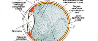
diagram of the structure of the eye
The following factors can affect the development of corneal opacities:
- Damage to the cornea. First, its surface layer is damaged, and then bacterial microflora joins, which corrodes the cornea and leads to the formation of ulcers. After some time, they turn into a thorn.
- Viral infection. Just viral conjunctivitis can cause the development of corneal clouding. But only if there is an inflammatory process in the cornea.
- Operations. The inside of the cornea is lined with endothelial cells. Their recovery after damage may not occur. With age, the number of these cells decreases. Surgery may result in an initial deficiency of endothelial cells decompressing the cornea. Due to this process, diffuse corneal dystrophy and complete clouding occur.
You can find out what eye phrd is here.
The doctor will be able to prescribe treatment only after the underlying cause of the disease has been eliminated. Otherwise, the relief that comes will be temporary and complications will arise after some time.
Classification
The cataract is classified according to its size characteristics and severity of turbidity. There are 5 categories of eyesores:
- 1st category. Avascular formations with a diameter of up to 6 mm. The lesion occupies a small area. The cloudiness is located in the center. The contours are not clear. Often the formation is transparent and does not cause a decrease in visual acuity. Diagnosis of the development of pathology is possible only through an ophthalmological examination.
- 2nd category. Avascular opacification with a diameter of more than 6 mm. Single formations of synechiae are observed. The pressure inside the eye cavity is normal. There is no decrease in picture quality.
- Category 3. Vascular cataracts with varying intensity. Characterized by different diameters. Synechiae are completely absent or present in isolated cases.
- Category 4. Belma with varying intensity. The category includes avascular and vascular opacities. The formations are flattened. As a result of the disease, corneal ectasia occurs. Synechiae develop on the anterior chamber and lens. This group includes cataracts that occur with aphakia, partial growth on the cornea or conjunctiva of the eyeball.
- Category 5. It includes leukomas, which have complications in the form of glaucoma. The formations occupy more than half of the cornea. Buphthalmos, fistula and staphyloma are also present.
According to genetic predisposition and the nature of provoking factors, the following forms of pathology are distinguished:
- congenital;
- acquired.
Depending on the degree of damage and the form of formation, 3 types of pathology are distinguished:
- cloud;
- spot;
- a real thorn.
Based on the scale of the cataract damage, there are:
- partial;
- subtotal;
- total forms.
Depending on the presence of a vascular network in the formation:
- without vascularization;
- with vascularization.
Types of turbidity
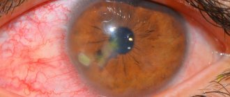
clouding of the cornea due to cataracts
Today, doctors distinguish 3 types of disease, differing in size and intensity:
- Little cloud. These are opacities that resemble a gray smoky spot. They are difficult to see and their area is limited. If the clouds are concentrated in the center of the cornea, they can affect visual acuity. The disease night blindness occurs after 40 years of age.
- Stains. They form in the center or on the periphery of the cornea. They are distinguished by intensity, durability, and have a significant effect on vision. A spot formed in a child can lead to the development of strabismus.
- Belma. These are persistent darkenings that occupy most of the cornea. They are formed as a result of scar changes. The thorn looks like frosted glass, with germination of blood vessels. If it is located in the center of the cornea, then vision is reduced to light perception. The thorn, called convex leukoma, can be congenital in nature, as it was obtained as a result of intrauterine inflammation. But most often it is an acquired pathology that develops against the background of injury or infection.
You can find out what to do when there is pain and burning in the eye here.
Most often, the disease is diagnosed in people over 55 years of age, which can result in complete blindness.
Symptoms
The main symptoms of the disease are:
- the appearance of grayish areas on the membrane of the cornea;
- hyperemia of the eyeball;
- photophobia;
- decreased level of vision;
- intense secretion of tear secretion;
- feeling of the presence of foreign particles.
If two symptoms are present simultaneously, consultation with an ophthalmologist is necessary to prevent the progression of the disease and its complications.
Diagnostic methods
If there are cloudy areas on the cornea and if the patient complains about visual acuity, it is impossible to immediately make an unambiguous diagnosis of ocular leukoma. At a time when there were no instrumental methods for diagnosing this disease, many doctors mistook cataracts and cataracts for the same disease. To clarify the diagnosis of ocular leukoma, the following studies must be performed:
- ophthalmoscopy (determines the level of transparency of the cloudiness and the reaction of the visual organ to light);
- biomicroscopy (determines the cause of cloudy eyes, the location of the spot and its edges).
Treatment
Therapy to cure the disease is based on the stage of the lesion and its extent. Several areas of treatment are used:
Drug therapy
Prescription and administration of medications is carried out only at the initial stage of the disease. The ophthalmologist prescribes:
- Hormone-based drugs. Thanks to medications, the inflammatory reaction is eliminated and the severity of symptoms is reduced.
- Ethylmorphine hydrochloride and potassium iodide injections. Additionally, it is possible to prescribe medications based on mercury and proteolytic enzymes. Biogenic stimulants are injected simultaneously into the skin.
- Vasodilator medications are prescribed to increase local blood circulation.
- Physiotherapeutic procedures are carried out with hormonal or natural components. Electrophoresis or phonophoresis is used. Among the natural ingredients used is aloe vera.
Surgical intervention
Cataracts of especially large sizes require immediate surgical intervention. To eliminate the disease, laser correction or keratoplasty is used. During the manipulation, the affected area of the cornea is replaced.
Cosmetic correction
In case of contraindications to the manipulation, cosmetic removal of a pronounced cataract is carried out. To do this, the patient wears a contact lens. Since it is impossible to cure a severe course of the disease without manipulation.
Folk remedies
Among the traditional methods, treatment of the disease is carried out using recipes:
- Honey and onion juice. To obtain a solution, pour the juice of the onion with a glass of hot boiling water. Dissolve 1 tablespoon of honey in the resulting mixture. To reduce the intensity of symptoms, 1 or 2 drops are instilled into the eyes.
- Rye bread. Condensation obtained from freshly baked baked goods treats the thorn. A hole is made in the bread into which a glass is placed. Condensation will appear on the walls of the container. After collecting the liquid, it is applied to the membranes of the eyes.
- Eyebright. Pour boiling water over a tablespoon of the herb and leave to steep for about 2 or 3 hours. Lotions are applied with the solution 2 to 3 times a day. If there is irritation after instillation, you can dilute the mixture with boiling water.
Folk remedies to help
As an adjuvant therapy to the main treatment, many patients resort to traditional methods. Such treatment is not a panacea and cannot act as a basis, but it still helps to slow down the progression of the disease and improve the general condition of the patient. Let's look at a few effective recipes:
- Recipe No. 1
You will need 10 g of onion pulp, 100 ml of hot milk. Mix, set for 8 hours, filter. From the prepared solution, make a lotion on the sore eye or instill 1 drop 2 times a day.
- Recipe No. 2
Fir resin is instilled 1 drop into the affected eye. There may be a slight burning sensation afterwards, which is normal.
- Recipe No. 3
To prepare, you need to combine honey and grain 50:1, stir. Apply the prepared drug to the affected area 2 times a day.
- Recipe No. 4
You need to pour boiling water over 30 g of dill seeds, put on low heat for 20 minutes, then cool, strain and instill 3-4 drops into the conjunctiva or use for rinsing.
These are not all the recipes that traditional medicine offers. However, ophthalmologists are quite skeptical about such treatment and believe that it can do more harm than good. That is why if a person decides to carry out treatment using traditional methods, it is necessary to consult with the attending physician. Self-medication can cause irreversible harm to health.
Video: What to do if the cornea of the eye is damaged
Prevention
Preventive measures to prevent the development of pathology are:
- protection of the organs of vision from ultraviolet radiation;
- prevention of mechanical eye injuries;
- eliminating the influence of harmful factors on the visual apparatus;
- when working at a monitor for a long time, perform eye exercises;
- balance the diet;
- maintain the body's immunological strength;
- follow the rules of hygiene procedures;
- take breaks from prolonged work in front of a monitor or with small particles.
Eyesore (corneal clouding)
A few decades ago, it was much more common to see a person with a noticeable eyesore than it is today. This fact alone should inspire serious and justified optimism, clearly indicating the significant successes of ophthalmology in eliminating this optical and cosmetic defect.
Useful video
When a cataract appears, it is necessary to consult a doctor, since in advanced stages the pathology is quite difficult to cure. Surgical intervention does not always give the desired effect. Sometimes it is possible to eliminate the manifestations of a cataract only cosmetically using a special lens.
Author's rating
Author of the article
Alexandrova O.M.
Articles written
2031
about the author
Was the article helpful?
Rate the material on a five-point scale!
( 1 ratings, average: 5.00 out of 5)
If you have any questions or want to share your opinion or experience, write a comment below.