Moles, or nevi on the eyeball, are quite rare. Depending on the location, nevus of the conjunctiva is distinguished - in the visible area of the eye, nevus of the choroid - in the fundus. The reason for their formation is the same as on the skin of the body - excessive accumulation of melanin in the cells. Isn't this dangerous for your eyesight?
By its nature, a nevus is a benign formation and is not dangerous. The phenomenon is truly rare: according to statistics, only 1% of people on Earth live with moles on their eyes. A nevus does not affect the quality of a person’s vision, even if it is located very close to the pupil. However, any such formation is fraught with hidden danger and, under certain factors, can develop into a malignant tumor. Let's tell you more about the two types of moles on the eyes.
What's happened
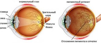
The choroid itself is a choroid that runs under the sclera towards the back of the eye. Its function is to ensure stable intraocular pressure, normalize the nutrition of the rods and cones of the retina.
The choroid consists of several layers that contain pigment cells. The formation of a nevus occurs from the cells of the supravascular outer plate; further development promotes penetration into deeper tissue layers, which leads to the manifestation of unpleasant symptoms.
The formation of the disease begins at birth, but it has no characteristic signs and is therefore not detected. Pigmented coloration appears in adolescents during puberty (from 11 to 14 years).
On this topic
- Organs of vision
How to get rid of wen on the eyelid
- Natalya Gennadievna Butsyk
- February 27, 2021
Subsequent accumulation of pigment is observed in people of mature age, therefore, after the age of 30, the color of the pathology acquires a brighter shade. However, such changes are observed only in 70-80% of patients; in the rest, coloring is not acquired.
The provoking factors have not been fully studied; the male and female half of the population suffer in equal numbers. More often, the pathology affects only one eye.
Useful video
Causes of birthmarks on the choroid of the eye. Effective treatment of this type of nevus.
Author's rating
Author of the article
Alexandrova O.M.
Articles written
2031
about the author
Was the article helpful?
Rate the material on a five-point scale!
( 2 ratings, average: 3.00 out of 5)
If you have any questions or want to share your opinion or experience, write a comment below.
Forms
There are stationary and progressive forms of pathology. Sometimes divided into the following types:
- Atypical - no pigment and halo-nevus with cancerous symptoms are present.
- A typical one is a flat, oval-shaped neoplasm with no signs of growth.
- Suspicious - the manifestation of pigment is observed.
To determine the stage of the disease, an expanded classification is used.
Stationary
A flat or slightly bulging rounded neoplasm measuring 1-6 mm. It has a gray or greenish-gray pigment color, the color is uniform.
The stationary form of the nevus does not impair vision; the formation has clear boundaries, only in some situations the edges are slightly blurred. During the inspection, it is possible to determine the accumulation of metabolic products.
Progressive
Determined by the ability to increase in size. During growth, the tumor loses clear boundaries, a change in the shape of the tumor and uniform distribution of pigment are observed.
Dystrophic disorders and compression of choroidal vessels are observed in the retina. The retina often detaches.
The pathology provokes visual impairment, the appearance of cloudy spots, and image distortion. This form has an unfavorable prognosis because it is prone to degeneration into cancer. Various complications often arise.
Depending on the location, several types are distinguished.
Retinal nevus
It is benign in nature, the lesion is located on the posterior choroid, and has no specific signs. The characteristic features are the flat shape of the formation, clear boundaries and retention of size over time.
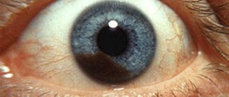
Nevus of the iris
The color of the tumor depends on the amount of melanin contained in the lining of the iris.
Nevus of the lacrimal caruncle
It is usually formed in an adult.
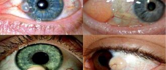
Conjunctival nevus
It is diagnosed in only 5% of cases. Due to its location, it is clearly distinguishable against the background of the eyeball, which facilitates diagnosis. Here, nevus growths, pigmented and cystic nevus are distinguished.
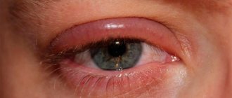
Periocular nevus
It is often observed in medical practice. Moles located on the eyelids and regional integument are easily diagnosed. The color of the new growths varies from light to dark brown.
Atypical nevi are characterized by the absence of pigment; they are located in the area of atrophy of the pale vascular membranes. Histology indicates the presence of a cancerous tumor.
The meaning of moles on the eye
Some people believe that any pigmented growth on the body has a symbolic meaning, and moles in the eye are no exception. There is an opinion that their location can tell a lot about the fate and character of a person. Those who believe in this will be interested to know what the appearance of a nevus in the eye means.
It is believed that people with such a new formation are endowed with wisdom and developed intelligence; they learn easily and do it with pleasure. Usually, owners of moles in the eye strive for self-improvement and constant development, but if this is not done, over the years, memory and thinking abilities will become less developed.
Also, the presence of a pigmented spot on the conjunctiva usually means that its owner is a good family man. He is able to treat his soul mate with reverence and love, never thinking about betrayal. Other family members also play an important role in his life, he quickly and firmly becomes attached to them and is always ready to help, regardless of the situation.
If we talk about character, then the owners of moles in the eye are usually soft, kind and non-conflicting. Such people are always ready to come to the aid of their neighbors free of charge, which some may even take advantage of. People with a pigmented spot in the eye usually do not cause trouble; in childhood it is easy for parents to raise them, and in adulthood they are non-conflict subordinates or wise, fair bosses.
Causes
The choroid is located under the sclera of the choroid, which stabilizes eye pressure and nourishes the retina. Since there are no nerve endings, there are no unpleasant pain sensations; they appear only when deeper layers are affected.
On this topic
- Organs of vision
What symptoms can be used to recognize optic nerve glioma?
- Natalya Gennadievna Butsyk
- September 3, 2021
The reason for the development of nevi lies in the increased concentration of melanin; this condition can be provoked by the following factors:
- Disturbances at the hormonal level.
- Infectious processes.
- Severe stressful situations.
- Inflammatory reactions.
- Genetic predisposition.
- Use of hormonal contraceptives .
People with fair skin and hair are more prone to the appearance of nevus.
Is it possible to avoid the appearance
To avoid the appearance of a birthmark on the eye, you should strictly adhere to some simple rules:
- When exposed to direct sunlight, it is recommended to use sunscreen.
- Do not go to the beach between 10 am and 5 pm. At such times, ultraviolet radiation is extremely dangerous, especially for people with light skin tones.
- To protect your eyes from the negative effects of ultraviolet rays, it is recommended to wear dark glasses.
- If a nevus appears on the eye, self-medication is prohibited. Such an intervention can cause a number of negative complications for the eye and its functions.
- When the first signs appear, you should immediately seek help from an ophthalmologist.
If an eye nevus appears: what it is and what measures to take can only be explained by a qualified doctor. Ophthalmologists recommend undergoing an examination every six months and diagnosing the eyeball.
Symptoms
The development of a stationary nevus does not reveal itself by any symptoms and is not detected during a visual examination; only an ophthalmologist can detect it.
As the pathology progresses, a number of signs appear:
- Deterioration of vision.
- Feeling of a foreign body in the eye area.
- Reducing the boundaries of the visual field.
Most often, a choroidal nevus is located behind the widest area of the eye, less often observed in the part of the equator.
On this topic
- Organs of vision
All about ocular sarcoidosis
- Natalya Gennadievna Butsyk
- September 3, 2021
Visually it can be determined by the following indicators:
- Flat shape or has a slight convexity.
- Size from 0.1 to 0.6 mm.
- The boundaries are clearly defined, only sometimes blurred in places.
- color .
- The retina is not subject to changes.
- On the surface, drusen - layers of waste products of retinal cells.
Progressive pathology is determined by the following symptoms:
- Increase in size.
- Edges lose definition.
- Squeezing of cherioid vessels.
- Formation of a yellow halo around the tumor.
- change .
If any of these manifestations are detected, it is necessary to contact a specialist in order to prevent any irreversible changes.
be careful
The presence of papillomas, warts, condylomas, moles and spines on the body is the first sign of malignant melanoma!
We hasten to warn you that most medications “treat” warts, papillomas, moles, etc. - this is a complete deception of marketers who make hundreds of percentage points on drugs whose effectiveness is zero. They do not cure the disease, but only mask the symptoms.
The pharmacy mafia makes huge money by deceiving sick people.
But what to do? How to treat if there is deception everywhere? Doctor of Medical Sciences Anatoly Makhson conducted his own investigation and found a way out of this situation. In this article, the Doctor also told how to 100% protect yourself from melanoma, for only 149 rubles! Read the article in the official source following the link.
Diagnostics
The initial diagnosis involves taking a medical history. If hemorrhages occur on the mucous membrane, a sensation of the presence of a foreign body, or a cosmetic defect, the doctor prescribes additional measures:
- Examination of the mucous membrane.
- examination using colored lenses. Using a red lens makes it possible to clearly determine the presence of a nevus. When replaced with green, the boundaries of the formation disappear; only with progressive pathology can some violations be identified.
- Color duplex study - assesses the state of general blood flow.
- Fluorescein angiography - makes it possible to identify the development of pathology at the initial stages; the presence of retinal angioma is determined. As the disease progresses, effusion of fluorescein is observed.
- Echography - indicates the exact location of the lesion.
- Ophthalmoscopy – assesses the condition of the retina and nerve fiber using a light beam.
- Ultrasound examination - the condition of the deeper layers of the eyeball is determined.
Each diagnostic measure makes it possible to determine all aspects of the pathology and select the correct therapy.
Types and states
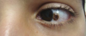
On the mucous membrane of the eye, moles are always visible - on the inner or outer part of the white. And for some reason they never appear on the mucous membrane of the eyelids.
They are divided into three types:
● non-pigmented, otherwise called vascular. Only a slightly pinkish spot is visible on the squirrel;
● cystoid, consisting of small cysts that, under a microscope, resemble a honeycomb;
● pigmented, usually yellowish to light brown, rarely black.
All these types of birthmarks can be either in a calm state, that is, frozen in their development, or in an activated state - growing, changing color.
Treatment
The need for treatment and the use of therapeutic measures directly depends on the type of pathology.
If a typical form is diagnosed, then therapy is not prescribed, but the patient should regularly visit an ophthalmologist for timely changes in the clinical course of the disease. The examination must be carried out at least once every six months.
If the growth of a neoplasm is detected, the specialist prescribes adequate measures, for this purpose the exact location and size of the pathology is clarified.
Laser treatment
It is used for hard-to-reach tumor localization. This measure is less traumatic compared to others. If urgent measures are required, microsurgery should be performed.
Electroexcision
It is carried out using an electric scalpel. The method is characterized by excision of the nevus, eliminating all defects that formed at the location of the lesion. At the same time, the patient undergoes plastic surgery for defects of the cornea and conjunctiva.
Surgery
The use of surgery is necessary for large pathologies.
Surgical therapy is necessary when malignant tumors are detected, as well as in cases where visual acuity deteriorates. Surgical methods are considered safe, and the rehabilitation period is relatively painless.
In childhood, therapeutic measures are prescribed when the pathological process progresses rapidly. The use of traditional medicine recipes often remains ineffective, so resorting to their help is not recommended.
Kissed by the sun
There is such a beautiful legend. Day after day, the son of the Sun God Ray secretly watched a beautiful girl who came to the source to draw water. He was about to open his heart to her, but then God called his son to him. In parting, Ray touched the girl’s cheek and left his mark on her - a mole. He flew away to his father, and at this time the daughter of the Wind God, Cloud, who wanted to get the young man as her groom, persuaded his younger brothers to fly around the earth and, fooling around, kiss all the women they met.
Possible complications
In the absence of adequate therapy, various types of complications can occur:
- detachment .
- Compression of the choroidal vessels, which leads to deterioration in the nutrition of rods and cones.
- Visual impairment .
- Degeneration into a cancerous tumor.
Pigmented nevus causes distortion of the visual image, the formation of “cloudy spots” in the visual field.
The most dangerous complication is considered to be degeneration into a malignant formation. In this case, the doctor observes a large dark spot under the conjunctiva; it is possible that this is a cancerous formation. Initially, it can be confused with hemorrhage or nevus of the conjunctiva of the eye.
Degeneration into cancer occurs in approximately 1% of all patients. Choroidal melanoma develops from a benign tumor. Metastases occur in a small percentage of cases.
The only method is to remove the affected eye. It is for this reason that in order to identify pathology at the initial stage of development, it is very important to undergo a timely examination by an ophthalmologist.
Locations
Birthmarks form in different areas of the eye:
- external and internal areas of the protein;
- tearful month;
- semilunar fold;
- iris or retina;
- limbo.
Location of conjunctival nevi
An ocular nevus forms on the inner and outer parts of the conjunctiva. Birthmarks are detected on the inner corner of the eye, the periphery of the cornea, the lacrimal month, and the semilunar fold. Sometimes neoplasms appear on the inside of the eyelid. Although moles are located near the pupil, they do not block the view or impair vision .
Localization of choroidal nevi
Tumors appear:
- on the back of the eyeball;
- on the fundus;
- at the ocular equator.
The spots are invisible to others; they are discovered by a doctor who performs diagnostics using specialized equipment. But an ophthalmologist is not always able to identify a nevus of the choroid of the eye. Diagnosis is difficult if the tumor lacks pigment.