Dry eyes are a symptom that most of us have experienced. This condition is also known as dry eye syndrome or xerophthalmia. Dry eyes occur when the lacrimal gland does not produce enough fluid. As a result, the composition of the tear film on the surface of the cornea is disrupted, its properties change, causing serious discomfort in a person: the illusion of “sand in the eyes,” pain, burning, a feeling of “tightness,” inflammation and decreased vision.
Previously, it was believed that dry eyes were a disease that primarily affected older people. However, recently dry eye syndrome has become significantly younger. Young people who spend a lot of time at the computer complain about dry eyes. Also, the problem is often encountered by those who use contact vision correction, especially in conditions of increased dry air or strong wind.
The structure of the tear film
Content:
- The structure of the tear film
- Causes of decreased tear production
- Reasons for decreased tear film stability
- Symptoms of xerophthalmia
- How is dry eye diagnosed?
- Drug treatment of xerophthalmia
- Surgical treatment of dry eye syndrome
- Treatment of complications of xerophthalmia
- Folk remedies for treating xerophthalmia
- Prevention of xerophthalmia
- Xerophthalmia and contact lenses
- What causes dry eyes?
The essence of xerophthalmia is insufficient hydration of the surface of the eye with the help of the tear film, which is formed by the tear fluid. Due to the fact that the film becomes less dense and its composition changes, the cornea of the eye dries out. The result is a feeling of dryness and irritation. The eyes begin to react painfully to dry air, wind, and cigarette smoke. There can be many reasons for disruption of normal eye moisture.
Recall that the tear film consists of three layers. The main one is the middle one, which consists of 98% water and also includes salts and proteins. This layer is called aqueous and it is, in fact, what we call tears that appear in the eyes when crying or laughing. The lacrimal glands, which are located at the outer edge of the eye, create a watery layer. They constantly secrete small amounts of tear fluid. When blinking, this fluid is distributed over the entire surface of the eye.
At the edges of the eyelids there are small meibomian glands that secrete fats (lipids). They form the outer layer of the tear film. Lipids prevent moisture from evaporating from the aqueous layer, acting as a natural protective barrier. In addition, it is thanks to them that the surface of the tear film becomes smooth and light rays, when passing through it, are refracted at the correct angle.
Finally, the thin transparent membrane of the conjunctiva, which covers the inner surface of the eyelid and the sclera of the eye, secretes a mucous substance that forms the so-called mucin layer. It helps smooth out all the micro-irregularities on the surface of the cornea and helps the tear fluid to be evenly distributed over the surface of the eye. After the tear fluid washes the surface of the organs of vision, through the lacrimal canaliculi it enters the lacrimal sac, and then into the nasal cavity.
There are many factors that lead to dry eyes. Traditionally, they are divided into two groups depending on the pathogenetic mechanism that “triggers” the development of “dry eye syndrome”. The first group are pathological conditions associated with a decrease in tear production or secretion, and the second are factors that lead to a decrease in the stability of the precorneal tear film.
Dry eye syndrome as a special case of dystrophic processes (AMD) and its friend - the computer
The number of diagnoses of dry eye syndrome around the world is growing along with the development of technology. The main factors: deterioration of atmospheric air, in particular near industrial zones, air-conditioned air, side effects of many medications, radiation from LEDs (especially those that are super bright in the blue part of the spectrum, including AMOLED screens and the like). Signs of the disease: itching, redness, fatigue, dryness in the eyes and eyelids. The reason is a violation of tear production.
If you identify the syndrome at an early stage, when a regular clinic does not yet diagnose it, you can avoid a whole sea of problems. Let's start with a short table to help you figure out whether this applies to you or is worth passing on.
What to pay attention to
Now we will talk about subjective assessment methods, that is, those that need to be confirmed by analyzes or other special tests. But, if you have a couple of these complaints, read on. No - just skip the post, you're in luck.
- Burning or itching in the eyes.
- Feeling of dryness in the eyes.
- Redness of the eyes.
- Feeling of a foreign body in the eyes.
- Feeling of eye fatigue, especially when working at a computer.
- Swelling and redness in the eyelid area.
- Unstable, fluctuating vision.
- Spontaneous lacrimation.
- Photophobia.
- Increased sensitivity to tobacco smoke.
If you are aged 45 years or older, or have rheumatoid diseases, thyroid diseases, previous herpes, mononucleosis, trigeminal nerve surgery, eye injuries and eye surgeries, or with frequent use of beta blockers, analgesics, anticholinergics, antidepressants, oral contraceptives, estrogens, anti-migraine drugs, the presence of botulinum toxin injections for cosmetic purposes in the paraorbital area, wearing soft contact lenses, swimming in chlorinated water, the risk increases, and complaints must be monitored especially carefully.
But let's move on to a slightly more scientific description of the syndrome.
About terms
The symptom
is a subjective feeling of dryness (objectively it may not exist).
The sign
is an objective decrease in fluid secretion (subjectively it may not be felt).
Syndrome
- symptoms and signs (up to blepharospasm)
Disease
- a clinical picture in which the most important manifestation is the syndrome. Typical situations: menopause, autoimmune exocrinopathy, vitamin A deficiency.
In Ancient Greece, the term “xerophthalmia”, that is, “dry eye”, was first introduced. But then they called it corneal blindness, associated with complete drying of the surface of the eye. In the nineteenth century, the meaning of the term shifted to the professional designation of Sjögren's syndrome and a number of keratitis. Only half a century ago the term took on its current meaning: von Rött named quantitative criteria for tear deficiency. The diagnosis is now divided into water deficiency, mucodeficiency and lipodeficiency, depending on which component of the tear is deficient.
Degrees of dry eye syndrome
The degree is not the stage of development of the disease, but the level of severity and possible consequences. For example, as a result of injury or chemical exposure, you can start immediately from the severe stage and quite quickly reach the terminal stage. But it is better, of course, to avoid this.
- Subclinical degree - no obvious symptoms; the patient sometimes complains of a feeling of dryness when using a hairdryer, in dry air, in strong winds or when wearing contact lenses for a long time, or when working at a computer for a long time.
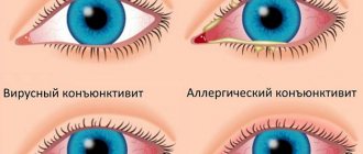
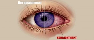
- Mild - symptoms are pronounced, itching also appears, less often - photophobia, even less often - fogging, very rarely - slight blepharospasm. A diagnosis of infectious or allergic conjunctivitis is often made.
- Moderate - all symptoms + new signs (erosion, punctate keratopathy, filamentous keratopathy, conjunctival hyperemia). At an average level, all changes are reversible.
- Severe - corneal ulcers, cataracts, corneal neovascularization, adhesions and other changes that are irreversible are added.
- Terminal - significant visual impairment is added due to obstruction of the optical field (for example, an ulcer). The end of this stage is the complete loss of the ability to distinguish objects with the eye.
Diagnostics
Any ophthalmology office can take your medical history, look at your eyes through a microscope, and do tear production tests.
The latter are especially important in diagnosing the early stages of dry eye disease, which is why they are included in most proper routine eye examinations. If the doctor has suspicions, he can also prescribe special diagnostic methods (vital staining with dyes; tear crystallography; cytocompression study; ultrasound examination of the lacrimal gland; laboratory tests of the gland), which require quite specific equipment in the laboratory or diagnostic center.
As a rule, most cases of dry eye syndrome are diagnosed by impaired tear production or associated changes in the eye, in particular blepharitis.
Association with inflammation of the eyelids (blepharitis)
The peculiarity is that patients complain of a symptom, and usually there is a picture of the disease.
But at the same time, diagnostics quite often does not identify signs and does not prescribe therapy. There is even an anecdote about this in ophthalmology: - What to do with a patient with blepharitis? - Send him to a competitor!
So, dry eye syndrome and blepharitis are two related problems. If dry eye disease was not caused by blepharitis, it can cause inflammation of the eyelid. If dry eye syndrome was caused by blepharitis, in any case, treatment of one requires treatment of the other. The main point is that you can replace tears for as long as you like, trying to combat dry eye syndrome with supportive treatment, but this will not give any results. It is necessary to cure blepharitis (or prevent it from developing in a scenario where it was not the reason for the initiation of the DES). Plus solve a number of accompanying problems related to deterioration of trophism. If the doctor prescribes only a tear substitute or a similar remedy, at first the patient simply endures it, and then the development of the syndrome begins with concomitant irreversible organic disorders of the tear-producing apparatus and superficial structures of the eye, up to ulceration of the cornea.
Hence two rules:
- If you have dry eyes, you need to get it diagnosed, and not wait for it to go away.
- If you were diagnosed with dry eye syndrome and only tear replacement was prescribed, but the exact reasons that caused it were not identified, you need to look for them and treat them. And already start preventing problems that will develop during the course of dry eye syndrome.
Here is a sample cause and effect diagram.
Treatment
Treatment is carried out not only by protecting the eye from fluid loss, but also, most importantly, by removing the cause of the disease.
More on this later. Be careful for now. It is better to be examined earlier (the test for tear production takes 10 minutes) than to join those for whose sake we are launching our bioreactors to produce gel. A little later I will talk about the tear and how it works and works.
Causes of decreased tear production
This group includes several reasons: disruptions in the functioning of the immune system (Sjögren's syndrome, transplant rejection, complications after radiotherapy of the head and neck); diseases of the hematopoietic system (malignant tumors, anemia, etc.); endocrine disorders (hypothyroidism, diabetes, menopause), infectious diseases (HIV, cholera, tuberculosis, etc.), dermatological problems (neurodermatitis, dermatitis, etc.).
Autoimmune diseases
The essence of autoimmune diseases is that there is a failure in the process of recognition by cells of the immune system of body tissues, which begin to be perceived as foreign. As a result, an abnormal immune response develops, the target of which becomes healthy tissues and organs.
The most common condition associated with xerophthalmia is Sjögren's syndrome or disease. It can be either primary or secondary. Primary Sjögren's syndrome is manifested by autoimmune damage to the salivary and lacrimal glands, and in the secondary syndrome, which manifests itself several years after the onset of the disease, problems with connective tissue arise (systemic lupus erythematosus, primary biliary cirrhosis, etc.).
Sometimes dry eye syndrome develops as a complication after radiation therapy of the neck and head due to the fact that the lacrimal gland tissue is also irradiated. Its structure undergoes changes, and the immune system begins to attack it as foreign.
In addition, an autoimmune response may develop after corneal transplantation from a donor.
Diseases of the hematopoietic system
Scientists have been able to establish that there is a connection between a decrease in the functions of the lacrimal gland and a number of diseases of the hematopoietic system. In particular, tear production is reduced in hemolytic anemia, lymphoma, lymphosarcoma, lymphocytic leukemia and other diseases.
Endocrine disorders
The purpose of the endocrine system is to maintain the stability of the internal environment of the body through the release of hormones into the blood. A “breakdown” in the operation of this system always causes disruptions in the functioning of a number of organs.
Dry eyes can occur with diabetes, hypothyroidism, and also in women during menopause. Thus, in diabetes mellitus the secretion of the lacrimal glands decreases. Insufficient secretion of tear fluid leads to a feeling of discomfort and “sand” in the eyes.
Hypothyroidism is insufficient production of thyroid hormones. It is accompanied by a decrease in the secretion of the exocrine glands: lacrimal and salivary glands. This is why patients with hypothyroidism often complain of dry eyes.
Finally, menopausal syndrome occurs in women after the body stops producing estrogen and progesterone. It is often accompanied by mood swings, insomnia, general malaise, “jumping” blood pressure, as well as impaired secretion of the lacrimal glands.
Infectious diseases
Infectious diseases are usually accompanied by a syndrome of general intoxication. As a rule, the body temperature rises and dehydration may begin. In this case, the reserves of the lacrimal gland are depleted and its secretion is reduced.
Dermatological diseases
Xerophthalmia is a symptom accompanying a number of skin diseases. These are congenital ichthyosis, herpetic dermatitis, neurodermatitis, etc.
Congenital ichthyosis is manifested by thickening of the skin, which begins to peel off in plates resembling fish scales. In addition to changes in skin texture, itching of the eyes and impaired secretion of the lacrimal glands are noted.
Neurodermatitis is a disease of an allergic-immunological nature that affects the mucous membranes and skin. In addition, the functioning of the autonomic nervous system, which takes part in the functioning of the lacrimal glands, is disrupted. As a result, the secretion of tear fluid is significantly reduced.
Herpetic dermatitis is damage to the skin and mucous membranes by the herpes simplex virus. If herpes blisters are localized in the eye area, they can spread to the conjunctiva and lacrimal gland.
Symptoms of “dry eyes” in diabetes: the absence of complaints does not mean that everything is fine
Diabetes is tricky. The disease can progress, affecting more and more new systems, and symptoms are scanty or absent altogether. In this respect, diabetes mellitus and dry eye syndrome are similar. Patients notice lacrimation, dryness, pain, “sand” and pain; they do not tolerate windy weather or air-conditioned air, and complain of pain and pain when instilling regular drops. But these complaints may not exist. The longer the patient is sick, the weaker the symptoms, and the situation progressively worsens, up to ulcers and scars on the cornea.
Reasons for decreased tear film stability
A decrease in the thickness of the tear film and a decrease in its stability can be caused by a number of reasons: both related to a person’s lifestyle, and caused by disturbances in the functioning of internal organs and body systems.
Corneal scars
Often, as a result of the presence of foreign bodies in the eye or as a result of corneal tuberosity associated with the presence of postoperative scars, the tear film becomes thinner and ruptures, resulting in xerophthalmia.
Neuroparalytic keratitis
This is an inflammation of the cornea, which leads to its sensitivity being reduced. In a normal state, a person blinks, wetting the eye when he feels irritation of the cornea due to the thinning of the tear film. If the cornea loses sensitivity, the patient's brain does not receive the signal to blink, and the surface of the eye remains dry for a long time. As a result, inflammation, clouding of the cornea and a significant decrease in vision develops.
Incomplete closure of eyelids
Sometimes the eyelids simply do not match the size of the eye. This condition is called lagophthalmos and can be either congenital or appear after surgical intervention. In addition, there is also exophthalmos - protrusion of the eyeballs, which also provokes incomplete closure of the eyelids. This condition is often observed in patients with hyperthyroidism, and can also be a consequence of injury or a tumor process.
In patients suffering from incomplete closure of the eyelids, even during sleep, part of the cornea remains open, which provokes the development of xerophthalmia.
Allergy
An allergy is a sharp reaction of the immune system to the body’s contact with any substance. Allergens most often include plant pollen, citrus fruits, pet hair, dust mites, etc.
When an allergen gets on the mucous membrane of the nose or eyes, swelling develops. The patient complains of a feeling of “sand” in the eyes, discomfort, and the illusion of a foreign body on the mucous membrane.
Stagnation of tear fluid
In a normal state, the tear fluid, having been on the surface of the cornea and moisturizing it, when blinking, moves to the inner corner of the eye and enters the nasal cavity through the system of lacrimal canaliculi. If the functioning of these tubules malfunctions due to an inflammatory process, stagnation of tear fluid occurs and its chemical composition changes. Bacteria begin to multiply in it and dust particles settle, which irritate the mucous membrane, causing drying of the eyes.
Using air heaters
Under normal ambient temperature and humidity conditions, moisture evaporates from the surface of the eyes in approximately 10 seconds. After this, the person blinks, closing the eyelids and moistening the eyes with tear fluid. However, if the air temperature rises and its humidity decreases, moisture from the surface of the eyes begins to evaporate faster. Therefore, people often experience discomfort when they are in a room where an air heater or air conditioner is operating.
Working at a monitor for a long time
Scientists have proven that when a person works at a monitor, he blinks at least half as often. As a result, the cornea inevitably dries out and dry eye syndrome develops.
Using contact lenses
Contact correction involves placing lenses on the cornea that help improve visual acuity. Ideally, lenses should completely match the size of the cornea and follow its shape. However, contact correction agents often provoke passive irritation of the conjunctiva, causing drying of the eyes. This is fraught with the development of keratoconjunctivitis.
Using low-quality cosmetics
Unscrupulous manufacturers of low-price cosmetics often use substances that are similar in their characteristics to those used to produce expensive analogues, but differ from them in quality characteristics. As a rule, such cosmetics have a number of side effects. In particular, poor quality mascara, eyeliner and eye shadow can cause the development of contact dermatitis or conjunctivitis and provoke the development of “dry eye syndrome”.
Pregnancy
Often xerophthalmia develops in women in an “interesting position.” Why this happens is not yet completely clear to doctors. Changes in hormonal levels and increased temperature are considered one of the reasons.
Side effects of some medications
Sometimes medications can trigger xerophthalmia. A number of medications, when used for a long time, can cause a feeling of dryness of the surface of the eye or aggravate existing discomfort. These medications include diuretics, some antidepressants, oral contraceptives, antihistamines, beta blockers, peptic ulcer medications, etc.
Causes of redness in the corner of the eye
There are several types of reasons why this symptom appears:
- Mechanical irritation . for example, exposure to dust, dirt, aerosols, smoke, foreign substances, strong winds, exposure to excessively bright light (for example, welding), prolonged eye strain, injuries;
- Physiological reasons - expansion of the blood vessels of the eye, but without any disruption of its functioning; this can happen with fatigue, drinking alcohol, severe sneezing, physical exertion, eye irritation with contact lenses or glasses if they are incorrectly selected;
- Eye pathologies - can be inflammatory or non-inflammatory in nature;
- Pathological changes in the functioning of other organs - for example, allergic diseases, diabetes mellitus, intoxication with toxic substances, hypertension, etc.
Redness of the outer corner of the eye
It often looks similar to the consequences of mechanical impact (as if the eye was rubbed), and there may be peeling of the skin, sometimes with pain . Redness of the outer corner of the eye is less common than the inner corner, and the redness is more often localized on the skin of the eyelid. It can be caused by both an allergic reaction to cosmetics and diseases.
Diseases
- Angular conjunctivitis - affects the corners of the eyes, can be allergic and bacterial, accompanied by a feeling of dryness, a foreign body in the eye, the flow of tears, and sometimes purulent discharge . In this case, the skin may become covered with small cracks, and the pain intensifies while blinking.
- Ocular herpes - accompanied by swelling of the eyelid, pain, fear of light .
- Regional blepharitis - this also causes thickening of the upper eyelid, swelling, burning and itching, and crust formation .
This is interesting! There are several forms of blepharitis. In the scaly or seborrheic form, the disease is combined with dermatitis, causing loss of eyelashes and, in rare cases, eversion of the eyelid. The ulcerative form is determined by ulcers on the eyelash line, where scars form over time. Demodectic mange is caused by mites of the genus Demodex, living at the roots of the eyelashes, and allergic mange is usually combined with conjunctivitis.
Redness of the inner corner of the eye
This unpleasant phenomenon can be caused, in addition to those already mentioned, by a number of diseases.
Diseases
- Disruption of the tear duct, which is located closer to the inner corner, or its inflammation - canaliculitis, accompanied by redness of the eyelids, severe discomfort in the corners of the eyes . There are similar signs of obstruction of the tear ducts, with the addition of severe lacrimation.
- Dacryocystitis is an inflammation of the lacrimal sac, with pus secreted from the lacrimal openings and swelling of the skin .
- Ingrown hair - causes redness and pain due to the ingrowth of eyelash hairs under the skin . Unfortunately, it is impossible to cope with this trouble on your own; you won’t be able to see the hair to remove it, and you will have to contact a specialist.
Inflammation of the corners of the eyes in children
Children's eyes are more sensitive than those of adults; their redness begins suddenly and often has physiological causes . such as overexertion, crying or sneezing, exposure to dust, colds.
This is interesting! A common pathology in infants is blockage of the tear ducts; the fact is that in the eighth month of pregnancy, a septum forms inside the fetus between the tear ducts and the nasal cavity. With the first cry of the newborn, it ruptures, but this does not always happen, and in this case, excess fluid may accumulate inside the tear ducts. This is the so-called dacryocystitis of newborns.
With decreased immunity or allergies, children often experience conjunctivitis or blepharitis, but there is another serious disease that can lead to blindness - uveitis, or inflammation of the vascular membranes of the eyes.
Note! Uveitis is a very serious disease and requires hospital treatment.
Inflammation of the corners of the eyes in adults
In addition to the above-mentioned diseases, there are disorders caused by an unhealthy lifestyle. Modern adults tend to overload their eyes with computer work, resulting in the formation of diseases such as dry eye syndrome and computer vision syndrome, accompanied by eye pain . which makes it difficult to look at the screen of an electronic device.
Dry eye syndrome, in addition to unpleasant sensations in the corners of the eyes, is also accompanied by a strong reaction to light, up to the inability to be in the sun .
Photo 2: Sometimes pain in the corners of the eyes is caused by uncomfortable shaped glasses with improperly adjusted nose pads. Source: flickr (Benjamin Thorn).
Symptoms of xerophthalmia
Dry eyes are a problem that manifests itself with many symptoms.
- Feeling of discomfort. It may seem to the patient that there is some kind of foreign body in the eye (“sand” and “mote”). Irritation, burning, pain, and photophobia also occur.
- The person complains that from time to time the image seems to become fuzzy and blurry. Most often, the quality of vision decreases towards the end of the day.
- Photophobia. Bright light causes discomfort, lacrimation, and a burning sensation.
- Poor tolerance to wind, smoke, air flow from air conditioning, etc.
- Patients who resort to contact vision correction complain that the lenses suddenly become uncomfortable. They begin to be perceived as a foreign body and may even cause pain.
- “Sticking” and slow opening of the eyelids.
How is dry eye diagnosed?
An ophthalmologist usually makes a diagnosis of dry eyes based on existing symptoms. At the same time, given the fact that “dry eye syndrome” may indicate another disease (for example, Sjögren’s syndrome), the doctor may conduct a more detailed examination and medical history.
In order to confirm that a person really suffers from “dry eye syndrome,” a special study is carried out - the Schirmer test.
This test is also known as the Schirmer test.
The test is carried out as follows. The doctor takes a strip of filter paper measuring 5x50 mm, bends the end of it and places it behind the edge of the lower eyelid. The patient is asked to close his eyes. After this, the strip should absorb the precorneal tear film and fluid from the tear duct within five minutes. If the patient suffers from dry eyes, the amount of tears absorbed by the paper may be negligible or nonexistent. If the tear film is significantly thinned, the Schirmer test will show an extremely low result. In this case, the norm is to wet the paper by 10 mm in 5 minutes.
It is noteworthy that this test can be performed with or without local anesthesia. If the doctor suspects the patient has “dry eye syndrome,” the paper, in contact with the conjunctiva, can cause reflex lacrimation, which “masks” this problem. To prevent this possibility, an anesthetic is instilled into the eye a few minutes before the test, as a result of which reflex lacrimation is eliminated.
Another test method for diagnosing dry eye syndrome is the Norn test. A 0.2% fluorescein solution is instilled onto the upper area of the patient's eyelids. After this, the patient should blink once. Next, it is examined, recording the time between the opening of the eyelids and the rupture of the precorneal tear film. If it ruptures earlier than after 10 seconds, we can talk about the patient’s predisposition to dry eyes.
An ophthalmologist may also recommend laboratory tests that will help definitively identify the cause of dry eyes. Thus, cytology of a scraping or imprint of the conjunctiva, immunological study of tear fluid and its crystallography, which helps to identify the type of eye disease, can be performed.
The doctor may also insist on conducting highly focused studies. They will help identify diseases, one of the secondary symptoms of which is dry eyes. This may be a general blood and urine test, which will help identify anemia, inflammatory processes and kidney disease; determination of immune complexes circulating in the blood, an increase in the number of which is a sign of autoimmune diseases; carrying out rheumatic tests; determining the level of thyroid hormones to exclude hypothyroidism; blood sugar test to exclude diabetes; determination of antibodies to the herpes virus.
An ophthalmologist can also use instrumental research methods, which make it possible to study the structure and properties of tear fluid. So, thiascopy can be performed - studying the structure of the tear film, the density of the mucous, aqueous and lipid layers. Tear osmolarity, which is an important factor affecting the strength of the tear film, can also be examined.
Treatment for dry eyes can be either medication or surgery. Surgical intervention is used when medical methods do not bring results. As a rule, surgical treatment is aimed at correcting defects of the cornea or eyelids, as well as eliminating complications resulting from dry eye syndrome.
Drug treatment of xerophthalmia
Drug therapy is the first stage of treatment for xerophthalmia. Its duration depends on how effective the treatment is. During drug therapy, a number of drugs are used.
Artificial tears
To compensate for the deficiency of your own tear fluid, “artificial tear” preparations can be used. Their density and composition vary. If the disease is mild, the use of eye drops is recommended.
In the event that the form of the disease is moderate and severe, the drug should be on the cornea for a longer time. Therefore, it is recommended to use medications in the form of ointments or gels, for example, Oftagel.
Oftagel is a drug with carbomer in maximum concentration. It has a long-term moisturizing effect, eliminates lacrimation and does not require frequent instillation. Moreover, the gel can be used once at any time of the day, which eliminates the need to use drops several times a day. Oftagel is suitable for people with complaints of periodic eye dryness and irritation.
Anti-inflammatory drugs
Anti-inflammatory drugs are one of the most widely used groups of drugs in the treatment of eye pathologies. By stopping the inflammatory process, they at the same time prevent the development of “dry eye syndrome”. Many of these drugs are combined, combining anti-inflammatory and antibacterial effects.
Metabolic drugs
Metabolic drugs are usually ointments that should be placed behind the lower eyelid several times a day. These products help increase the content of pantothenic acid in the eye tissues, which takes part in metabolism, increases the regenerative properties of cells and helps normalize the production of tear fluid.
Antihistamines
Antiallergic drugs help stop an allergic reaction in the body, which can also manifest itself as discomfort and a feeling of “sand” in the eyes.
Antibiotics
Antibacterial drugs are used in ophthalmology quite often, because even if the inflammatory process is not bacterial in nature, there is always a risk of a bacterial infection. Most often, antibiotics are used in the form of topical ointments.
Eye problems with diabetes
If you have diabetes, you should visit your ophthalmologist regularly to avoid eye problems. High blood sugar (glucose) levels increase the risk of developing eye problems due to diabetes. In fact, diabetes is the leading cause of blindness in adults aged 20 to 74 years.
If you have eye problems or diabetes, you should not buy a new pair of glasses if you notice blurred vision. This may be a temporary eye problem that develops quickly due to high blood sugar levels.
High blood sugar levels in diabetes cause swelling of the lens, which changes your ability to see. To correct this type of visual impairment, you must return your blood sugar to the target level (90-130 milligrams per deciliter (mg/dL) before meals and less than 180 mg/dL one or two hours after meals or 5-7.2 mmol/L and 10mmol/l, respectively). From the time you begin to have good control over your blood sugar levels, it will take up to three months for your vision to fully recover.
Blurred vision due to diabetes may also be a symptom of a more serious eye problem. People who have diabetes can develop three main types of eye problems: cataracts, glaucoma and retinopathy.
Cataracts and diabetes mellitus
A cataract is a darkening or clouding of the eye's lens, which is normally clear. The lens allows us to see and focus on an image, like a camera. Although anyone can develop cataracts, people with diabetes may develop these eye problems at an earlier age and the condition progresses more quickly than people without diabetes.
If you have diabetes and have developed cataracts, your eye may not be able to focus on a light source and your vision will be impaired. Symptoms of this problem in diabetes include blurred or glare-free vision.
Treatment for cataracts usually involves surgical removal and then placement of a lens implant, and may require glasses or contact lenses to further correct vision.
Glaucoma and diabetes mellitus
When normal drainage of fluid inside the eye stops, it accumulates, increases pressure and develops another eye problem that can occur with diabetes called glaucoma. The pressure damages the nerves and blood vessels of the eye, causing vision changes.
In the most common form of glaucoma, there may be no symptoms until the disease becomes severe and significant vision loss occurs. In more rare cases of this eye problem, symptoms may include headaches, eye pain, blurred vision, watery eyes, glaucomatous halos around lights and vision loss.
Treatment for this diabetes eye problem may include special eye drops, laser treatments, medications, and surgery. You can avoid serious eye problems from diabetes by getting an annual glaucoma screening test from your ophthalmologist.
Diabetic retinopathy
The retina is a group of specialized cells that convert light passing through the lens into images. The ocular or optic nerve carries visual information to the brain.
Diabetic retinopathy is one of the vascular (blood vessel related) complications resulting from diabetes mellitus. This diabetic eye disease occurs due to damage to small vessels and is called microangiopathy. Kidney disease and nerve damage due to diabetes are also microangiopathies. Damage to large blood vessels (also called macroangiopathy) includes complications such as heart disease and stroke.
Numerous studies have proven the connection between microangiopathies and high blood sugar levels. You can reduce your risk of developing these eye problems by improving your blood sugar control.
Diabetic retinopathy is the leading cause of irreversible blindness in developed countries. The duration of diabetes is the single most important risk factor for developing retinopathy. The longer you have diabetes, the more likely you are to develop this serious eye problem. If retinopathy is not detected early or treated, it can lead to blindness.
People with type 1 diabetes rarely develop retinopathy before puberty. In adults with type 1 diabetes, retinopathy also rarely develops in the first five years of the disease. The risk of developing retinal damage increases as diabetes progresses. Intensive control of your blood sugar levels will reduce your risk of developing retinopathy. DCCT, a large study of people with type 1 diabetes, found that people with diabetes who achieved tight control of their blood sugar levels using an insulin pump or multiple daily insulin injections were 50-75% less likely to develop retinopathy. nephropathy (kidney disease) or nerve damage (these are all microangiopathies).
People with type 2 diabetes usually have signs of eye problems at the time of diagnosis. In this case, controlling blood sugar, blood pressure and cholesterol levels play an important role in slowing the progression of retinopathy and other eye problems.
Types of retinopathy in diabetes mellitus:
- Background retinopathy.
Sometimes there is damage to the blood vessels but no visual impairment. This is called background retinopathy. At this stage, you need to carefully monitor your diabetes to prevent underlying retinopathy from progressing into a more serious eye disease. - Maculopathy.
During the maculopathy stage, a person develops damage in a critical area called the macula. Because it occurs in an area that is critical to vision, vision can be significantly reduced with this type of eye problem.
- Proliferative retinopathy.
New blood vessels begin to grow on the back wall of the eye. Since retinopathy is a microangiopathic complication of diabetes mellitus (disease of small blood vessels), this type of retinopathy develops due to an increasing lack of oxygen in the affected vessels of the eyes. The vessels in the eyes become thinner and clogged and begin to remodel.
Next »
Surgical treatment of dry eye syndrome
Surgical intervention is resorted to in situations where drug therapy has exhausted its reserves and has proven insufficiently effective.
There are several types of surgical interventions that are used for dry eye syndrome.
Blockage of the tear ducts
To prevent tear fluid from accumulating in the arches of the eyelids, the lacrimal ducts may be blocked. As a result, when you blink, the cornea begins to be washed with tears more effectively. Most often, this surgical intervention involves blocking the lacrimal openings with special plugs, as well as their coagulation using a laser beam or an electric scalpel.
Reducing the area of tear fluid evaporation
Sometimes doctors resort to stitching the edges of the eyelids, as a result of which the palpebral fissure narrows. This helps reduce the area of tear fluid evaporation. As a rule, this intervention is carried out if simply blocking the lacrimal ducts does not help normalize the secretion of the lacrimal glands.
Implantation of additional lacrimal glands
An extremely complex, but at the same time effective method of treating xerophthalmia is the transplantation of additional mucous glands. They are implanted from the patient’s mouth into the soft tissue of the appendages of the organs of vision. The effectiveness of this surgical intervention depends mainly on the qualifications of the surgeon.
Folk remedies for treating xerophthalmia
For the treatment of “dry eye syndrome”, some remedies from the arsenal of traditional medicine have proven themselves well. However, it should be remembered that such methods cannot increase the secretion of tear fluid and are not able to heal organic eye defects. The remedies offered by traditional medicine have an antiseptic and metabolic effect on the epithelium of the eye, relieving symptoms and discomfort. Therefore, they can be used in parallel, but not instead of traditional drug treatment.
The most popular remedy to help get rid of the feeling of “sand” in the eyes is rinsing with decoctions of sage, chamomile and calendula. They are prepared in a water bath at the rate of one tablespoon of raw material per glass of boiling water.
You can also prepare a tincture of sheepsfoot. To do this, mix one tablespoon of herb with a glass of water and bring the mixture to a boil. The broth is filtered and made into eye baths.
Honey drops have proven themselves to be quite good. They are prepared by mixing a teaspoon of honey (only natural honey can be used) with 500 ml of distilled water. The honey should completely dissolve. The product is used twice a day, one drop in each eye. Store the prepared composition in the refrigerator.
Prevention of xerophthalmia
As you know, preventing a disease is always much easier than treating it later. Xerophthalmia is no exception. Statistics show that there are a number of factors that contribute to the development of this problem. Thus, the eyes dry out with increased visual tension, prolonged work at the computer and reading; as a result of low air humidity; in conditions of high ambient temperature; in the presence of constant directed air flows, for example, from a fan or air conditioner; due to irritating factors (toxins, allergens, dust).
In order not to disturb the balance between the secretion of tear fluid and its evaporation, you should limit the time you spend at the computer, and also take breaks regularly, once every thirty minutes, during which you close your eyes.
If there is an air conditioner or fan in the room, the air flow should not be directed towards the face.
When working in high temperature conditions, you should use moisturizing drops - “artificial tears”.
Xerophthalmia and contact lenses
Best materials of the month
- Coronaviruses: SARS-CoV-2 (COVID-19)
- Antibiotics for the prevention and treatment of COVID-19: how effective are they?
- The most common "office" diseases
- Does vodka kill coronavirus?
- How to stay alive on our roads?
Dry eyes are one of the most common reasons for not using contact vision correction. Most often, when using lenses, patients complain of discomfort towards the end of the day, as well as when working at a monitor.
If the use of contact lenses is associated with constant discomfort, you should discuss with your doctor the possibility of switching to another type of contact correction or changing the solution used to care for the lenses. In addition, the use of moisturizing drops may be recommended to reduce unpleasant symptoms.
Please note that many eye drops cannot be used while wearing contact lenses. As a rule, existing restrictions are indicated in the instructions for the medicinal product. Also, do not use eye ointments while using lenses.
Preventing dry eyes
If you have initial symptoms of dry eyes, you should do the following for prevention:
Blink more often
When using a computer, smartphone or other digital device, we tend to blink less often. As a result, dry eyes may develop. Make a conscious effort to blink frequently when using these devices. In addition, you can periodically close your eyelids completely and close your eyes to renew the tear film.
Take frequent breaks while using the computer
Make it a habit to look away from your screen every 20 minutes and look at something that is at least within your reach for at least 20 seconds.
Following this simple rule will not only prevent the development of dry eyes but also preserve your vision.
Avoid aggressive environmental factors
A person with dry eyes should avoid anything that may worsen these symptoms. For example, being in an excessively hot room, in the wind. Smoking causes the development of dry eye.
Tears evaporate faster in a hot room, like any other liquid. You can take steps to prevent evaporation. In winter, during the heating season, be sure to use a humidifier.
www.vseozrenii.ru